
Abstract
Background:
Each year, more than 800,000 vascular and cardiac surgeries are performed therefore, there is a great need for suitable material for bioprosthetic operations. Porcine pericardium is a double-walled sac that covers the heart and can be used in vascular and cardiac thoracic surgery.
Objective:
The aim of the present study was to evaluate the decellularization process and biomechanical properties in porcine pericardial tissue after the decellularization treatment.
Methods:
A detergent based protocol was used for the decellularization of porcine pericardium. Histological analysis and contact cytotoxicity assay were performed. Additionally, biomechanical testing and in vivo biocompatibility by implantation into Wistar Rats were performed.
Results:
The histological analysis showed the preservation of the extracellular matrix, without any observable cellular remnants. No toxic effects were noticed when contact cytotoxicity assay performed. The decellularized tissues, after implantation in Wistar Rats, remained for up to 12 weeks without being rejected. Finally, the biomechanical testing showed no significant differences between native and decellularized tissues.
Conclusion:
In this study, the decellularization of the porcine pericardium produced a non toxic scaffold, free of any cellular remnants, thus serving as an alternative material for tissue engineering applications including heart valve and vascular patch development.
Keywords
Introduction
The decellularization of porcine pericardium and its potential use as a natural scaffold is a promising approach in the field of tissue engineering and regenerative medicine. Porcine pericardium can be used in tissue engineering, as a biomaterial for the construction of heart valves, vascular grafts and patches [1]. It is known, that pericardial tissue acts as a protective barrier of the mammalian heart, containing pericardial fluid, contributing to the physiological function of the heart. The native structure of porcine pericardium consists of three layers: (1) the serosa which is the inner thin layer, (2) the fibrosa, which is the thicker layer and (3) the epipericardial connective tissue layer, an outer layer that is partly continuous with the pericardiosternal ligaments [1–4]. Its extracellular matrix (ECM) contains collagen type I, arranged with various structures, such as fibrils, fibers, fiber bundles and laminates. Also it consists of elastic fibers and glycosaminoglycans (GAGs) like dermatan sulfate, chondroitin sulfate and hyaluronan [1–6].
Worldwide over 300,000 heart valve replacements and over 570,000 arterial bypasses are performed, therefore there is a great need for developing bioprosthetic scaffolds [7]. The availability to obtain pericardial tissue from animals, attracted the attention of cardiac surgeons. Cross-linked bovine pericardium with glutaraldehyde is commonly used in vascular surgery as patch for longitudinal arteriotomy [5]. Furthermore, it is used in cardiac and thoracic surgery as a material for repairing intracardiac and diaphragmatic defects and also for the development of bioprosthetic heart valves [8]. Despite its advantages, two major limitations have been observed including degeneration and calcification when implanted in the human body. In this way, a more suitable pericardial material is needed for the applications of tissue engineering [9–11]. Decellularization has been proven that can reduce calcific degeneration and provide a cellular free material [12,13]. Also, previous studies have shown that decellularized pericardial tissues had lower antigenicity and the three dimensional structure was preserved adequately [5,10].
The decellularization of native tissues has been proven effective however, there is little information on tissue mechanics and matrix composition after decellularization treatment in porcine pericardium [9–11]. In the present study we evaluated the impact of decellularization treatment on ECM structure and the biomechanical properties of porcine pericardium. Porcine pericardium was decellularized according to a previously described detergent based protocol [14]. The current protocol is composed with the zwitterionic detergent 3-[(3-cholamidopropyl)dimethylammonio]-1-propanesulfonate (CHAPS) and the anionic detergent sodium dodecyl sulfate (SDS) [15]. It is proven that SDS has toxic effects in the remaining ECM after the decellularization. In order to assess the toxic effects of SDS in decellularized pericardial tissues, we performed cytotoxicity assay. Also the in vivo stability of the decellularized tissues were tested, through implantation experiments in Wistar Rats. Our results showed that decellularized porcine pericardium might provide an alternative material for tissue engineering applications and its potential use in humans.
Methods
Tissue procurement
Porcine pericardia were obtained from Clinical, Experimental Surgery, and Translation Research of Bioacademy Research Foundation Academy of Athens (BRFAA), under sterile conditions, rinsed with distilled water to remove blood and body fluids and dissected to remove the external fat.
Pericardium decellularization
According to decellularization protocol, the pericardial tissues (
Histological analysis
Segments of non decellularized (
Contact cytotoxicity assay
Decellularized pericardial tissues were cut into 1 × 1 cm patches and attached to the center of the wells of a six-well culture plate (Orange Scientific, Belgium). Human mesenchymal stem cells passage 3 (hMSCs-P3) from the Wharton Jelly were seeded into each well at a density of
Implantation of porcine pericardial tissues in Wistar Rats
To evaluate the efficiency of decellularization, decellularized pericardial tissues (
Biomechanical testing of pericardium
The purpose of mechanical testing was to investigate the stress–strain behavior of porcine pericardium before and after decellularization process. Before testing, the pericardia were visualized under polarized light to identify the direction of collagen bundles. Pericardial tissues strips (1.5 × 0.2 mm) were dissected based on collagen fibers orientation in horizontally and in vertically way. The strips were mounted under zero strain onto a purpose-built holder and subjected to uniaxial tensile testing. All testing was conducted at room temperature (23°C), and during testing, the samples remained hydrated by continuously being sprayed with PBS 1× (Gibco, Thermo Fischer Scientific, USA). During testing, load data from the load cell and specimen compression data from the stroke of the cross-head of the testing machine were acquired at a rate of 20 Hz. To obtain an accurate measure of the tissue gauge length, the testing machine was set to produce a specimen preloading of 0.01 N before the operating program started to acquire any data. Therefore, zero compression was taken at the point where a load of 0.01 N was detected. The final gauge length (Lo) of the specimen was calculated as the initial gauge length minus the compression required to produce the specified preloading. Failure was taken to occur when the first decrease in load was detected during compression. The recorded load (F, in Newtons) and specimen compression (
Statistical analysis
The stress–strain curves for each specimen were analyzed and used to obtain the following biomechanical parameters: (a) Elastic phase slope (E1-E), (b) Collagen phase slope (Coll-E), (c) Transition stress (
Results
Morphological analysis of decellularized pericardial tissue
Histological analysis showed that decellularized pericardial tissues retained the ECM components without any evidence of cellular and nuclear material (H&E, Masson’s, Trichrome, Elastic van Gieson and Toluidine Blue). Staining of pericardial tissues with Masson’s Trichrome and elastin van Gieson indicated that the collagenous and elastic components of the extracellular matrix were well preserved following the decellularization treatment. The GAGs of the decellularized pericardial tissues were preserved as indicated by Toluidine blue staining (Fig. 1).
Contact cytotoxicity assay
Microscopic images showed that hMSCs P3 were expanded and being attached with the decellularized pericardial tissues in 6-well plates after 24 h and 48 h of incubation (Fig. 2). No sign of cell cytotoxicity has been observed, thus exhibiting the same morphology as the negative control. When hMSCs P3 were cultured with cyanoacrylate glue, no evidence of cells was found.
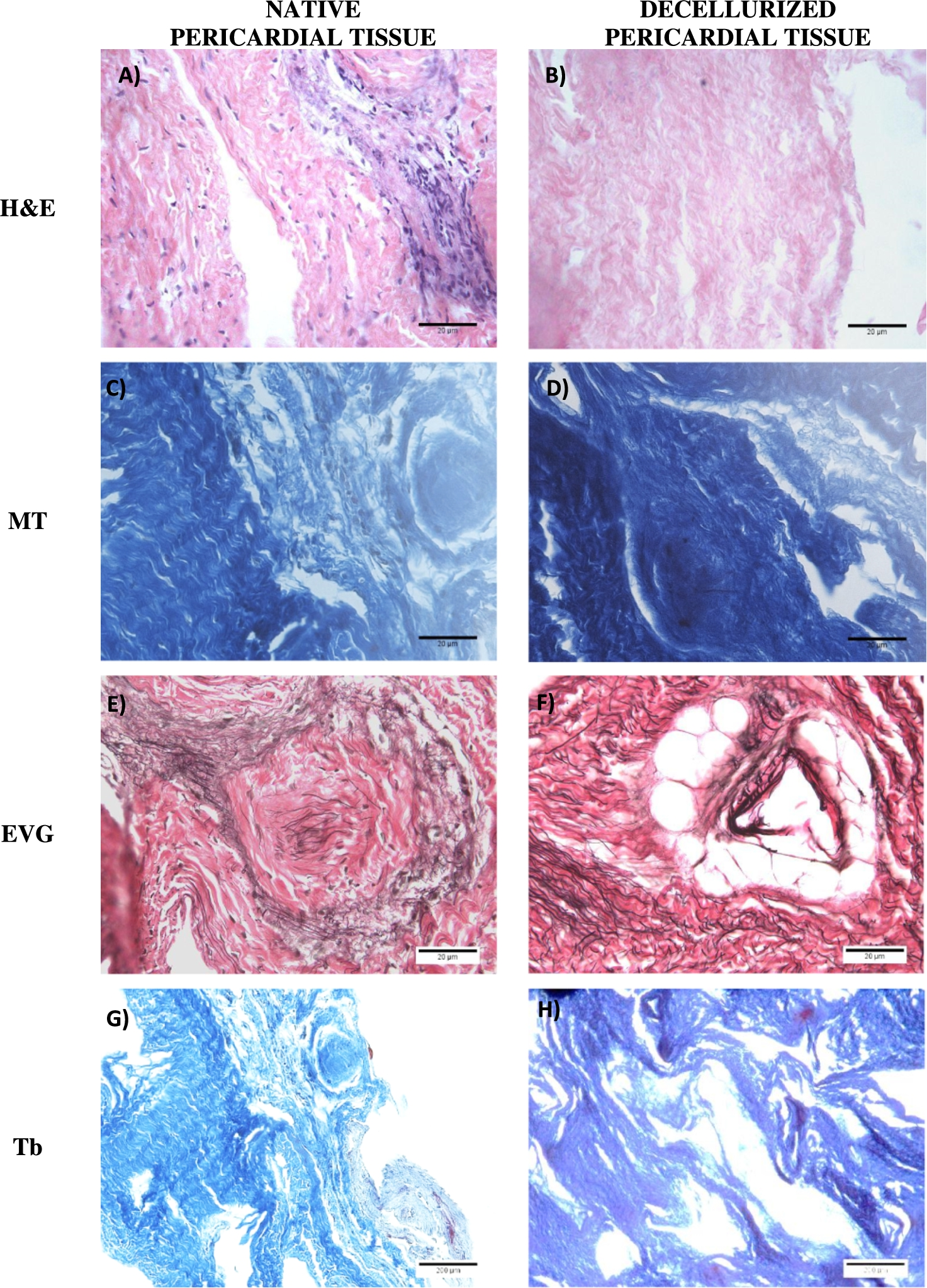
Native and Decellularized Pericardial Tissue stained with H&E, Masson’s Trichrome, Elastic Van Gieson and Toluidine blue. Native Pericardial Tissue with H&E (A), Masson’s Trichrome (C), Elastic Van Gieson (E) and Toluidine blue (G) stainings. Decellularized Pericardial Tissue stained with H&E (B), Masson’s Trichrome (D), Elastic Van Gieson (F) and Toluidine blue (H). Original Magnification 40×, scale bars 20 μm.

Contact Cytotoxicity assay of decellularized porcine pericardial tissue seeded with hMSCs. (A) Decellularized pericardial tissue with hMSCs following 24 h incubation. (B) Decellularized pericardial tissue with hMSCs following 48 h incubation. (C) Cyanocrylate glue with hMSCs P3 (positive control) and (D) hMSCs-P3 alone (negative control). Original magnification 10×, scale bars 100 μm.
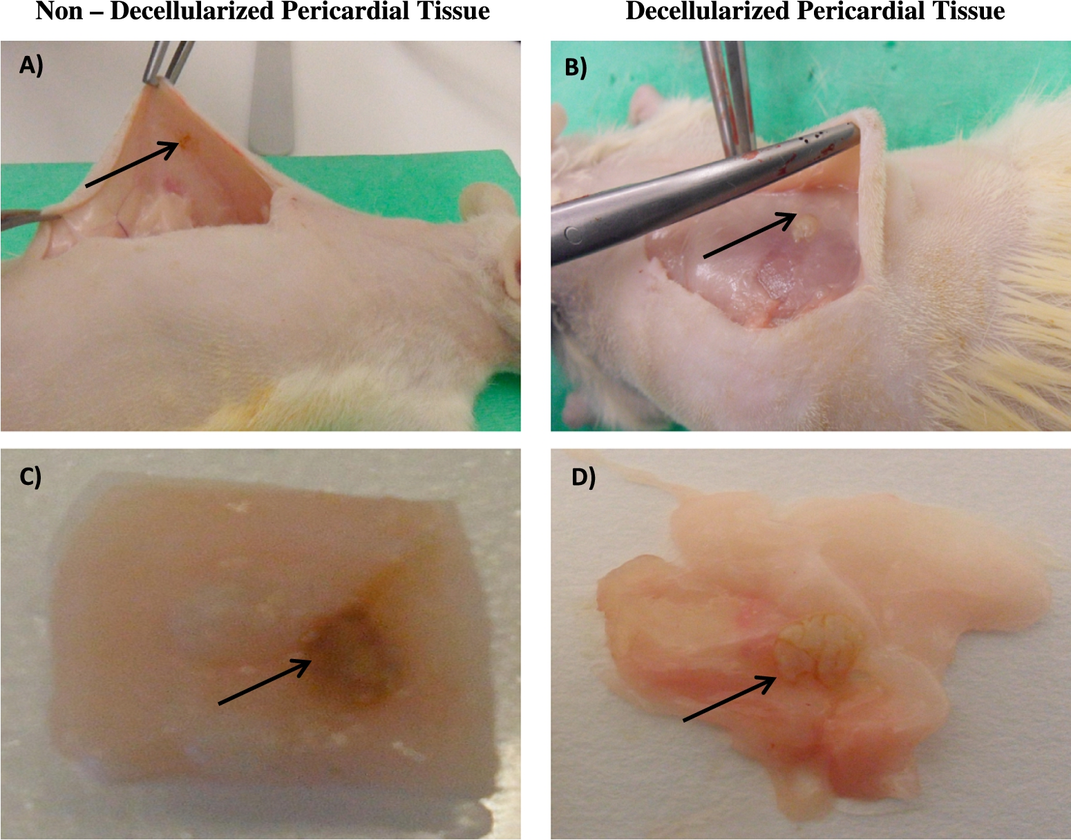
Examination of non-decellularized and decellularized pericardial tissue into Wistar Rats after 12 weeks of implantation. (A) Non Decellularized pericardial tissue implanted in the right scapular region of Wistar Rats. The arrow indicated the graft of non decellularized pericardial tissue, that was implanted in Wistar Rats. (B) Decellularized pericardial tissue implanted in the right scapular region of Wistar Rats. The arrow indicated the graft of the decellularized pericardial tissue, that was implanted in Wistar Rats. (C) Non Decellularized pericardial tissue after excision from Wistar Rats. Extensive necrotic tissue was observed in the graft of non decellularized pericardial tissue after 12 weeks of implantation. (D) Decellularized pericardial tissue after excision from Wistar Rats. No necrotic tissue was observed in decellularized pericardial tissue after 12 weeks of implantation.

Histological analysis of non-decellularized and decellularized grafts following 12 weeks postoperatively. (A) Non-decellularized pericardial tissue, (B) decellularized pericardial tissue stained with H&E, original magnification 20×, scale bars 100 μm. Immunostaining for CD3, CD4 and CD11b in non decellularized pericardial tissue (C, E and G) and in decellularized pericardial tissue (D, F and H) respectively, original magnification 10×, scale bars 200 μm.
The efficiency of decellularization in pericardial tissues was evaluated by implantation in the right scapular region of Wistar Rats. Non decellularized pericardial tissues served as controls. All rats that were implanted with the non-decellularized and decellularized grafts survived and successfully recovered after the surgery. The grafts remained within the Wistar Rats for 12 weeks (Fig. 3). Twelve weeks following the surgery, infiltration of fibroblasts and macrophages was indicated in all grafts except from the control groups. The presence of fibroblastic cells in decellularized grafts suggested the potential of graft remodeling over a long-term implantation.
Non-decellularized grafts were infiltrated by macrophages mostly as shown by H&E staining (Fig. 4). Immunostaining for CD3, CD4 and CD11b indicated a lower infiltration of macrophages and monocytes in decellularized pericardial tissues, comparing to non-decellularized tissues.
Biomechanical assessment
The biomechanical assessment of the native and decellularized pericardia was performed in two different ways. The 12 samples were analyzed biomechanically based on the orientation of the collagen fibers. For this purpose, native and decellularized pericardia were analyzed in vertically and horizontally way.
In the vertical orientation of collagen fibers, there was not any significance difference between native and decellularized pericardia in the average elastic phase slope, average collagen phase slope and ultimate tensile strength (Table 1). Although, statistically significant differences (
On the other hand, the horizontally oriented collagen fibers showed statistically significant differences (
Discussion
The aim of the present study was the validation of the biomechanical properties in porcine pericardium after decellularization treatment with a previously described protocol [12,14]. The ideal graft should be free of any cells, present adequate resistance and be able to tolerate long term mechanical stress [15]. The decellularization process aims to preserve the ECM of the tissue while removing all the cellular materials [15]. However, this process cannot be achieved at 100%, and the remaining cell or cell debris can promote calcification and tissue rejection. The effect of decellularization method on the properties of animal tissues should be analyzed carefully due to differences in compositional and structural characteristics. It has been described previously that decellularization of porcine heart valve with SDS achieved to maintain the biomechanical properties of heart valve leaflets, while when SDS used in bovine pericardium decellularization failed to preserve the GAGs content, thus altering its biomechanical properties [5,16].
In this study, histological analysis (H&E, MT, EVG and Tb) of the decellularized porcine pericardium revealed no presence of cells while maintaining its 3D histoarchitecture. Furthermore, treatment of pericardial tissues with the current decellurization protocol, achieved to preserve the most of the GAGs content in the decellurized samples as shown in Fig. 1(G) and 1(H). It is well known that SDS is an ionic deterging, capable of binding to collagen fibers and GAGs, thus removing them and promoting swelling of the tissue, caused by a potential break in hydrogen bonds of the collagen fibers [5,15]. Based on this fact, Courtman and his colleagues proposed that using SDS as decellularization reagent could possible alter the thermal stability of the collagen and GAGs content [17]. In contrast to Courtman’s study which used 1% (w/v) SDS, in our study only 0,05% (w/v) SDS was used for the treatment of pericardial tissues following by several washes in order to be removed from the tissue. In this way may be explained the differences in the results between the two studies. Furthermore, the decellularized tissue did not show any cytotoxicity effect when seeded in vitro with hMSCs P3 after 24 h and 48 h of incubation, confirming the successful removal of SDS from the decellularized tissues.
The in vivo biocompatibility of the decellurized porcine pericardial tissues was assessed by implantation to the right scapular region of the Wistar Rats. The results of the immunohistochemistry stain revealed only a minimum infiltration of macrophages to the decellularized tissues after 12 weeks of implantation. The insignificant infiltration of macrophages after 12 weeks, confirmed the success of decellurization process, and further it was observed that decellularized tissues could be a part of the natural in vivo remodeling process depending on the needs of the recipient’s body.
The present study also involved the comparison of the biomechanical properties between native and decellularized pericardial tissues. For better understanding of the mechanical properties, the pericardial tissues were analyzed based on the orientation of the collagen fibers. Our results showed no significant differences between native and decellularized tissues in the average elastic phase slope, average collagen phase slope, and ultimate tensile strength at vertically oriented collagen fibers. On the other hand, when the same parameters were analyzed based on the horizontally oriented collagen fibers, there was statistically significant differences in average phase slope, transition strain, ultimate tensile strength and failure strength. These results are in accordance with the study of Oswal and his colleagues [5]. Oswal analyzed the alterations in biomechanical properties which caused after decellularization using 0.1% (w/v) SDS and cross linked with glutaraldehyde treatment [5,18–20]. Although no significant differences were occurred in biomechanical properties between these groups, we achieved the same results using less quantity of the ionic detergent (0.05 w/v SDS). Furthermore, previous studies that used glutaraldehyde as a cross-linking agent failed to achieve endothelization of the tissue surface [14,21]. Glutaraldehyde is well known to associate with altered mechanical properties, calcification and cell cytotoxicity. Also, it has been noted from several groups that glutaraldehyde treated tissues failed to achieve complete immunosuppression [21,22]. Therefore, the decellularization of porcine pericardium represents a more appropriate and feasible approach for cardio thoracic scaffolds.
Biomechanical properties of native and decellularized pericardia tissue
Biomechanical properties of native and decellularized pericardia tissue
Values are mean ± SD. Significant difference,
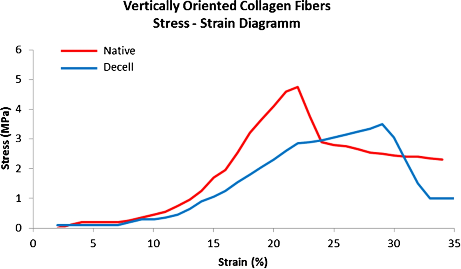
Mean stress–strain behavior of the native and decellularized pericardia with vertically oriented collagen fibers (
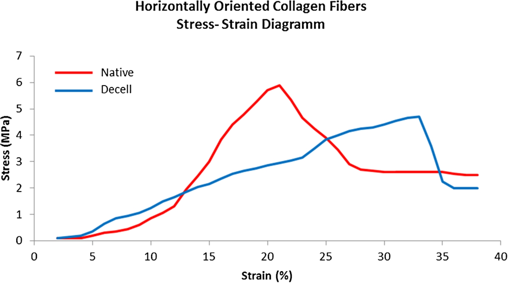
Mean stress–strain behavior of the native and decellularized pericardia with horizontally oriented collagen fibers (
To summarize, porcine pericardium was decellularized successfully and maintained its original mechanical properties without the need of any cross-linking agent. Thus, we propose the porcine pericardium as a potential biological scaffold capable for heart valve or vascular patch development. A further assessment of the decellularized pericardium for the presence of a-Gal epitopes is indicated, in order to demonstrate the potential use of the scaffold in humans.
Conflict of interest
The authors have no conflict of interest to report.