
Abstract
BACKGROUND:
To enhance calcium silicate cements (CSCs) towards a specific clinical application of endodontics and vertebroplasty, the addition of oxide dopants (Bi2O3, SrO, ZnO, ZrO2) as radiopacifiers allows for tailoring of material properties.
OBJECTIVE:
Effects of oxide dopants on the in vitro physicochemical properties and osetogenic activity of CSCs were investigated.
METHODS:
The setting time, compressive strength, radiopacity, and osteogenic ability of the cements were evaluated. The ability of cement samples to support MG63 attachment, proliferation, differentiation, and mineralization was assessed.
RESULTS:
The greater the oxide amount, the higher the setting time and radiopacity were in the cement. The effect of the oxide dopants on radiopacity followed the order Bi2O3 > ZrO2 > SrO > ZnO, which were greater than 3 mm of Al recommended by ISO 6876: 2001 standards. ZrO2 could reinforce compressive strength of the control cement, while ZnO remarkably reduced the strength. The adverse effect of Bi2O3 and ZrO2 was found on cell number, alkaline phosphatase (ALP) activity and mineralization of MG63 cells. SrO supported cell attachment, proliferation and differentiation, and significantly increased cellular mineralization compared to the control.
CONCLUSIONS:
The 20 wt% SrO-containing CSCs may be applied to endodontic treatment and vertebroplasty surgery.
Introduction
Over the last decade calcium silicate-based materials have attracted great scientific interest because of favorable biocompatible properties that may support their clinical use as bone defect repair and regeneration [1–4]. Studies have shown that newly formed bone tissue can grow on the surface of the calcium silicate-based materials, in addition to the deposition of a bone-like apatite layer at the tissue-material interface [5,6]. With the increasing popularity of minimally invasive techniques, the development of calcium silicate cements (CSCs) that mold to the shape of the bone cavity and harden when injected in situ has attracted a great deal of attention. However, improving upon the inherent shortcomings of CSCs, such as poor radiopacity, is imperative for achieving clinical use in endodontics and vertebroplasty, which require good visualization during injection and post-treatment [7]. Several additives can be admixed to the powder of cements in order to improve or establish certain qualities. Bismuth oxide (Bi2O3) is commonly used to improve radiopacity due to its high molecular weight. It is questioned if it would be the best radiopacifier to be associated with CSCs [1,8–10]. This is because Bi2O3 not only extends the setting time of the cement [1], but is also responsible for the reduction in cell growth [10]. Zinc is an essential trace element that can increase bone mass [11]. Zinc deficiency is associated with skeletal growth retardation and alterations in bone tissue calcification [12]. Zirconia is currently used in the femoral head of hip prostheses and dental restoration because of high mechanical strength and toughness, and good abrasion resistance and chemical stability in vivo [13]. Silva et al. suggested that ZrO2 may be a good alternative for use as radiopacifier in substitution to Bi2O3 in term of immune response [14]. Strontium is a bone-seeking element, of which 98% in the human body can be found in the skeleton [15]. Strontium-containing materials stimulate osteoblast differentiation and bone formation [16,17]. These oxide additives may tailor the physicochemical and biological properties of CSCs making them suitable for broader clinical applications. However, the addition of radiopacifiers might be detrimental to some of the physical, mechanical, and biological properties of biomaterials [7,10,18].
The present study systematically examined the effects of oxide dopants in equal doping proportions on the physicochemical properties and osteogenic activities of CSCs. The major characterization techniques included setting time, radiopacity, compressive test and in vitro osteogenic activity.
Materials and methods
Specimen preparation
Reagent-grade tetraethyl orthosilicate (Si(OC2H5)4; Sigma-Aldrich, St. Louis, MO, USA) and calcium nitrate (Ca(NO3)2 ⋅ 4H2O; Showa, Tokyo, Japan) were used as precursors for SiO2 and CaO, respectively. The catalyst was 2 M nitric acid, and absolute ethanol was used as the solvent. The molar ratio of Ca(NO3)2 ⋅ 4H2O to Si(OC2H5)4 was 3:2. General sol-gel procedures, such as hydrolysis and aging, were adopted. A detailed description of the calcium silicate powder’s fabrication can be found elsewhere [19]. A variety of oxides including Bi2O3, SrO, ZnO and ZrO2 at 10 wt% or 20 wt% were added to the calcium silicate powder using a conditioning mixer (ARE-250, Thinky, Tokyo, Japan) for 10 min at 1500 rpm. The cement specimens were prepared by hand mixing the powder with distilled water at liquid-to-powder ratios of 0.5 mL/g. The cements were placed in a cylindrical Teflon mold to form cylindrical specimens; the specimens were stored in an incubator at 100% relative humidity and 37 °C for 1 day to set, with the exception of the setting time measurements. The specimen codes “CS control”, “CSBi10” and “CSBi20” represented cements containing 0, 10, and 20 wt% Bi2O3, respectively, as listed in Table 1.
Dopant content, setting time, compressive strength, and radiopacity of various calcium silicate cements, along with radiopacity of some commercially available endodontic products for comparison purpose
Dopant content, setting time, compressive strength, and radiopacity of various calcium silicate cements, along with radiopacity of some commercially available endodontic products for comparison purpose
Mean values followed by same superscript letters were not significantly different (P < 0.05) from CS.
The setting time of the cement was tested using a 400-g Gillmore needle with a flat end, 1 mm in diameter, according to ISO 9917-1:2003. Each material was mixed and placed in a cylindrical Teflon mold (diameter = 6 mm and height = 6 mm). The tests were performed in an incubator maintained at 37 °C with a relative humidity of at least 90%. The setting time was recorded as the time that elapsed between the end of mixing and the time when the needle failed to create an indentation of 1 mm in depth in three separate areas of the cement. The average setting time was calculated from night replicate specimens.
Phase composition and morphology
To investigate the phase composition, the cement specimens were characterized using X-ray diffraction (XRD; Bruker D8 SSS, Karlsruhe, Germany) with Ni-filtered CuK𝛼 radiation operating at 40 kV, 100 mA and a scanning speed of 1°/min. The morphology was observed by field-emission scanning electron microscopy (FESEM; JEOL JSM-7401F, Tokyo, Japan). The specimens were coated with gold using a JFC-1600 (JEOL, Tokyo, Japan) coater and examined by FESEM operating in the lower secondary electron image mode at 3 kV accelerating voltage.
Compressive strength
To examine compressive property the specimen size of 6 mm in diameter × 12 mm in length was used. The compressive strength (CS) as measured at a crosshead speed of 0.5 mm/min using a static mechanical testing machine AG-1000E (Shimadzu, Kyoto, Japan) with a 10 kN load cell. The strength value of each cement specimen was calculated using the relationship defined in the equation CS = P∕𝜋r 2, where P is the peak load (Newtons, N) and r is the radius (mm) of the specimen. The maximal compression load at failure was obtained from the recorded load–deflection curves. Ten specimens were tested for each composition.
Radiopacity
The radiopacity of various cement specimens were determined according to the method in ISO 6876: 2001. The radiopacity was measured by irradiating specimens alongside an aluminum step wedge (10 steps, 1 mm per step). The radiographs were taken using a Belmont Belray 096 Dental X-ray unit (Takara Belmont Corp., Osaka, Japan), operating at 70 kV and 10 mA for 0.33 sec exposure time and a focus-surface distance of 200 mm. A standard curve of gray-level values versus thickness of aluminum was established to determine the radiopacity value of each specimen using ImageJ software (National Institutes of Health, Bethesda, MD, USA). The corresponding gray-level value for each specimen was superimposed on the standard curve and the equivalent thickness of aluminum was recorded. Three parallel experiments were performed with the data of every group.
Cell culture
The human osteoblast-like cells line MG63 (BCRC 60279, Hsinchu, Taiwan) were used to evaluate osteogenesis of the cement (6 mm diameter × 1 mm thick). The cement without the oxide dopant was used as a negative control. The cells were suspended in Dulbecco’s modified Eagle medium (DMEM; Gibco, Langley, OK, USA) containing 10% fetal bovine serum (FBS) (Gibco) and 1% penicillin/streptomycin solution (Gibco) in 5% CO2 at 37 °C. Before cell incubation, specimens were sterilized by soaking in a 75% ethanol solution and exposure to UV light overnight.
Cell attachment and proliferation
MG63 cell viability was examined using the MTT (3-(4,5-dimethylthiazol-2-yl)-2,5-diphenyltetrazolium bromide; Sigma-Aldrich) assay. To assess attachment, cells were cultured for 6 h, 12 h, and 1 day. Proliferation was assessed at days 3 and 7. Cell suspensions (5,000 cells per well) were seeded on specimen surfaces in a 48-well plate. Before the end of the incubation period, 100 μL of MTT solution and 900 μL of DMEM containing 1% penicillin/streptomycin were added to each well followed by 3 h of incubation. After incubation, supernatants were removed, and the 500 μL of 2-Propanol (Sigma-Aldrich) were added to each well. The plates were then shaken until the formazan crystals had dissolved, and 150 μL of the solution from each well was transferred to a new 96-well plate. Plates were read in a Sunrise microplate reader (Tecan, Salzburg, Austria) at 570 nm, with a plate wavelength of 650 nm. The results were reported in terms of absorbance form three independent measurements.
Alkaline phosphatase activity
To evaluate the effect of oxide content on early cell differentiation, ALP activity of MG63 at a density of 5000 cells per well on the various specimens was measured after 7 and 14 days of incubation. ALP catalysed the hydrolysis of the colourless organic phosphate ester substrate (p-nitrophenyl phosphate; pNPP) to p-nitrophenol (a yellow product) and phosphate. ALP activity was measured using the TRACP & ALP assay kit (Takara, Shiga, Japan) according to the manufacturer’s instructions. Three measurements were carried out.
Calcium quantification
The mineralized matrix synthesis was analysed using an Alizarin Red S staining method, which identifies calcium deposits. After culturing for 14 and 21 days, MG63 cells were washed with PBS and fixed in 4% paraformaldehyde (Sigma-Aldrich) for 10 min at 4 °C. This was followed by staining for 10 min in 0.5% Alizarin Red S (Sigma-Aldrich) in PBS at room temperature. The stained cells were completely washed with PBS to reduce nonspecific Alizarin Red S stain. To quantify matrix mineralization, the calcium mineral precipitate was destained with 10% cetylpyridinium chloride (Sigma-Aldrich) in PBS for 30 min at room temperature. The absorbance of Alizarin Red S extracts was measured at 560 nm using a Sunrise microplate reader. Mean absorbance values were obtained from six independent experiments.
Statistical analysis
One-way analysis of variance statistical analysis was used to evaluate the significance of the differences between the mean values. Scheffé’s multiple comparison testing was used to determine the significance of the deviations in the data for each specimen. In all cases, the results were considered statistically significant at a P value of less than 0.05.
Results
Phase composition and morphology
Figure 1 shows the XRD patterns of the CSCs with and without 20 wt% dopants. It indicated a poor crystalline material with well-defined peaks for the CSC control. The diffraction peak near 2𝜃 =29.4° corresponded to a calcium silicate hydrate phase structure overlapping with calcite (CaCO3) because of the hydration reaction, and incompletely reacted inorganic component phases of 𝛽-Ca2SiO4 at 2𝜃 between 32–34° was found [20]. It therefore seems reasonable to suspect that other diffraction peaks were ascribed to phase composition of the oxide dopants. SEM micrographs of the hydrated cement samples with and without 20 wt% oxides are presented in Fig. 2. The as-hardened control (Fig. 2a) essentially appeared rather smooth looking with particle entanglement and several micropores. After the addition of various oxides in the cement (Fig. 2b–e), the surfaces also presented similar microstructure.

X-ray diffraction patterns of the dicalcium silicate cements with and without 20 wt% oxide after mixing with water.
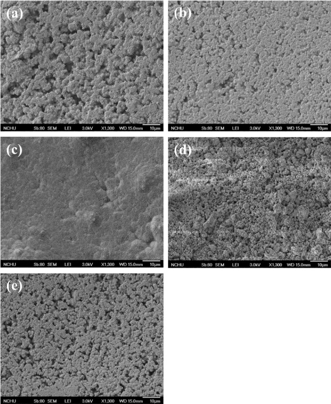
SEM micrographs of (a) the set calcium silicate cements with 20 wt% (b) Bi2O3, (c) SrO, (d) ZnO and (e) ZrO2.
The radiopacity of the control without oxide was recorded as 2.1 mm of Al (Table 1). The cement containing 10 wt% oxides had a radiopacity slightly higher than the recommended 3 mm of Al (ISO 6876: 2001 standards), while 20 wt% dopants presented a remarkable radiopacity that can be accepted in the clinical applications. Not surprisingly, the radiopacity increased with increasing dopant content. Among the oxide dopants, 20 wt% Bi2O3 produced the significantly (P < 0.05) higher radiopacity of the cement with values of 9.4 mm of Al compared to the other groups; in contrast, ZnO elicited a lower radiopacity value. The oxide-dependent radiopacity value decreased in the order: Bi2O3 > ZrO2 > SrO > ZnO in the equal doping amount.
Setting time
The setting time of the control cement was about 15 min, as listed in Table 1. Similar to the result of radiopacity, the higher the dopant content in the cement, the great the setting time was found. When the cement contained 20 wt % Bi2O3 and SrO, the setting values became about 25 min, whereas 20 wt% ZrO2 resulted in the greater setting time of 37 min. Interestingly, ZnO had a setting time similar to that of the control.
Compressive strength
The relationship between the dopant content and compressive strength of the cements is also shown in Table 1. The type and content of the oxide dopants appreciably affected the strength value of the cements. The strength of the control had a value of 19 MPa, which was significantly (P < 0.05) higher than 20 wt% SrO group (12.6 MPa) and ZnO group (1.4 MPa). ZrO2 had higher strength values (P < 0.05) of 29.7 and 32.5 MPa, respectively, for 10 and 20 wt% content compared to the control. The strength values of Bi2O3–containing cements were comparable to that of the control.
Cell attachment
To elucidate the effects of various dopants on osteogenic activities, the biological functions of MG63 cells cultured on cement specimens with various dopant contents were evaluated. First of all, the numbers of initially attached cells for the CS control and radiopaque cements were different, as shown in Fig. 3. It can be clearly seen that the attachment of MG63 cells cultured on ZrO2-containing cement surfaces was significantly (P < 0.05) lower than that on the surface of the cement control at all culture time points, while ZnO and SrO did not adversely affect the cell attachment. The 20 wt% Bi2O3 also led to the significantly (P < 0.05) lower cell attachment on the cement surface compared to the control.
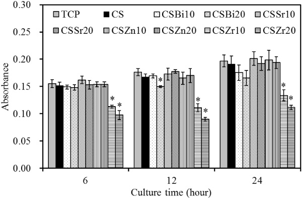
MTT assay for MG63 cell attachment cultured on the cement specimens at various time points. ∗Statistically significant difference (P < 0.05) from CS.
Figure 4 shows the increased absorbance value steadily for all of the samples on Days 3 to 7, revealing increasing numbers of viable cells. There was no significant difference (P > 0.05) in the absorbance between the control and SrO and ZnO-containing cement following culture for 3 and 7 days, while 20 wt% Bi2O3 reduced the cell proliferation. The cells seeded on the ZrO2 had the worst metabolic activity. On day 7, the absorbance value for 20 wt% ZrO2 cement was approximately 43% lower than that of the control.
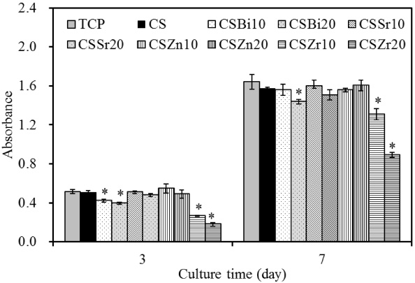
MTT assay for MG63 cell proliferation cultured on the cement specimens at various time points. ∗Statistically significant difference (P < 0.05) from CS.
The intracellular ALP level is measured to observe the differentiation activity of cells, as shown in Fig. 5. The ALP level decreased with increasing Bi2O3 and ZrO2 content of the cements at all incubation times, while both SrO and ZnO cements had a similar ALP expression to the control.
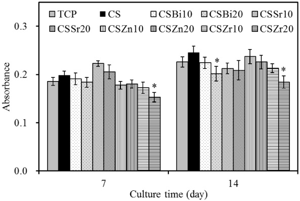
ALP assay on MG63 cells presented as optical density for cell differentiation on cement specimens after 7 and 14 days of culture. ∗Statistically significant difference (P < 0.05) from CS.
Quantification of calcium mineral deposits by the Alizarin Red S assay showed that on day 14, less mineral deposition was found in MG63 cells cultured on the ZrO2 cement surfaces, while SrO had the greatest mineral deposition (Fig. 6). With increasing culture time, mineral deposition increased for the cells cultured on all cements. By day 21, the mineral deposition of the cells seeded on ZnO cement was comparable to the control. In contrast, a significant 37% increment (P < 0.05) of calcium content was measured for the 20 wt% SrO cement (CSSr20) compared to the CS control. Of note, 20 wt% Bi2O3 was also deleterious effect on cell mineralization.

Quantification of calcium mineral deposits by Alizarin Red S assay of MG63 cells cultured on cement specimens after culture for 14 and 21 days. ∗Statistically significant difference (P < 0.05) from CS.
When used in endodontic and vertebroplastic treatment, the cement materials should have sufficient radiopacity to be distinguished from the peripheral anatomical structures. Calcium silicate cement lacks sufficient radiopacity; therefore, radiopacifier needs to be added for reaching an acceptable level of radiopacity [21]. In this work, we focused our investigation on the CSCs with different oxide dopants (Bi2O3, SrO, ZnO and ZrO2) to enhance cement performance. Although the effects of radiopacifiers such as Bi2O3 and ZrO2 on the physical properties, sealing ability, and biocompatibility of calcium-based cements have been documented [1,8,10], there have been few systematic studies on their effects of physicochemical properties and in vitro osteogenic activities.
The types and varying amount of dopant can have impact on the overall physicochemical properties, mechanical strength, and cell–material interactions. The present results showed that oxide-doped CSCs differed in their material composition evidenced by XRD, accordingly, different properties were found. More importantly, the dopant-dependence has been observed in radiopacity, which was the focus in this study. The CSC control exhibited a low intrinsic radiopacity (equivalent to 2.1 mm of Al) that was insufficient for distinguishing the cement from tooth and bone, consistent with previous studies [10,22]. Not surprisingly, the radiopacity increased with increasing dopant content. The cement containing 10 wt% oxides had a radiopacity slightly higher than the recommended 3 mm of Al in the ISO 6876: 2001 standards. After the addition of 20 wt% oxide, the resulting radiopacity of all cement systems increased to a value significantly greater than the minimum recommended value. Among the used dopants, the radiopacity value of the Bi2O3 cement was notably enhanced and achieved the highest because it had a greatest molecular weight. Apart from Bi2O3, efforts have been put into developing other radiopacifiers. ZrO2 added to the CSC may increase the radiopacity level over ISO standard, and may potentially be applied as alternative to commonly used Bi2O3 radiopacifier, as suggested in a previous report [9]. Nevertheless, it is worthwhile to refer the radiopacity of the commercially available endodontic materials for comparison purposes before the clinical applications. The white-colored ProRoot mineral trioxide aggregate (WMTA) containing 20 wt% Bi2O3 had the radiopacity values ranging from 5.7 to 9.3 mm of Al [10,21,23]. MTA-Angelus demonstrated a radiopacity of 3–3.3 mm of Al [24]. For the ZnO-eugenol cement used for filling materials, IRM and Super EBA were equivalent to 5.7 mm and 3.3 mm of Al, respectively [24]. Sealer 26, epoxy resin-based endodontic sealer, presented a value of 5.9 mm [24]. In addition, the radiopacity of gutta-percha was 7.2 mm of Al equivalence [25]. No matter which the developed CSC was used either a filler or sealer, the results of the present study revealed that all tested oxide dopants might potentially be added to CSC as radiopacifiers to enhance the radiopacity values higher than 3 mm of Al, but other properties such as setting time and osteogenesis were necessary to be examined before the clinical use.
Given that the setting time is an important factor for satisfying clinical requirements, a long setting time could cause problems clinically because of the cement’s inability to maintain its shape and support stresses within this time interval. It was found that Bi2O3 prolonged the setting times of CSCs, in agreement with reports from other researchers [1,14]. Increasing the content of dopants significantly prolonged the setting time of the cement. Except the ZnO dopant, SrO and ZrO2 retarded the setting reaction of CSCs, possibly because the oxide dopants acted as inert filler and did not participate in the hydration reaction of the cement [26].
Another concern was whether the incorporation of various dopants into CSCs declined mechanical strength. Interestingly, the addition of Bi2O3 to CSC slightly increased the compressive strength value. The mechanical properties of the hardened cements resulted from the reaction of the liquid phase, such as water, with the cement components [10]. The incorporation of ZnO into the cements could change the microstructure of the cement by acting as flaws within the cement matrix [9], which may exacerbate existing cracks and adversely influenced the mechanical properties. In contrast, the inherently tough ZrO2 dopant can be acted as reinforcement agent to enhance the mechanical properties of the cement.
Further research regarding the in vitro osteogenic activity is needed from the viewpoint of clinical aspects in order to establish their prospective applications. SrO and ZnO have received special interest in recent years due to their potential benefits enhancing cell functions. Regarding the cell viability, the numbers of initially attached cells seeded on the CS control and radiopaque cements were different. When MG63 cells were seeded on the Bi2O3 and ZrO2 cement surfaces, an inhibitory effect was observed on attachment and proliferation. Bi2O3 has been shown to interfere with cell growth [27] and promote inflammatory reaction on subcutaneous tissue [14]. Although ZrO2 is known to be compatible, excessive amount might be with a cytotoxic response in cell culture, as confirmed by the current results.
Cell differentiation studies, like cell proliferation assay results, showed a significant impact of Bi2O3 and ZrO2, with an emphasis on the importance of material composition. ALP activity decreased with increasing Bi2O3 and ZrO2 content. ALP is expressed during the post-proliferative period of extracellular matrix maturation and initiates matrix mineralization [19,28]. To more fully assess the role of oxide dopants in cell function, mineralization ability was also examined. Alizarin Red S staining is a common histochemical technique used for detecting calcium deposits in mineralized tissues and cultures [29]. The ability of cells to produce a mineralized matrix and nodules in materials is important for bone regeneration. Recently, we also found 20 wt% calcium silicate component appreciably improved the biological functions of the ZrO2 implant materials [29]. It is reasonable to consider that the in vitro osteogenic activity of ZrO2 is inferior to the calcium silicate materials. Thus, the presence of ZrO2 can lead to the lower osteogenic activity of cells seeded on the composite cements. Of note, the presence of Bi2O3 and ZrO2 in the cement had a dose-dependent negative effect on the osteogenic activity of osteoblastic cells. On the other hand, incorporation of ZnO into CSC did not change the mineralization level expressed by MG63 cells compared to the control, whereas no effect was also observed on the cell attachment and proliferation. Contrarily, the SrO surfaces could provide a superior environment for osteoblasts to function normally. Calcium deposit formation was most noticeable on the SrO cement surface, speculating that this composite cement will produce more mineralized tissue formation upon in vivo implantation. Although the base materials or species of cultured cells are different, previous studies show that the Sr-containing materials can up-regulate osteoblastic cell viability, ALP activity, and exert a stimulatory effect on bone formation [16,17,30], as found in our current investigation. Hence, SrO seemed to be a potential alternative radiopacifier for CSC because it showed excellent osteogenesis and provided satisfactory radiopacity and physicochemical properties.
Conclusions
The study sheds light on the effect of oxide dopants on the radiopacity, setting time and in vitro osteogenic activity of calcium silicate cement. The results indicated that oxide dopants at an appropriate fraction could significantly enhance radiopacity, although the addition of oxides increased the setting time of CSC. Taken together, SrO can be used to replace the commonly used Bi2O3 in calcium silicate materials in term of superior osteogenic activity. The bioactive SrO cement with enough radiopacity may be applied to the endodontic treatment and vertebroplasty surgery.
Footnotes
Conflict of interest
None to report.