
Abstract
BACKGROUND:
The scaffold for head and neck reconstruction needs mechanical strength to maintain specific forms. Hydroxyapatite (HA) enhances the mechanical strength of hydrogel and is routinely used for cartilage regeneration. However, there is a demand for hydroxyapatite that controls chondrogenic cell behavior.
OBJECTIVE:
Our aim was to regulate HA morphology through a hydrothermal process using organic acid and enhance chondrocyte proliferation and differentiation using shaped-regulated HA.
METHODS:
HA was synthesized from dodecanedioic acid (DD:HA) and oleic acid (OA:HA) by a hydrothermal method and then coated onto glass plates. Surface properties of the samples were compared by various techniques. Surface roughness and contact angles were calculated. Proliferation and differentiation of chondrogenic cells were measured by MTT assays and Alcian Blue staining, respectively, after various incubation periods.
RESULTS:
The morphological structures of DD:HA and OA:HA were different; however, the crystallinity and chemical structures were similar. Surface roughness and hydrophilic behavior were higher on DD:HA. DD:HA enhanced chondrogenic cell proliferation over time. The differentiation of ATDC5 cells was also increased on the DD:HA surface compared with those in other groups.
CONCLUSIONS:
DD:HA enhanced cell viability to a greater extent than OA:HA did, indicating its excellent potential as an inorganic material compatible with chondrocyte regeneration.
Introduction
Chondrogenic cells are the most popular cells to engineer cartilage for head and neck reconstruction. Although these cells can be applied as cell suspension or cartilage sheet, owing to the demand for specific forms such as an auricle, trachea, or nose, engineered scaffold with strong mechanical properties is essential to repair cartilage defects in head and neck reconstruction [1]. The hydrogels/hydroxyapatite (HA) composites show high mechanical strength and have potential as scaffolds for cartilage regeneration [2]. However, because of the high biocompatibility, osteoinductivity, and osteoconductivity of HA, high content of HA in hydrogel induces calcification [3].
HA with distinct surface topographies affects the bone cell activities. For example, coating the surface of rough stainless steel with HA can inhibit osteoblast proliferation [4]. HA with nanorod morphology enhances cell proliferation and osteogenic differentiation [5]. Nathanael et al. found that HA nanowires with specific surface properties enhance the osteoblast response [6]. Moreover, the nanoscale structures combined with micro/submicro scale roughness of titanium improved the osteoblast differentiation and provided for the implant osteointegration [7]. Based on the above information, controlling the chondrogenic cells behavior using HA with different morphology is also attracted to provide advanced clinical approaches for head and neck reconstruction.
The hydrothermal method is useful for producing different forms of HA powders with excellent crystal quality [8]. Synthesis conditions have been reported to affect HA morphology. In particular, HA structure can be altered by the reaction pH, temperature, rate, and method [9–11]. Composites containing metal ion-doped HA show different irregular shapes, such as rod, plate, whisker, and dandelion [12]. We previously reported that plate-like HA can be produced in different sizes to control the morphologies of growing crystals; this feature is important for bone mimetic preparations as orthopedic devices [13].
ATDC5 cells are well-established as chondrocytes cell lines. Chondrogenic ATDC5 cells differentiate into mature chondrocytes by insulin treatment in vitro and produce cartilage-like extracellular matrix [14]. Furthermore, ATDC5 cells can be regulated by biomaterial properties such as surface topography, crystallinity, and chemistry. Octacalcium phosphate (OCP), a hydroxyapatite precursor, has been reported to suppress chondrogenic differentiation of ATDC5 by the inhibition of Sox6 mRNA after adhering to OCP [15]. Charge densities of hydrogels also regulate ATDC5 cell proliferation and differentiation [16]. Furthermore, electrospun chitosan/poly(vinyl alcohol) (PVA) with carbonated calcium provides a suitable environment for ATDC5 cell growth in comparison with chitosan/PVA with apatite [17]. In addition, surface morphology has been reported to control osteoblast cell behavior. Wide HA nano-sheets enhanced cell proliferation [18]. The rod-like nano-HA promoted spreading and growth of MC3T3-E1 osteoblast cells compared to the flake-like HA [19]. The porous scaffold design of HA also provides for cell proliferation, differentiation, and cell growth. HA on annealed surface shows better attachment of MC3T3 cells than on as-deposited surface [20]. HA coated on polystyrene cell culture plates by alternate soaking process enhanced osteogenic differentiation but not as much chondrogenic differentiation [21]. However, little is known about the effect of HA structure on ATDC5 cell behavior.
In the current study, we evaluated the behavior of chondrogenic cells grown on glass coated with HA synthesized using the hydrothermal method. We hypothesized that different surface topographies would alter the activities of chondrogenic cells.
Material and methods
Synthesis and preparation of HA from dodecanedioic acid and oleic acid
The precursor calcium oleate was synthesized by separately adding 0.1 M CaCl2 (95%, 10 mL) and 1.2 M NaOH (97%, 10 mL) dropwise into a mixture of ethanol (EtOH, 99%, 6.0 g) and oleic acid (OA; technical grade, 90%, 6.0 g) under magnetic stirring (300 rpm). Under continuous stirring for an additional 30 min, 0.2 M NaH2PO4 ⋅ 2H2O (99%, 5 mL) was added in a dropwise manner to the white slurry containing the precipitated precursor. The mixture was then transferred into a 50-mL polytetrafluoroethylene-lined stainless-steel pressure vessel (DAB-2; Berghof Products + Instruments, Eningen, Germany) autoclave and heated at 150 °C for 18 h at a heating rate of 5 °C/min. To synthesize HA from dodecanedioic acid (DDDA; 99%), the same procedure was used with 2 mmol DDDA rather than OA. All reagents were purchased from Wako Pure Chemical Industries (Osaka, Japan). NaH2PO4 ⋅ 2H2O was purchased from Kanto Chemical Co., Inc. OA was purchased from Sigma-Aldrich (St. Louis, MO, USA). Cyclohexane (99.5%) was purchased from Tokyo Chemical Industry, Japan.
Drying and glass coating of HA
The resulting mixture was washed twice with EtOH and dried at 60 °C overnight. Dry HA powder was collected. The HA powder synthesized from DDDA (0.1209 g) was added to 0.15 M OA (2.547 mL) prepared in 50 mL cyclohexane (99.5%). After mixing by ultrasound for 1 h, the mixture was centrifuged and washed twice with EtOH for 2 min. EtOH (45 mL) was added before dropping the HA into highly purified water. The glass plates were cleaned with acetone, ethanol, and purified water by ultrasonication for 5 min. The two types of HA slurries (HA from DDDA, HA from OA) were dropped into water in different beakers. HA that was suspended over the water surface was transferred onto the glass substrate (15 mm diameter) by dip-coating. Glass plates coated with HA (DD:HA and OA:HA) were dried on a hot plate at 60 °C and heated at a sintering temperature of 350 °C for 1.5 h.
Surface characterization
Scanning electron microscopy (SEM), X-ray diffraction (XRD), Fourier transform infrared spectroscopy (FTIR)
The surface topography was investigated by SEM (Model S-3400 NX; Hitachi, Tokyo, Japan). Powder XRD (D8 Advance; Bruker AXS GmbH, Billerica, MA, USA) with nickel-filtered Cu Kα radiation was conducted to identify the phase produced in the resulting HA-coated glass plates (DDDA and OA) at a constant voltage and current of 40 kV and 40 mA, respectively. The lattice parameters and d-spacing values of HA were calculated by previous FTIR (FT-IR4700; Jasco Corp., Oklahoma City, OK, USA) was used to monitor structural changes in the coatings over a wavelength range of 40–4000 cm−1.
Surface roughness
Glass plates were washed with 70% ethanol in a 10-cm dish. A LEXT OLS4100 laser microscope (Olympus, Tokyo, Japan) was used to measure the surface roughness of HA. Three random positions on the sample surface were tested to determine the roughness value (Ra).
Contact angle
The contact angle of the glass-coated HA was measured to evaluate the wettability of the samples by static contact angle measurement with a contact angle meter (DMs-301; Kyowa Interface Science, Saitama, Japan). Ultrapure water (drop size: 2 μL) was used as the wetting liquid. Ten measurements were performed for each surface treatment, and the average value of the contact angle was calculated.
Cell culture
Chondrogenic cells (ATDC5 cells) were cultured in 6-cm cell culture dishes in maintenance medium to maintain the cells in a prechondrogenic stage. The medium consisted of Dulbecco’s modified Eagle’s medium/Ham’s F-12 medium (Wako), supplemented with 5% fetal bovine serum, 1% penicillin and streptomycin, and 10 μg/mL human transferrin, and the cells were grown in a humidified atmosphere of 5% CO2 at 37 °C. The culture medium was refreshed every 3 days.
After the cells reached 80% confluence in the dishes, adherent cells were removed from the substrate by adding 0.25% trypsin. Twelve wells (n = 4) were maintained in growth medium as negative controls for chondrogenesis. Differentiation medium was composed of ATDC5 medium and 10 μg/mL bovine insulin. The remaining wells were induced with chondrogenic differentiation medium as a positive control. Cells were incubated in a humidified atmosphere for up to 28 days, and the medium was changed twice per week.
Analysis of cell proliferation by 3-(4,5-dimethylthiazol-2-yl)-2,5-diphenyl-tetrazolium bromide (MTT) assay
Attached cells on the sample surface were quantified by MTT assays (Dojindo Laboratories, Kumamoto, Japan). Cells at a density of 5 × 10³/cm2 were seeded onto the sample surface and then incubated in a CO2 incubator for cultivation. After incubation for 1, 3, 7, 14, 21, and 28 days, the medium in the wells was removed by aspiration. Then, ATDC5 cell medium and 100 μL MTT solution was added to each well, and the plates were further incubated for 3 h at 37 °C. The samples were changed to new dishes and washed with phosphate-buffered saline. Next, 200 μL dimethyl sulfoxide was added to each well, and the samples were transferred to 96-well plates. The optical density of the resulting solution was measured with a microplate reader at a wavelength of 570 nm.
Cell differentiation by Alcian Blue staining
Differentiation of ATDC5 cells cultured for 21 or 28 days in each sample group (n = 4) was assessed by Alcian Blue staining. After 21 or 28 days of culture, the cells were washed with phosphate-buffered saline and fixed in 95% ethanol containing 0.1 M HCl for 10 min. Next, 3% acetic acid was added for 3 min at room temperature. Alcian Blue staining (Sigma) was performed to detect proteoglycans. Samples were stained with 1% Alcian Blue, washed with 3% acetic acid, and then transferred to new plates containing 8 M guanidine HCl. The plates were then incubated overnight. The optical density was determined with a microplate reader (Model 680, Bio-Rad Laboratories, Hercules, CA, USA) at 595 nm, using 415 nm as the reference wavelength.
Statistical analysis
Data from experiments characterizing the surface properties of the substrates are shown as means ± standard deviations (SDs) of all measurements from different samples. Statistical analyses were performed by one-way analysis of variance (ANOVA) followed by multiple comparisons (Dunnett T3). Results with p values of less than 0.05 were considered statistically significant.
Results
Surface characterization
SEM
The surface morphologies of glass coated with HA from DDDA (DD:HA) and glass coated with HA from OA (OA:HA) determined by SEM are shown in Fig. 1. As shown in Fig. 1(a), DD:HA was composed of small plate-like structures. Long, fiber-like HA characteristics were observed for OA:HA, as shown in Fig. 1(b).

SEM images of glass coated with HA synthesized using the hydrothermal method from different materials. (a) DDDA, (b) OA.
The XRD patterns of DD:HA (curve a) and OA:HA (curve b) are shown in Fig. 2. The patterns revealed that the crystal structure of DD:HA was similar to that of OA:HA. The lattice parameters of HA calculated from the XRD patterns were a = 9.148 Å and c = 6.884 Å. The d-spacings for the (001), (211), and (300) lattice planes were 3.44, 2.81, and 2.72 Å, respectively.
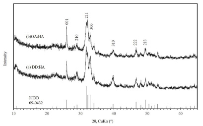
XRD patterns of the HA powder synthesized by the hydrothermal method from two different materials. (a) DDDA, (b) OA.
The FTIR spectra of HA showed absorption due to vibrational modes from phosphate and hydroxyl groups. The FTIR spectra of synthetic DD:HA and OA:HA are presented in Fig. 3. The presence of peaks at 3600 cm−1 for DD:HA and 3590 cm−1 for OA:HA were representative of HA hydroxyl groups, whereas the peak at 1090 cm−1 was attributed to the P-O stretching mode. The absorbance bands at 625 cm−1 for DD:HA and 636 cm−1 for OA:HA represented HPO4 2− ions.
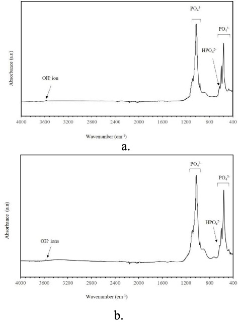
FTIR transmission spectra of HA powder synthesized by the hydrothermal method from two different materials. (a) DDDA, (b) OA.
The results obtained by the sessile drop method on the different samples are shown in Fig. 4. The results are presented as means ± SDs of at least 10 measurements (one per sample). The water contact angle measurements gave values of 12.99 ± 1.5° for DD:HA and 10.323 ± 5.196° for OA:HA. The contact angles measured for the DD:HA and OA:HA samples were significantly smaller than those of the positive control. HA surfaces were hydrophilic in nature. The roughness R a of the control, DD:HA, and OA:HA, shown as the means ± SDs in Fig. 5, had values of 0.014 ± 0.004, 0.186 ± 0.020, and 0.292 ± 0.036 mm, respectively. The surface of the OA:HA sample was rougher than that of the DD:HA and control samples.

Variations in contact angle values in glass coated with HA at a sintering temperature of 350 °C (n = 7). ∗∗∗ p < 0.0005.

Surface roughness of the DD:HA and OA:HA samples (n = 3). Ra values are shown as means ± standard deviations. ∗ p < 0.05, ∗∗∗ p < 0.005.
As shown in Fig. 6, optical density values in MTT assays increased significantly from week 1 to 4. This indicated that DD:HA and OA:HA were biocompatible and did not negatively affect chondrogenic cell viability or proliferation. Although the results were comparable on days 1 and 3, cell proliferation was enhanced over time. Cell proliferation on the DD:HA sample was significantly higher than on OA:HA after 1 week.

ATDC5 cell proliferation after 1, 3, 7, 21, and 28 days of incubation, as measured by MTT assay at an optical density of 570 nm. Values are presented as means ± SDs (n = 4). ∗ p < 0.05, ∗∗∗ p < 0.005, # p < 0.001.
As shown in Fig. 7, cell differentiation was enhanced from day 21 to 28, whereas activity in ATDC5 cells was significantly higher in the DD:HA group than in the other groups, suggesting enhanced chondrogenic cell differentiation. After 28 days of culture in differentiation medium, cells in the DD:HA group showed significantly lower differentiation than in the other groups.

Proliferation of ATDC5 cells, as measured by Alcain blue staining and extracted with 8 M guanidine-HCl. Data were measured at an optical density of 595 nm. The significance of all results was evaluated using ANOVA followed by multiple comparisons (Dunnett T3) for comparisons with corresponding control groups. ∗ p < 0.05 and ∗∗ p < 0.005.
In this study, we investigated the enhanced proliferation of chondrogenic cells on HA synthesized by the hydrothermal method. The surface topographies and chemical characterizations of HA were also evaluated. SEM imaging showed that glass coated with HA synthesized by the hydrothermal method exhibited different morphological structures; that is, DD:HA had a plate-like morphology, and OA:HA had a fiber-like structure. The morphological structure of DD:HA was similar to the plate-like structure of HA previously reported by Horiuchi et al. [13].
The XRD pattern of the HA powder synthesized from different materials showed many sharp peaks, suggesting that the samples were well-crystallized. All XRD patterns indicated that the sample was pure HA with no other impurities based on reported data (JCPDS: 09-0432). Additionally, the HA powders synthesized by each material both had a single-crystalline phase. In contrast, all HA powders from different materials (DDDA and OA) were composed of single-crystalline phase HA. The intensity of the 300 reflection, which was related to the a, b plane of the HA crystal from DDDA, was increased to a greater extent than that of the OA peak. Thus, although the morphology of the HA differed, there were no significant difference in the crystal lactic structure between DD:HA and OA:HA. Two groups of HA were well-crystallized with a single crystalline phase. The crystalline intensity of DD was increased to a greater extent than that of the OA peak, but there were no significant differences in the crystal structure.
Strong bands with a pronounced peak in the 1110–970 and 600–480 cm−1 ranges represented absorption peaks of PO4 3− ions. A weak band at 3590 cm−1 in the spectrum of DD:HA was assigned to the vibration of OH groups. Additionally, the spectra of DD:HA showed small peaks at 2100–2400 cm−1 due to C-H stretching vibration. Some slight vibrations were found because of the presence of impurities and absorption of atmospheric CO2 during sample preparation.
According to previous reports, the surface topography and roughness of HA affected cell adhesion and proliferation [7,22,23]. In this study, the Ra value of the OA:HA surface was significantly higher than that of the other groups. Thus, we assumed that lower chondrogenic cell proliferation occurred on the rougher OA:HA samples. Based on our results, although the hydrophilic surface promoted cell proliferation, higher surface roughness decreased the cell proliferation.
Previous studies have shown that the wettability of material surfaces affects cell proliferation [24,25]. Additionally, Yang et al. found that high hydrophilicity enhanced osteoblast proliferation [26]. Low contact angle values result in high hydrophilicity on sample surfaces. According to our water contact angle results, the surfaces of HA were more hydrophilic than the control sample surface. These conditions are thought to promote cell proliferation.
Chondrogenic cell proliferation in vitro was increased on glass-coated DD:HA surfaces, particularly after 7 days. DD:HA stimulated significantly higher cell proliferation than the other surfaces. Thus, the proliferation of ATDC5 cells was enhanced after 1 week on DD:HA samples. Moreover, ATDC5 cell differentiation was significantly higher on the DD:HA surface, but decreased in differentiation medium on the OA:HA surface. Notably, the surface roughness of HA affects cell differentiation [22,23,27]. Ponsonnet et al. demonstrated that higher surface roughness led to lower cell proliferation [25]. Various theories have been proposed to explain how HA morphology improves cell viability. HA coating may improve osteoconductivity [28], enhance cell attachment [29], and increase protein absorption [26]. Further studies are needed to evaluate factors such as total DNA and gene expression in chondrogenic cells grown on different surfaces.
HA has been widely used as a bone substitute in biomedical applications [30–32]. The osteoconductivity and biocompatibility of HA have been investigated previously [21,33]. In this study, glass coated with HA synthesized from DDDA and OA by a hydrothermal method was evaluated to determine its physicochemical and biological properties. DD:HA showed lower surface roughness and higher hydrophilicity than OA:HA. Cells grown on DD:HA showed enhanced proliferation compared with those grown on OA:HA. These findings suggest that DD:HA promoted cell viability, supporting the potential biomedical applications of DD:HA.
Conclusions
In this study, we characterized HA synthesized using a hydrothermal method and the chondrogenic cell response during growth on glass coated with HA. Use of DD:HA resulted in better cell viability than that of OA:HA. Notably, DD:HA enhanced chondrogenic cell proliferation and differentiation in vitro. The results suggested that chondrogenic cell behavior was affected by the HA coating. Hence, coating with HA may be beneficial during cartilage regeneration.
Footnotes
Acknowledgements
We would like to thank the instructors at the Department of Inorganic Biomaterials and Department of Pediatric Dentistry at Tokyo Medical and Dental University for providing assistance with the study. This study was partially supported by a JSPS Grant-in-Aid for Scientific Research (B) (grant no. 16671149).
Conflict of interest
None declared.