
Abstract
BACKGROUND:
The heart is surrounded by a membrane called pericardium or pericardial cavity.
OBJECTIVE:
In this study, we investigated the pericardial fluid (PF) for coating polycaprolactone (PCL) scaffolds. PFS, which is a PF component, was used for the coating material. In addition to using PFS for surface coating, MED and fetal bovine serum (FBS) were also used for comparison.
METHODS:
Pericardial fluid cells (PFSc) isolated from PF were cultured on coated PCL scaffolds for 1, 3, and 5 days. Cell viability was determined using 3-(4, 5-di-methylthiazol- 2-yl)-2, 5-diphenyltetrazolium bromide (MTT) assay.
RESULTS:
The MTT assay results showed that the viability of cells on PCL scaffold coated with PFS increased over time (P < 0.005), and cell viability was significantly different between PCL scaffolds coated with PFS and non-coated PCL scaffolds. However, cell viability was significantly higher in the PCL scaffolds coated with PFS than non-coated and coated with FBS, MED, and PCL scaffolds. Scanning electron microscopy (SEM) microscopy images and MTT assay indicated that PFSc are attached, proliferated, and spread on PCL scaffolds, especially on PCL scaffolds coated with PFS.
CONCLUSIONS:
These results suggest that PFS is a biocompatible material for surface modification of PCL scaffolds, which can be used as a suitable material for tissue engineering applications.
Introduction
The heart is located in a sac known as the pericardium, which is a serous membrane that encloses the heart. The pericardium sac contains fluid that has two layers, an outer layer (fibrous pericardium) and an inner layer (serous pericardium). The fibrous pericardium consists of connective tissue, which is the outer layer of the pericardium. Serous pericardium is the pericardium’s inner layer and is divided into two layers: visceral and parietal pericardium. Between these two layers a pericardial cavity in a sac filled fluid is called pericardial fluid [1].
Genetic and environmental factors destroy the cardiovascular system, which causes cardiac diseases. The treatment of cardiac diseases is an essential issue all over the world. After cardiac damage, such as a heart attack, heart tissue must be regenerated quickly. Tissue engineering studies aim to treat damaged tissue as cardiac through scaffold or scaffold-free applications. The damaged tissue can be replaced with tissue engineering organs or tissues developed in vitro to support tissue functions and repair the damaged tissue directly in an in vivo environment. Tissue engineering methods for producing a new tissue/organ are performed respectively to derive cells from a patient to culture on a three-dimensional (3D) scaffold and then replace them with damaged tissues. The scaffold is designed to mimic the natural extracellular matrix (ECM) for supporting cell adhesion, migration and proliferation, cell-cell communication, and reforming natural ECM [1].
Electrospinning is one of the most attractive methods and has been used to produce a scaffold that mimics the extracellular matrix of native tissue for two decades. This method is an easy scaffold fabrication method because supporting cell-cell communication, fiber and pore size, and the polymer’s degradation time are controllable [2]. The electrospun scaffolds are made of polycaprolactone (PCL) and blended with polyvinyl pyrrolidone (PVP), which gives practical information about regulating cell behavior. It showed that low PCL fiber density positively affects cell movement. Also, the cell migration is affected by the hydrophobicity and porosity of the scaffold. The electrospun scaffolds have been extensively studied in tissue engineering because they are similar to natural ECM. This scaffold has been used for clinical applications in medical implants, drug delivery carriers, and regenerative matrix for defective tissue. Also, scaffolds promote and guide cell proliferation, differentiation, and tissue growth [3]. Some studies have been performed to enhance cell attachment of the PCL scaffolds. In a study by Mijovic et al., cell attachment on PCL scaffold treated with NaOH solution was compared with PU electrospun, fibrin, and amniotic membran scaffolds [4]. The hydrophobicity of the PCL scaffold was converted to hydrophilicityusing cold and non-thermal plasma. After the PCL scaffold’s surface modification, the fibroblast cells’ behavior was observed to support adhesion and proliferation of fibroblast cells with the cold atmospheric plasma technique [5]. In this study, pericardial fluid availability was researched for surface modification for the first time in the literature.
Pericardial fluid is a plasma ultrafiltrate. It is difficult to define the cell population of pericardial fluid. In normal conditions without the disease, pericardial fluid volume is changeable according to body size; for example, in rabbits, the volume is 0.4–1.9 mL. In adult humans, the volume is about 20–60 mL [6]. The cell population of pericardial fluid in healthy humans is heterogeneous. These are mesothelial cells with a high percentage of lymphocytes, granulocytes, macrophages, eosinophils, and basophils [7,8]. In pericardial fluid, the concentration of K+ is higher than the plasma fluid. The concentration of the other ions is lower in pericardial fluid. The protein contents of pericardial fluid were albumin, globulins, macroglobulins, and fibrinogen [9,10]. The PF is a suitable environment for cell accommodation and contains mesenchymal stem cells (MSCs) [11].
In this study, we hypothesize that PF has also been contained as a structure like ECM. A natural polymer of pericardial fluid-structure (PFS) and synthetic polymer PCL was used for scaffold materials to study this. Pericardial fluid cells isolated from pericardial fluid were used to show cell attachment on these scaffolds. Also, we developed a novel scaffold protocol using pericardial fluid for the improvement of cell attachment. To the best of our knowledge, this is the first in vitro study which shows that pericardial fluid, including PFS, plays a critical role in pericardial cell accommodiation providing perfect surface modification.
Materials and methods
In this study, a novel approach for enhancing cell adhesion of PCL scaffolds has been revealed. For this aim, besides the well-known surface modification method, pericardial fluid was used for the first time. Fetal bovine serum (FBS) and MED were used to compare the effect of PFS on surface modification, and as a negative control, non-coated PCL was used. The study is summarized in the flowchart (Fig. 1).

Flowchart of the study methods.
PF samples were obtained from slaughtered bovines from a slaughterhouse immediately after slaughter. Firstly, the thoracic cavity was opened with care not to damage the pericardium. The pericardial fluids were aspirated from the pericardial cavities without removing any organs, by the side of the heart apex with 50-mL sterile syringes, maintained at room temperature, and transported to the laboratory, as seen in Fig. 2. The PF (liquor pericardium) was obtained from bovines aged between 2–3 years and weighing between 350–450 kilograms.
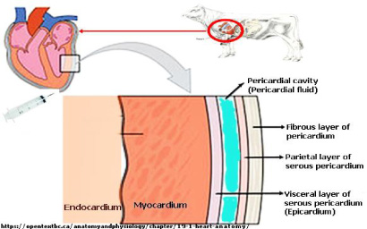
Pericardial fluid and its structure [12].
Pericardial fluid samples of approximately 50–100 mL were obtained from bovine hearts. The samples were centrifuged twice. The first centrifugation was done at 400xg for 5 minutes to remove particulates in the pericardial fluid. The pellet containing particulates was discarded, and the supernatant was transferred to fresh 50 mL falcon tubes. The second centrifugation was done at 4500xg for 5 minutes, and the supernatant was transferred to fresh 50 mL falcon tubes and filtrated with a 0.22 μm cell strainer. This sterile and acellularized supernatant was coded as PFS and then placed in −20 °C storage to use coating PCL scaffolds. The pellet, which contained pericardial fluid cells, was coded as PFSc, suspended in 200 μL PFS, and then cultured in Alpha Modified Eagle’s minimum essential medium (Alpha-MEM) media supplemented with 10% fetal bovine serum (FBS), 250 U/mL penicillin, 250 μg/mL streptomycin and 25 mM glutamine, which were purchased from Thermo Fisher Scientific (USA), and 20 μg/mL bovine insulin (Sigma Aldrich, Germany) grown in 5% CO2 at 37 °C. For the two-dimensional (2D) monolayer culture, 1 × 10⁵ cells/cm2 were seeded in cell culture dishes. The 2D monolayer cells were grown to 95% confluence. Adherent cells were passaged with 0.25% Trypsin-EDTA solution purchased from Thermo Fisher Scientific when the cell population confluence was 95%. After trypsinization, the cells were counted with Muse TM Count&Viability Assay (Merck-Millipore, Germany) cell analyzer and ready to be used in further cell culture experiments.
Production of scaffold by using electrospinning method
Electrospinning was used for the production of the scaffolds. The PCL molecular weight was 80 kDa and purchased from Sigma-Aldrich. The chloroform and methanol were purchased from Merck Chemical Co. The PCL solution of 20% (w/v) was prepared in a 4:1 (V/V) mixture of chloroform and methanol by stirring for 1 hour.
The PCL solution was stirred at 25 °C and 250 rpm for 60 min. The PCL fibrous membranes were produced by electrospinning on a high voltage electrospinning system (Nanospinner, Inovenso, Turkey). The volume flow rate, needle tip to collector distance, and electrical voltage were 2 ml/h, 15 cm, and 20 kV. The PCL morphology was viewed using scanning electron microscopy (SEM; ls-10 Life Science Zeıss, Germany).
Sterilization and modification of PCL
The PCL scaffolds were cut into pieces the size of 0.5 × 0.5 cm2 using a sterile lancet, sterilized under UV light for 30 min, and then sterilized with 70% (v/v) ethanol for 30 min. The scaffolds were then rinsed with Dulbecco’s PBS (DPBS) purchased from Thermo Fisher Scientific (USA).
Surface modifications of PCL scaffolds and cell seeding
A semi-crystalline PCL is hydrophobic biodegradable polyester. For increased cell attachment, PCL scaffolds were treated with PFS, FBS, and MED for two hours to increase their hydrophilicity. PCL scaffolds without coating served as negative controls. Cell counts were assessed using Muse TM Count&Viability Assay cell analyzer. After counting the PFSc, they were seeded (1 × 10⁵ cells/cm2) on the scaffolds and maintained in a humidified atmosphere 5% CO2 at 37 °C. The culture medium was changed every second day.
MTT assay for cell viability
MTT assay was used for the evaluation of cell viability by measuring the mitochondrial activity. This assay works based on metabolically active cells reducing the tetrazolium salt of MTT (Sigma-Aldrich) to purple formazan. After counting cells with Muse TM Count&Viability Assay, cells (1 × 10⁵ cells/cm2) were seeded on the PCL scaffolds for 1, 3, and 5 days. After these days, the medium was removed, and the medium with the MTT solution (5 mg/mL in PBS) was added to each well, followed by incubation for 3 hours. After 3 hours, purple formazan was formed, and the medium was removed, and 1 mL dimethyl sulfoxide (DMSO, Sigma-Aldrich) was added to dissolve the formazan. 200 μL aliquots were then transferred to a 96-well plate, with three replicates per sample. DMSO was set as the blank. Absorbance was measured at a wavelength of 560 nm (Glomax Elisa Reader, Promega, USA). Cell proliferation on scaffolds was assessed using MUSE TM (Merck-Millipore) cell analyzer.
Imaging of scaffold spun with SEM
SEM was used for imaging the electrospun PCL membranes. Before the cell culturing, membranes were cut 0.5 × 0.5 cm2 and then mounted onto a microscope stub and coated with gold (3–6 nm). Images of SEM were taken at 500
After cell seeding on scaffolds, cell attachment was observed under SEM. Cell seeded scaffold samples were rinsed in DPBS and fixed with 2,5% (v/v) glutaraldehyde/DPBS solution for 30 min at room temperature. The scaffolds were rinsed twice with DPBS and subsequently dehydrated using an ascending alcohol series (30%, 50%, 70%, 90%, and 100%) for 2 min each. Finally, after removing ethanol, hexamethyldisilazane (HMDS) (purity >99.0%, Sigma-Aldrich) immersed for 5 minutes and dried at room temperature. After drying, scaffolds were mounted on aluminum stubs with the cell-loaded surface oriented upwards, sputter coated with gold, and morphologies of cells attached onto the surface were viewed using SEM.
Cell attachment on PCL scaffolds coated with PFS was compared with non-coated PCL and PCL scaffold coated with FBS and MED. The performance of PCL scaffolds coated with PFS was analyzed using MTT results. After surface modification with FBS, MED, and PFS of PCL scaffolds, PFSc were seeded on PCL scaffolds to observe cell attachment and viability. In Table 1, four different combinations of PCL scaffolds are seen.
PCL scaffolds with PFSc
PCL scaffolds with PFSc
Statistical analysis was performed using SPSS software (v.21.0; IBM Corp., Armonk, NY, USA), and each experiment was conducted at least three times and replicated six times. All the absorbances data were expressed as the mean ± standard deviation (SD) for n = 6. Comparisons between groups coated with PCL scaffolds were analyzed using variance (ANOVA) with Bonferroni’s multiple comparison test. P < 0.05 was considered statistically significant (Fig. 3). The changes over time of cell viability from day 1 to day 5 in each group were compared using the Wilcoxon signed-rank test (Fig. 4).
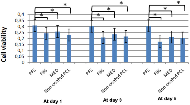
Effect of coating PCL scaffold with PFS. Cell viability was measured by MTT assay on days 1, 3, and 5. Values are mean ± SD; n = 6 analyzed by Bonferroni’s multiple comparison test to compare between the results of coated and non-coated PCL scaffolds groups (significance level (p < 0.05) is indicated by ∗).
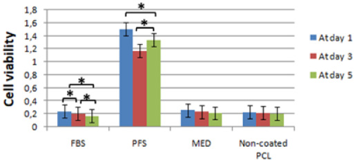
The changes over time of cell viability from day 1 to day 5 in each group were compared by Wilcoxon signed-rank test. Values are mean ± SD; n = 6 (significance level (p < 0.05) is indicated by ∗).
In this study, surface modification of PCL scaffolds was performed using FBS, MED, and PFS. Firstly, PFSc was seeded on petri dishes to observe the proliferation of the cells. For 3D culturing, PFSc were seeded on PCL scaffolds coated with FBS, MED, and PFS. After surface modification of the scaffolds, PFSc cells were seeded on PCL scaffolds coated with FBS, MED, and PFS to analyze proliferation and viability for 1 to 5 days. As a negative control, PFSc cells were seeded on non-coated PCL scaffolds.
There was no significant difference (p > 0.005) in PCL scaffolds non-coated and coated with FBS and MED on days 1, 3, and 5. In contrast, there was a significant difference (p < 0.005) in PCL scaffolds coated with PFS on all days. PCL scaffolds coated with MED and FBS were similar on all days (Fig. 3).
According to Fig. 3, PCL scaffolds coated with PFS significantly increased cell attachment and cell viability, analyzed with Bonferroni’s multiple comparison test. In all paired comparisons, it was seen that cell attachment and viability were highest (p < 0.05) in the PCL scaffold coated with PFS (Fig. 3).
SEM imaging of PCL scaffold
PCL scaffold morphology of PCL scaffolds was viewed using SEM. Figure 5 shows SEM images of PCL scaffolds before cell seeding. The SEM images on days 1, 3, and 5 show the cell attachment on coated and non-coated PCL scaffolds (Figs 6–8).
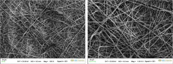
SEM images of PCL scaffolds at the magnification of 500× and 1.00 Kx before cell culture.
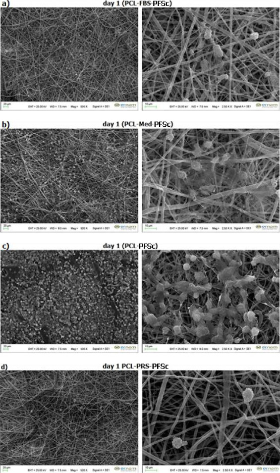
Scanning electron micrographs of PCL scaffolds non-coated and coated with FBS, MED, PFS for one day (24 h). Magnification at 500× and 2500×. (a) FBS, (b) MED, (c) non-coated, and (d) PFS.
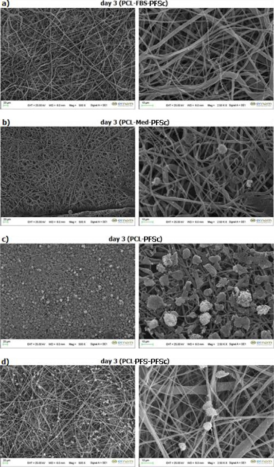
Scanning electron micrographs of PCL scaffolds non-coated and coated with FBS, MED, PFS for three days (72 h). Magnification at 500× and 2500×. (a) FBS, (b) MED, (c) non-coated, and (d) PFS.
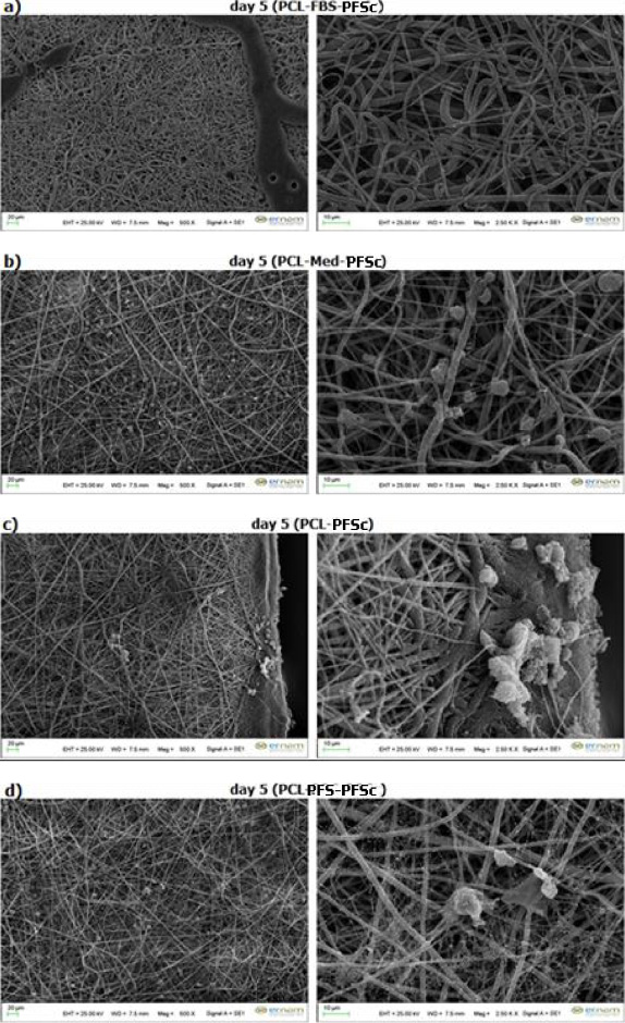
Scanning electron micrographs of PCL scaffolds non-coated and coated with FBS, MED, PFS for five days (120 h). Magnification at 500× and 2500×. (a) FBS, (b) MED, (c) non-coated, and (d) PFS.
According to obtained SEM images on days 3 and 5, cell attachment was not observed on the PCL scaffold coated with FBS. At the same time, cell attachment was observed on the PCL scaffolds non-coated and coated with FBS and MED; however, cell count was significantly decreased. On the other hand, on the PCL scaffold coated with PFS, suitable cell attachment was observed, and cell count was not decreased. Also, cell proliferation on the PCL scaffold coated with PFS was observed better than all other combinations of PCL scaffolds, as seen in Figs 6–8.
Although the PCL scaffold is widely used in biomedical research due to its biocompatible, mechanically durable, and biodegradable properties, its hydrophobic feature negatively affects cell adhesion. To solve this problem, many natural proteins, such as collagen, gelatin, and fibronectin, are used to increase cell adhesion with surface modification of the PCL scaffold [12–14]. In our study, PFS, which is the PF component obtained from the bovine heart, was used for the surface modification of the PCL scaffold. The effect of PFS on cell viability and cell adhesion was compared to PCL scaffolds coated with MED and FBS and non-coated PCL scaffolds. With SEM images and MTT analysis, it was concluded that cell viability and cell adhesion were better in PCL scaffolds coated with PFS on days 1, 3, and 5. The effect of PFS on cell viability and cell retention is because PF contains complex protein structures such as albumin, globulins, macroglobulins, and fibrinogen [9,15]. Studies have confirmed that the scaffold made of albumin protein, which is also in PF content, positively affects mesenchymal stem cells' (MSCs) growth and differentiation [16]. Thus, in our study, the coating of PCL scaffolds with PFS containing albumin had a positive effect on cells' attachment and viability. The effect of the PF on cell adhesion and viability is not only due to protein content PF contains growth factors such as basic fibroblast growth factor (bFGF), acidic fibroblast growth factor (aFGF), hepatocyte growth factor (HGF), vascular endothelial growth factor (VEGF), and prostaglandin and cytokine [17–21]. PF has a crucial effect on vascular smooth muscle cells, myocardial cell, and vascular endothelial cell growth and stimulation [22]. Because of PF, this content provides a suitable environment for mesenchymal stem cells (MSCs) [11].
MTT analysis and SEM images show that PFS isolated from bovine PF increased cell adhesion by containing protein components such as fibrinogen, albumin, globulins, growth factors, prostaglandin, and cytokine. In the later days of cell culture on the PCL scaffolds coated with PFS, it was observed that besides preserving cell viability and number, cells formed ECM-like network structures on the scaffold surface, as seen in SEM images (Fig. 8). Thus, the results showed that these cells could create their own ECM on the PCL scaffold coated with PFS.
The non-parametric analysis results showed statistically significant differences (p < 0.05) between the PFS coated PCL scaffolds and PCL scaffolds coated with FBS, MED, and non-coated PCL scaffolds. However, PCL scaffolds coated with PFS showed a better effect on the attachment and viability of cells.
We sought to develop a suitable material for coating biomaterials for increasing cell attachment. As a natural coating material for surface modification, PFS coated PCL scaffold has demonstrated suitable property for cell attachment in vitro. We have shown proof of concept for using acellularized PF as a coating material to increase cell attachment with these findings.
As a result of this study, pericardial fluid, which has not been illuminated yet, can also be a suitable scaffold candidate for tissue engineering applications, particularly vascular tissue engineering. In the next study of our study group, the effects of PCL scaffolds coated with PFS will be investigated for other cell lines cultured. We plan to use this PFS (ECM-like network) as an ECM structure for vascular tissue engineering studies.
Footnotes
Acknowledgements
This work was supported by the Scientific Research Projects Unit of Erciyes University with the project ÖNAP, FOA-2016-6692, Turkey.
We would like to thank Erciyes University Genom and Stem Cell Centre (GENKOK) and Nanotechnology Centre (ERNAM), and the Laboratory of Biomaterials and Tissue Engineering at Abdullah Gul University in Kayseri, Turkey. Additional thanks are given to Dr. Burak Açıkgöz from the Department of Cardiovascular Surgery at the Dr. Sadi Konuk Training and Research Hospital in İstanbul, Turkey, for his valuable contribution to our study.
Conflict of interest
The authors do not have any conflict of interest to report.