
Abstract
BACKGROUND:
Vascular smooth muscle cells (VSMCs) are one of the main components of arterial walls and actively remodel the arterial walls in which they reside through biomechanical signals applied to themselves. Contractile or differentiated VSMCs have been observed in normal blood vessels. In pathological vascular conditions, they become dedifferentiated from contractile to non-contractile or synthetic cells, and a similar change is observed when VSMCs are placed in culture conditions. The mechanisms regulating VSMC differentiation remain unclear at this stage.
OBJECTIVE:
In this paper we investigated the effects of substrate stiffness on the morphology, intercellular tension, and differentiation of VSMCs.
METHODS:
Rat VSMCs were cultured on polyacrylamide (PA) gels, with elastic moduli of 15 kPa, 40 kPa, and 85 kPa, and PDMS substrate with elastic modulus of 1 MPa, and their morphology, intercellular tension, and contractile differentiation were assessed.
RESULTS:
Using fluorescence microscope image-based analysis and nano-indentation imaging with atomic force microscopy, we found that cell spreading and stiffening were induced by substrate stiffening in VSMCs. Interestingly, VSMCs on PA gel substrates with medium stiffness (40 kPa) showed significant elongation and shape polarization, and their 𝛼-SMA with F-actin cytoskeleton expression ratio was significantly higher than those of cells on other substrates.
CONCLUSION:
The results indicate an existing optimal substrate stiffness for promoting VSMC differentiation, and also indicate that cell shape polarization might be a key factor for VSMC differentiation.
Keywords
Introduction
Vascular smooth muscle cells (VSMCs) regulate vascular contraction and dilation. They actively remodel the vascular wall in which they reside through biochemical and biomechanical signals [1–3], and maintain the mechanical hoop stress in the wall to a normal level via their contractility [4]. In normal vascular walls, mature differentiated VSMCs have a contractile phenotype. Contractile VSMCs have a bipolar elongated shape and are abundant in myofilaments, mainly composed of actin and myosin. VSMCs contract in response to mechanical and hormonal stimuli, have little synthetic ability, and seldom proliferate [5]. Under pathological conditions, such as hypertension and atherogenesis, VSMCs undergo dedifferentiation from the contractile state to the synthetic state. Synthetic VSMCs actively proliferate and migrate. A similar change in cell dedifferentiation is observed when VSMCs are removed from native aortic tissue and placed in culture conditions; VSMCs exhibit a less elongated morphology and spread randomly on the flat surface of culture dishes. The mechanism of their differentiation and dedifferentiation is important to understand smooth muscle pathophysiology in diseases, and develop tissue-engineered blood vessels.
From a biomechanical standpoint, the effects of the physical or mechanical characteristics of extracellular substrates have been the focus of recent studies. Some studies have examined cellular responses to the stiffness of different substrates, such as polyacrylamide [6], hydrogels based on polyethylene glycol [7], and polydimethylsiloxane (PDMS) [8]. Specifically, VSMCs spread well on stiffer substrates [9] and their proliferation also increases with substrate stiffness [10]. Such findings suggest that VSMC behavior and their differentiation could be modified due to changes in substrate stiffness.
Cell-substrate adhesion is crucial to cellular mechanotransduction. Cells generate mechanical tension through contraction of the actin-myosin cytoskeletons, and transmit this force via focal adhesions to the underlying substrates. Thus, cellular tension is believed to play a pivotal role in regulating various cellular events, such as cell proliferation [11], migration [12,13], differentiation [14], and remodeling of the extracellular matrix (ECM) [15]. These cellular forces are potentially affected by substrate stiffness, resulting in modulation of cellular differentiation.
Thus, in this study, we fabricated polyacrylamide (PA) gel substrates with different elastic moduli (15 kPa, 40 kPa, and 85 kPa) and PDMS substrates (∼1 MPa) to investigate the effects of substrate stiffness on the morphology, intercellular tension, and differentiation of VSMCs. We assessed the cellular mechanical responses and differentiation using fluorescent microscopy image-based analysis, and nano-indentation imaging with atomic force microscopy.
Materials and methods
Fabrication of PA gel and PDMS substrates with varying stiffness
Polyacrylamide (PA) gel substrates with varying stiffness (<100 kPa) were fabricated using different amounts of crosslinker, as described previously [16]. In short, the glass surface of 35 mm glass-bottom culture dishes (No. 0, Matsunami, Osaka, Japan) were treated with 3-aminopropyltrimethoxysilane and 0.5% glutaraldehyde. PA gel solution with the desired concentration of acrylamide and bis-acrylamide was allowed to polymerize, using ultraviolet (UV) radiation, to form ∼200 μm thick gels on the slides (Fig. 1). Sulfo-SANPAH was used to link fibronectin (0.5 μg/cm2) to the PA gel surface. In order to investigate the effects of stiffer substrates (∼1 MPa) on the cell responses, PDMS substrates were also used in this study. PDMS solution (Sylgard 184, Dow Corning) mixed at a 15:1 ratio of base monomer to curing agent were poured over 𝜙35 mm glass-bottomed culture dishes (No. 0, Matsunami) and were cured at 70 °C for 4 h. The surface of the PDMS membrane was exposed to oxygen plasma (5 mA, 10 Pa) for 2 min using a plasma generator (SEDE-P, Meiwafosis, Tokyo, Japan) to create a hydrophilic surface to link fibronectin.
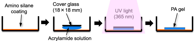
Process flow diagram for fabricating the polyacrylamide gel substrate.
To determine the elastic modulus of PA gel and PDMS substrates, atomic force microscopy (AFM) indentation measurements were performed using a NanoWizard IV AFM (JPK Instruments-AG, Germany) mounted on top of an inverted optical microscope (IX73, Olympus, Japan) equipped with a digital CMOS camera (Zyla, Andor). For AFM indentation measurements, we used the pyramidal tip of V-shaped silicon nitride cantilevers, equipped with a 5 μm borosilicate bead (CP-PNPS-BSG-A, sQube). The spring constant of each cantilever was determined before measurements, using the thermal noise method (Hutter and Bechhoefer, 1993), in water. The tip of the cantilever was placed over the PA gels or PDMS substrates and monitored using the optical microscope. Indentations were performed at 10 different points in each substrate with a constant indentation speed of 2 μm/s. Substrate elasticity values were calculated from the force-indentation curves by applying a Hertzian model [17] for spherical tips, assuming the sample is isotropic and linearly elastic.
Elastic moduli of the polyacrylamide (PA) gel and polydimethylsiloxane (PDMS) substrates measured by atomic force microscopy (mean ± S.D.)
The A7r5 rat embryonic VSMCs (CRL-1444, ATCC) were used as the test model. A7r5 cells were cultured in Dulbecco’s modified Eagle’s medium (DMEM, Invitrogen, USA) supplemented with 10% fetal bovine serum (JRH Bioscience, USA), penicillin (100 U/mL), and streptomycin (100 μg/mL, Sigma) at 37°C in 5% CO2. The cells were passaged repeatedly at a 1:4 split ratio when they reached ∼80% confluence. Then the cells were seeded onto the PA gel and PDMS substrates. The initial cell density was controlled to a relatively low density of ∼20 cells/mm2 to assess the effects of substrate stiffness on cell proliferation. VSMCs cultured for three days were used in subsequent experiments.
Measurement of the mechanical properties of the cells
AFM is a valuable tool for quantifying the mechanical properties and internal tension of single cells in many situations [18,19]. For AFM imaging of the surface topography and surface mechanical properties of cells, VSMCs on the PA gel and PDMS substrates were adapted to a CO2-independent medium (Invitrogen) for 30 min at room temperature (25°C). The AFM quantitative imaging (QI) mode was then used to obtain a force-displacement curve at each pixel of 128 × 128 pixels (100 μm × 100 μm of measured area), using a precisely controlled high-speed indentation test and rectangular-shaped silicon nitride cantilevers with a cone probe (BioLever-mini, BL-AC40TS-C2, Olympus, Japan), at a spring constant of 0.04–0.08 N/m and a nominal tip radius of 10 nm. These high-speed indentations were performed until reaching a pre-set force of 1 nN. This typically corresponded to cell indentation depths of 200–400 nm. Cell elasticity was calculated from the force-displacement curves obtained, by applying a Hertzian model for the cone tip [17], assuming that the sample is isotropic and linearly elastic. Elastic moduli could then be extracted by fitting all force-displacement curves with the following Hertzian model approximation:
To assess the morphological changes and smooth muscle differentiation of cells cultured on the PA gel and PDMS substrates, their F-actin cytoskeletons, nuclei, and major smooth muscle contractile differentiation proteins, 𝛼-SMA, were stained fluorescently as follows: VSMCs cultured on these substrates were fixed with phosphate-buffered saline (PBS(-), Nissui, Tokyo, Japan) containing 3.7% formaldehyde for 10 min, permeabilized with PBS(-), containing 0.1% Triton X-100 (ICN Biomedicals, Irvine, CA, USA), for 5 min, and rinsed with PBS(-), containing 1% bovine serum albumin (BSA), to block nonspecific protein binding. We also incubated the fixed cells with this blocking solution for 30 min before treatment with staining reagents. The samples were incubated with mouse antibodies against 𝛼-SMA (1:200 dilution, A2547, clone 1A4, Sigma) for 1 hour at room temperature. This was followed by incubation with secondary antibody (rabbit anti-mouse Alexa Fluor 488, 1:200 dilution, Invitrogen) for 1 hour at room temperature. All antibodies were diluted in PBS(-) containing 1% BSA. The fluorescent image of the stained cells was captured using an inverted fluorescence microscope (IX71; Olympus, Japan) equipped with an electron-multiplying charge-coupled device (CCD) camera (C9100-12, Hamamatsu Photonics, Japan), light-emitting diode (LED) light source (X-Cite XLED1; Olympus, Japan), with a 40× objective lens (LUCPlan FLN40x, NA = 0.60, Olympus, Japan). Optical components of the microscope, such as LED light source intensity, filters and iris diaphragms were kept under the same condition during the measurements. With these images, we assessed cell morphology, using the two-dimensional parameters of the cell area A and shape index S.I. (Eq. ((3))), where p is the cell perimeter.
Data are expressed as mean ± S.D. or mean ± S.E.M. Differences were analyzed for statistical significance using Student’s paired and unpaired t-tests in the statistical analysis program MEPHAS (in Japanese, http://www.gen-info.osaka-u.ac.jp/testdocs/tomocom/). P-values <0.05 were considered significant for all analyses.
Results
First, we analyzed the morphological changes of VSMCs using fluorescent imaging. VSMCs spread on all PA gel substrates and PDMS substrates, but the cells on the soft (1.5 kPa) substrate had a relatively small spreading area and blurred F-actin cytoskeletons (Fig. 2A), indicating their weak adhesion strength to the substrate. As the substrate stiffened, cells sufficiently spread on the PA gels and showed thick bundles of actin stress fibers (Fig. 2B and C). Cells on the medium (40 kPa) substrate exhibited elongated bipolar shapes (Fig. 2B), while cells on the stiffer (85 kPa) and hard (1 MPa) substrate showed relatively irregular shapes with randomly oriented actin cytoskeletons (Fig. 2C and D). Using these fluorescent images, we counted the cell numbers on each substrate, and observed that substrate stiffening promoted cell proliferation: the cell numbers of a certain area in the soft, medium, stiffer, and hard substrates were 29 ± 12 cells/mm2 (mean ± S.D., n = 8 substrates), 47 ± 19 cells/mm2 (n = 7 substrates), 56 ± 14 cells/mm2 (n = 7 substrates), and 63 ± 21 cells/mm2 (n = 7 substrates), respectively.
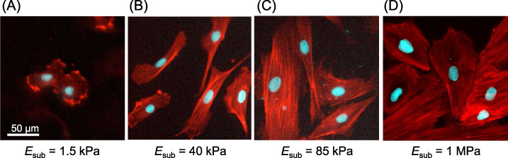
Typical examples of fluorescent images of F-actin (red) and the nucleus (cyan) in vascular smooth muscle cells cultured on the polyacrylamide (PA) gel (A–C) and PDMS (D) substrates. Thick bundles of actin stress fibers were observed clearly in cells on polyacrylamide gel substrates with medium stiffness (40 kPa), stiffer (85 kPa), and PDMS hard (1 MPa) substrates. The cell numbers also increased significantly with the increase in substrate stiffness.
Next, we analyzed the surface microstructure and mechanical properties of VSMCs grown on PA gel and PDMS substrates, using live cell AFM imaging (Fig. 3). The living VSMCs on the soft (1.5 kPa) substrates exhibited smooth surface (Fig. 3F), resulting in homogeneous surface mechanical properties (Fig. 3J). On the other hand, in the medium (40 kPa), stiffer (85 kPa) and hard (1 MPa) substrates, cell height decreased in substrate stiffening (Fig. 3G–I), and the cells clearly showed microstructures of thick actin stress fibers on the apical surface of the cells, which was represented as a linear distribution with higher elastic modulus (Fig. 3K–M), especially in the PDMS hard substrates (Fig. 3M).

An example of the atomic force microscopy (AFM) force-indentation curves of the cells described in J–M (A). The phase contrast images (B–E), surface topography (F–I), and mechanical properties (J–M) of vascular smooth muscle cells cultured on polyacrylamide gel (1.5 kPa, 40 kPa, 85 kPa) and PDMS (1 MPa) substrates. Precisely controlled, high-speed indentation tests were performed to obtain a force-indentation curve at each 128 × 128 pixel. The cell stiffness increased significantly with the increase in substrate stiffness.
Finally, we assessed the effects of substrate stiffness on vascular smooth muscle differentiation using fluorescent image analysis (Fig. 4). VSMCs on the soft (1.5 kPa) substrates showed weak fluorescence of 𝛼-SMA and its expression level was quite different in each cell (Fig. 4A). In contrast, thick bundles of 𝛼-SMA colocalizing on F-actin fluorescence were clearly observed in cells on the medium (40 kPa) substrates (Fig. 4B, white arrows). Although such 𝛼-SMA bundles were also observed in cells on the stiffer (85 kPa) and hard (1 MPa) substrates, their distribution was restricted at the center of the cells (Fig. 4C and D, white arrowheads), and the fluorescence intensities of 𝛼-SMA were significantly lower than those of their F-actin cytoskeleton (Fig. 4C and D).

Typical examples of the fluorescent images of F-actin (red), nucleus (blue), and 𝛼-SMA (green) in vascular smooth muscle cells cultured on polyacrylamide gel (A–C) and PDMS (D) substrates. Note that the 𝛼-SMA to F-actin cytoskeleton expression ratio in cells on substrates with medium stiffness (40 kPa) was significantly higher than those of cells on other substrates.
A summary of the results of our morphological and mechanical analysis is shown in Fig. 5. Cell spreading area, A, and surface elastic modulus, E, significantly increased in the medium (40 kPa) substrates and their increase became gently in the stiffer (85 kPa) substrates with no statistical significance (Fig. 5A and D). Both A and E of the cells on the PDMS hard substrates significantly increased compare to those on PA gels (Fig. 5A and D). Cell height, H, simply decreased with substrate stiffening (Fig. 5C). On the other hand, the S.I. of VSMCs on the medium (40 kPa) substrates was significantly smaller than those of the other groups (Fig. 5B), indicating that the medium stiffness substrate promoted cell elongation and shape polarization. Expression indices of F-actin, I F-actin × A, increased with substrate stiffening, while those of 𝛼-SMA, I 𝛼-SMA × A, reached plateau in the stiffer substrates (85 kPa) (Fig. 5E). F-actin expression particularly showed a two- and threefold increase in cells on the stiffer (85 kPa) and hard (1 MPa) substrates, respectively (Fig. 5E, red bars). However, the intensity ratio of 𝛼-SMA to F-actin, I 𝛼-SMA∕I F-actin, significantly decreased in cells on the stiffer (85 kPa) and hard (1 MPa) substrates and increased in cells on the medium (40 kPa) substrate than for other groups (Fig. 5F), indicating that the medium stiffness substrate facilitated vascular smooth muscle differentiation.

Changes in the projected cell area (A), shape index (B), cell height (C), whole cell stiffness (surface elastic modulus) (D), F-actin or 𝛼-SMA expression (E), and the expression ratio of 𝛼-SMA to F-actin (F) in vascular smooth muscle cells cultured on polyacrylamide gel (1.5 kPa, 40 kPa, 85 kPa) and PDMS (1 MPa) substrates. C and D were obtained by AFM measurements, the others were assessed with fluorescent images. Data were expressed as mean ± S.E.M.
Researchers have embraced the concept that substrate stiffness potentially affects cellular function, such as cell proliferation [11], migration [12,13], and differentiation [14], and several studies on the responses of VSMCs have been reported. Sazonova et al. [9] showed that the expression of proteins associated with cell-matrix adhesion was regulated by substrate stiffness, and a selective increase in cell density. Substrate stiffening also induced a contractile-to-synthetic phenotypic transition in VSMCs through the down-regulation of DNA methyltransferase expression [10]. However, many previous studies only focused on changes in biochemical factors such as protein or mRNA expression of cells during substrate stiffening. An advantage of this study is that we focused on the changes in cell morphology and intercellular tension that are deeply involved with cellular functions; we assessed the correlation between cellular mechanical responses and differentiation, using fluorescent microscopy image-based analysis and nano-indentation imaging with AFM.
We found that substrate stiffening from the soft (1.5 kPa) to the medium (40 kPa) induced F-actin cytoskeleton maturation (compare Fig. 2A and B) and cell spreading (Fig. 5A) in VSMCs. These phenomena with substrate stiffening have been considered a global response of cells, and have also been observed in previous studies using VSMCs [9,21] and other mesenchymal cells [22]. The significant increase of the cell spreading area was also observed in the PDMS hard (1 MPa) substrates, which was more than an order of magnitude stiffer than PA gels (Fig. 5A). However, in this study, no significant increase was observed in the spreading area and maturation of F-actin cytoskeletons (compare Fig. 2B and C) from cells on the medium (40 kPa) to stiffer (85 kPa) PA gel substrates. Furthermore, significant differences were also not observed in cell height and whole cell stiffness between the medium (40 kPa) and stiffer (85 kPa) PA gel substrates (Fig. 5C and D). On the other hand, cells on the stiffer (85 kPa) substrate clearly showed a linear distribution with higher elastic modulus (Fig. 3L), which was similar to those of cells on the PDMS hard substrates (Fig. 3M), compared with cells on the medium (40 kPa) substrate (Fig. 3K), indicating that internal tension on actin stress fibers significantly increased in VSMCs on stiffer (85 kPa) and hard (1 MPa) substrates. This suggests that the tensional response of individual actin stress fibers is quite sensitive to substrate stiffness.
Interestingly, the S.I. value was significantly decreased in VSMCs on substrates with medium stiffness (40 kPa) (Fig. 5B), indicating that cells showed remarkable elongation and shape polarization. Consequently, their 𝛼-SMA to F-actin cytoskeleton expression ratios were significantly higher than those of cells on other substrates (Fig. 5F). In contrast, cells on the stiffer (85 kPa) PA gel substrate showed irregular shapes with randomly oriented F-actin (Fig. 2C) which was similar to those of cells on the PDMS hard substrates (Fig. 2D), and their 𝛼-SMA to F-actin expression ratios were reduced compared to those of cells on the medium (40 kPa) substrates (Fig. 5F). Quinlan et al. [23] reported that the expression of 𝛼-SMA in valvular interstitial cells were upregulated on the 15–80 kPa substrate, but not on the glass (70 GPa) substrate. Scott et al. [24] investigated the response of adventitial fibroblasts on substrates with different stiffness (1 kPa, 3 kPa, 10 kPa), and revealed that 𝛼-SMA expression increased in cells cultured on the substrate with medium stiffness (3 kPa). These reports and the results of our study indicate that there exists an optimal substrate stiffness ranging from several kPa to several 10 kPa, which promotes VSMC differentiation, and that cell shape polarization might be a key factor for VSMC differentiation.
Incidentally, in vivo normal VSMCs keep their contractile phenotype and adhere to elastin lamina, whose elastic modulus was reported to be over 400 kPa [25]. A discrepancy exists between this in vivo value and the reported value of optimal substrate stiffness for VSMC differentiation obtained from in vitro studies, including our present study (40 kPa). Nagayama et al. [26,27] measured the mechanical properties of VSMCs, freshly isolated from vascular tissue, and they reported that the elastic modulus of freshly isolated VSMCs was ∼15 kPa in a relaxed state, and it increased to ∼90 kPa, following contractile activation. Considering the above, optimal substrate stiffness for VSMC differentiation might be equivalent to the stiffness of contractile VSMCs themselves.
Conclusion
We investigated the effects of substrate stiffness on the morphology, intercellular tension, and differentiation of VSMCs using PA gels and PDMS substrates, whose elastic moduli were 15 kPa, 40 kPa, 85 kPa, and 1 MPa. Using a combination of fluorescence microscopy image-based analysis and nano-indentation imaging with atomic force microscopy, we found that cell spreading and cell stiffening was induced by substrate stiffening in VSMCs. Interestingly, VSMCs on substrates with medium stiffness (40 kPa) showed significant elongation and shape polarization, and their 𝛼-SMA to F-actin cytoskeleton expression ratio was significantly higher than those of cells on other substrates. These results indicate that there exists an optimal substrate stiffness to promote VSMC differentiation and cell shape polarization, which might be a key factor for VSMC differentiation.
Footnotes
Acknowledgements
This work was supported in part by a Grant-in-Aid from the Ministry of Education, Culture, Sports, Science and Technology, Japan (nos 17H02077 and 19K22944); the Naito Foundation, Japan; the Takahashi Industrial and Economic Research Foundation, Japan; and AMED-CREST from the Japan Agency for Medical Research and Development (AMED) (nos JP19gm0810005 and JP20gm0810005).
Conflict of interest
The authors declare no conflict of interest.