
Abstract
BACKGROUND:
Nowadays, biomaterials used as a scaffold must be easy to deliver in the bone defect area. Extracellular matrix (ECM) hydrogels are highly hydrated polymers that can fill irregular shapes and act as bioactive materials.
OBJECTIVE:
This work aims to show the effects of ECM hydrogels derived from bovine bone (bECMh) on proliferation, cytotoxicity and expression of pro-inflammatory cytokines in three cells types involved in tissue regeneration, as well as biocompatibility in vivo.
METHODS:
In vitro, we used an extract of bECMh to test it on macrophages, fibroblasts, and adipose-derived mesenchymal stem cells (AD-MCSs). Cell proliferation was measured using the MTT assay, cytotoxicity was measured by quantifying lactate dehydrogenase release and the Live/Dead Cell Imaging assays. Concentrations of IL-6, IL-10, IL-12p70, MCP-1 and TNF-α were quantified in the supernatants using a microsphere-based cytometric bead array. For in vivo analysis, Wistar rats were inoculated into the dorsal sub-dermis with bECMh, taking as reference the midline of the back. The specimens were sacrificed at 24 h for histological study.
RESULTS:
In vitro, this hydrogel behaves as a dynamic biomaterial that increases fibroblast proliferation, induces the production of pro-inflammatory cytokines in macrophages, among which MCP-1 and TNF-α stand out. In vivo, bECMh allows the colonization of host fibroblast-like and polymorphonuclear cells, without tissue damage or inflammation.
CONCLUSIONS:
The results indicate that bECMh is a biocompatible material that could be used as a scaffold, alone or in conjunction with cells or functional biomolecules, enhancing proliferation and allowing the filling of bone defects to its further regeneration.
Keywords
Introduction
Work and sporting injuries, traffic accidents, degenerative diseases as well as infections can produce orthopedic trauma, most of which are bone and cartilage fractures that require surgery for their reconstruction and rehabilitation [1]. Regeneration is still dictated by three factors [2], that together are capable of restoring functions of affected or diseased organs [3–5]: extracellular matrix (ECM) which provides a scaffold, signaling biomolecules that regulate cell growth and differentiation, and stem cells required for the formation of tissue [6]. Thus, complete regeneration will be achieved if a structural scaffold is supplied, conferring mechanical properties, but also, stimulating migration of cell populations [7].
The use of autogenous graft is the optimal option to treat mineralized tissue lesions since it causes less immunological rejection [8]. However, the use of grafts from the same patient has some disadvantages, such as a second surgical intervention and the probability of infection. For this reason, a variety of materials have been developed for mineralized tissue engineering, being the most used isolated components of the ECM [9,10].
The ECM is the natural and ideal biological scaffold material. For this reason, in natural conditions, is custom designed and produced by the resident cells of each tissue and organ [11]. The main functions of ECM are to support and provide the scaffolding and signaling for cells to communicate with each other and with their external environment [8]. The composition of ECM has not been fully described, for this reason, the synthesis of an emulated ECM in vitro is not possible yet. However, isolation of ECM individual components such as collagen, fibronectin, laminin, and hyaluronic acid can be performed. Besides, ECM scaffolds can also be harvested from different tissues as bone, small intestine, liver, pancreas, skin, and urinary bladder, among others [11].
Nowadays, there is a demand for materials to let easy delivery by injection and that can be cross-linked in vivo to form a scaffold. These requirements are accomplished by hydrogel formulations giving, as a result, a physical and chemical cross-linked hydrophilic scaffold that can be used to deliver biological molecules and provides a 3D environment similar to native ECM [12]. Hydrogels are defined as highly hydrated polymer materials ( >30% water by weight). ECM hydrogels offer benefits such as injectability, the ability to fill an irregularly shaped space, and the sufficient bioactivity of a native matrix [13]. The polymer chains can be synthetic or natural as alginate, chitosan, collagen, hyaluronic acid. Synthetic and natural hydrogels can be applied to fill spaces, deliver bioactive molecules, drugs, and cells to stimulate tissue growth [14]. Natural hydrogels can be isolated from human, porcine, and bovine tissues [15,16], and can be used for proliferation, cell differentiation, and tissue formation stimulation [17–19].
Hydrogels have been considered the biomaterial of choice for tissue engineering applications [20] and evidence support that ECM hydrogels derived from bovine bone (bECMh) can promote proliferation, as well as osteogenic differentiation of cell lines and primary cells in vitro [15,21,22]. Nevertheless, most of the studies-perspective is based on the study of a single type of cell or tissue. Besides, there is a gap in determining the biocompatibility and effects of bECMh in vivo. Thus, the focus of the present article is to show the effects of bECMh on proliferation, cytotoxicity and expression of pro-inflammatory cytokines in vitro in cells involved in tissue regeneration such as macrophages, fibroblast and mesenchymal stem cells, as well as biocompatibility in vivo.
Materials and methods
Preparation of ECM hydrogel derived from bovine bone (bECMh)
Fresh bovine tibiae were collected from a certified trail. Cancellous bone was selected for ECM extraction following the methodology of Sawkins et al. [15]. Briefly, the cancellous bone was demineralized in 0.5 N HCl (25 ml/gr of bone) at room temperature for 24 h under stirring. Then, lipids were extracted with a 1:1 mixture of chloroform and methanol and then lyophilized. Enzymatic decellularization was performed with 0.25% trypsin and ethylenediaminetetraacetic acid (EDTA) solution (GIBCO Life Technologies, USA) at 37 °C and 5% CO2 under stirring for 24 h. Digestion and solubilization were carried out with pepsin (Sigma-Aldrich, USA) at a concentration of 1 mg/ml in 0.01 N HCl, on a stirring plate until solubilization, to form bovine-bone ECM (bECM) pre-gel. Then, the bECMh was obtained by mixing the bECM pre-gel with PBS, and NaOH (1 M) allowing to gel for 30 min at 37 °C and then it was stored at 20 °C.
Finally, to determine cellular content, samples before and after trypsin digestion were fixed in 10% formalin, embedded in paraffin, and cut into 7-μm sections. The slices were stained with H&E and images were taken at 10× and 40× using a TCS SP8 microscope system (Leica, Germany).
Protein analysis
For a qualitative analysis of collagen in bECMh, sodium dodecyl sulfate (SDS)-polyacrylamide gel electrophoresis (PAGE) was carried out, using 5% (w/v) polyacrylamide (Bio-Rad, USA) for the separating gel and 4% (w/v) polyacrylamide for the stacking gel. The bECMh and purified Collagen type 1 (COL-1) as a control (Roche, Germany) were prepared in 0.5 M Tris-HCl buffer (pH 6.8) containing 10% SDS, 30% glycerol, 1% 2-mercaptoethanol, and 0.02% bromophenol blue and then heated to 95 °C for 5 min. Next, 4 μL of a protein marker (Himark Unstained protein Standard, Thermo, USA), and 5 μL of the samples were loaded and electrophoresed at 100 V on vertical slab gels until the bromophenol blue had moved out of the gel. Polyacrylamide gels were stained for 2 h with 0.1% Coomassie blue R-250 in acetic acid/methanol/water 2:5:5 (v/v/v) and were destained in 7.5% acetic acid/15% methanol.
Cell culture conditions for biocompatibility assays
RAW 264.7 murine macrophages (TIB-71, ATCC), NIH/3T3 murine fibroblast (CCL-92, ATCC), and human AD-MCSs Adipose-Derived Mesenchymal Stem Cells (PCS-500-011, ATCC) cell lines and were grown with Dulbecco’s Modified Eagle’s Medium (GIBCO Life Technologies) supplemented with 10% heat-inactivated fetal bovine serum (GIBCO Life Technologies) and Penicillin-Streptomycin (10,000 U/mL) (GIBCO Life Technologies) at 37 °C with 5% CO2. Cells of the third passage were used at 80% of cell confluence for all experiments. Cells were harvested using 0.25% trypsin-EDTA (GIBCO Life Technologies).
bECMh-extracts preparation for biocompatibility assays
Soluble extracts from bECMh were prepared as follow: 1 ml of hydrogel (40 mg/ml) and 9 ml of DMEM were placed with 0.05 mm glass in a tissue disruptor for 1 minute at maximum speed (Disruptor Genie, Scientific Industries, USA) and incubated at 37 °C with constant slow orbital shaking for 24 h. Finally, samples were centrifuged at 13,000 rpm, the recovered supernatants were filtrated with 0.22 μm membrane (Cytiva Whatman, Uniflo Syringe Filters, USA), and then used for LDH, MTT, live/dead, and cytokine expression assays. Cells treated with DMEM were used as controls.
Cellular viability assay
Cell proliferation/viability was measured using the MTT assay (CellTiter 96 NON-Radioctive Cell Proliferation Assay, PROMEGA, USA). RAW 264.7, NIH/3T3, and AD-MCSs were cultured into a 96 well plate with supplemented medium DMEM placing 7.5 × 10³ cells/well. At 24 h media were replaced by bECMh-soluble-extract and after 24, 48, and 72 h of incubation measures were performed. Briefly, media were removed and the cells were incubated with 3-(4,5-dimethylthiazol-2-yl)-2,5-diphenyltetrazolium bromide. 15 μl of MTT solution was added to each well for 2 h. The violet formazan salts were then dissolved by adding 100 μl of stop solution. OD was measured at 570 nm in a microplate reader.
Cytotoxicity of bECMh assay
RAW 264.7, NIH/3T3, and AD-MCSs were cultured into a 96 well plate with supplemented medium DMEM placing 15 × 103 cells/well. At 24 h medium was replaced by bECMh-soluble-extract and after 24 h of incubation, cytotoxicity was measured by quantifying lactate dehydrogenase (LDH) release in supernatants by colorimetric assay following the manufacturer’s instructions (Sigma-Aldrich, St. Louis MO, USA). The optical density (OD) of the samples was measured at 490 nm in a microplate reader.
Live/Dead assay
RAW 264.7, NIH/3T3, and AD-MCSs were cultured into a 24 well plate with supplemented medium DMEM placing 10 × 104 cells/well. At 24 h media were replaced by bECMh-soluble-extract and after 24 h the cells were incubated with triazole orange 84 nM (Sigma-Aldrich) and propidium iodide 4.3 μM (Sigma-Aldrich) for 5 min at room temperature. The samples were then examined using a TCS SP8 microscope system at 40×. Relative fluorescence units (RFU) were measured, and percentages for cell viability were obtained.
Cytokine expression analysis
RAW 264.7, were cultured into a 24 well plate with supplemented medium DMEM placing 5 × 10⁵ cells/well. At 24 h media were replaced bECMh-soluble-extract and after 24 h of incubation, IL-6, IL-10, IL-12p70, and MCP-1 TNF-α were quantified in the supernatants using a microsphere-based cytometric bead array (CBA) according to manufacturer’s instructions (BD Bioscience, USA). Data were acquired using Accury C6 flow cytometer and quantification was done using FCAP Array software v3.0 (BD Bioscience). Cells treated with LPS were used as positive controls.
In vivo biocompatibility
3-month-old Wistar rats (250–300 gr) were anesthetized by placing 1 μL of sedative cocktail (70% Ketamine, 15% Xylazine, 15% injectable water) per mg of weight, intramuscularly. The back was shaved and disinfected with 70% alcohol. With a insulin syringe, 100 μl of bECMh, positive control (chloroform, Sigma-Aldrich) and negative control (PBS 1X) were inoculated into the sub-dermis, taking as reference the midline of the back, and the specimens were sacrificed at 24 h. Subsequently, 2 cm in diameter of tissue were cut around the inoculation area. Gross Examination was carried out, then, tissue was fixed with 1X PBS with 4% paraformaldehyde, and histological evaluations (H&E) were carried out to observe the inflammatory reaction. Ethical issues use of animals was approved according to the signed statement of the Animal Ethics Committee of the Faculty of Biological and Chemical Sciences, Universidad Autónoma de Sinaloa.
Statistical analyses
Experiments were performed three times by triplicate, and results were represented as mean ± SD. ANOVA and Dunnet’s post hoc test were used for 3 or more parametric variables. p < 0.05 was considered as statistically significant.
Results and discussion
bECMh obtained from DBM is mainly a cell-free collagen-rich scaffold
After demineralization of bone using HCl and lipid extraction, enzymatic decellularization with trypsin/EDTA was assessed by imaging and analysis of (H&E) stained section (Fig. 1A and 1B). Demineralizated bone ECM (DBM) without trypsin treatment was used as control showing cellular content with observable nuclei (black arrows). On the other hand, the absence of nuclei and the presence of acellular lacunae is evident in bECM after trypsin/EDTA treatment (red arrows), demonstrating that Sawkins et al. [15] methodology is enough to obtain a cell-free scaffold.

bECM decellularization and electrophoretic protein profile. (A) DBM shows cellular content before treatment with trypsin/EDTA (black arrows). (B) Free-cell regions with the absence of nuclei and the presence of acellular lacunae (red arrows) are evident in bECM after trypsin/EDTA treatment. (C) To analyze qualitatively the presence of collagen in bECMh an SDS-PAGE was runned using COL-1 as a control. The molecular weight (MW) of bands of α1, α2, β, and γ were calculated based on the values of retention factor (Rf). bECMh showed a high content of α1 and α2 chains when compared to COL-1. Histological slides (H&E) magnification 40×, scale bar 100 μm.
Then, to analyze qualitatively the presence of collagen in bECMh an SDS-PAGE was runned using COL-1 as a control. The molecular weight (MW) of bands of α1, α2, β, and γ were calculated based on the values of retention factor (Rf). bECMh showed a high content of α1 and α2 chains when compared to COL-1 (Fig. 1C). This is relevant since type 1 COL exerts very important functions working as a scaffold for bone cells, maintaining bone strength [23], promoting bone formation, and regulating collagen fibrillogenesis [24]. This characteristic of bECMh would enable the interaction with other collagenous and non-collagenous proteins to assemble fibril bundles and fibers [25] that are needed to form an ideal niche for tissue regeneration as is required for hydroxyapatite (HA) deposition [26].
To analyze proliferation induced by bECMh extract, an MTT assay was developed in macrophages, fibroblasts and AD-MSCs. All cell lines showed increased cell viability from 24 to 72 h with both, bECMh and DMEM (Fig. 2A, B, and C). Interestingly, fibroblast showed an increased proliferation at 48 h when compared with DMEM control (p = 0.02) and this behavior was maintained until 72 h. Meanwhile, macrophages and AD-MSCs and macrophages showed a higher proliferative behavior enhanced by DMEM stimuli. Previous studies have already shown that proliferation in different cell lines is positively affected by the presence of DBM hydrogels. For instance, mouse primary calvarial cells (mCPs) proliferation is enhanced by pepsin-digested and solubilized bDBM hydrogel [15]. bECMh is obtained eliminating all cellular components which facilitate rapid degradation of scaffolds and it is followed by replacement with site-appropriate functional host tissue and cells. The presence of cellular remanents has been linked with macrophage polarization profile to M1 and pro-inflammatory phenotype [27]. In our case, the lack of cellular content can explain the low macrophages proliferation, avoiding macrophages polarization and the resultant pro-inflammatory phenotype. On the other hand, fibroblast proliferation is needed for de novo ECM formation [28]. The induction of fibroblast proliferation is key since stromal cells can serve as a source of growth factors and cytokines to support and enhance regeneration [29]. Recent cell-based therapies are based on the use of fibroblast, ECM, and cryopreserved placental tissue to induce tissue regeneration [30]. Nevertheless, is also known that failure in tissue regeneration most commonly results in chronic inflammation and/or fibrosis (orchestrated by fibroblasts), which leads to damaged neoformation of nonfunctional tissue [31]. This can be remedied in real conditions, by the presence of MSCs in the zone, since they have an antifibritic effect that is related to their anti-inflammatory and angiogenic properties [32]. Besides, MCSs secrete cytokines that inhibit fibroblasts [33] and factors such as Hepatocyte growth factor released by MSCs that can downregulate the expression of TGF-β1, COL-1, and COL-3 in fibroblasts, and upregulate the expression of matrix metalloproteinases 1, 3, and 13, thus promoting the turnover of ECM [33]. The combination of these cell types in the niche is important, and there is evidence showing that ist co-habitability minimizes scarring in the regeneration zone [34].
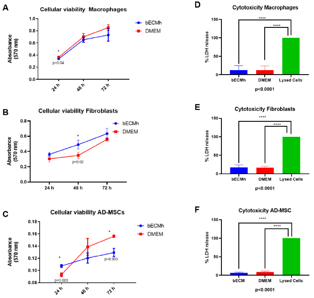
To analyze proliferation induced by bECMh extract, MTT assay was developed in macrophages (A), fibroblasts (B) and AD-MCSs (C). All cell lines showed increased cell viability from 24 to 72 h with both, bECMh and DMEM. To evaluate cytotoxicity, a total of 4 mg/mL were incubated in contact with macrophages (D), fibroblasts (E) and AD-MCSs (F). Interestingly, we found that LDH release patterns were similar among the different cell lines when compared to the DMEM group (BECMh versus DMEM) and statistical differences were only found when OD was compared to LDH release of control (lysed cell,p < 0.0001).
Besides, for evaluating cytotoxicity of the soluble bECM hydrogel extract, a total of 4 mg/mL were incubated in contact with macrophages, fibroblasts, and AD-MSC. Interestingly, we found that LDH release patterns were similar among the different cell lines when compared to the DMEM group (bECMh versus DMEM). Optical densities (OD) were higher in macrophages (0.135 ± 0.08 versus 0.102 ± 0.04 OD) in comparison to fibroblasts (0.072 ± 0.03 versus 0.067 ± 0.02 OD) and AD-MCSs (0.048 ± 0.02 versus 0.062 ± 0.01 OD) and statistical differences were only found when OD was compared to LDH release of control (lysed cell, p < 0.0001).
As cytotoxicity is a basic evaluation to analyze biocompatibility of materials and an important parameter for using a biomaterial as scaffold we also evaluated cell viability of macrophages, fibroblast and AD-MSCs in contact with bECMh extract by using LIFE/DEATH Cell Viability Assay by confocal microscopy. We found a very similar behavior, cell viability did not show significant differences (bECMh versus DMEM). Percentages of live cells of macrophages (93.7% versus 94.3%), fibroblast (92.3% versus 93.3%) and AD-MSCs (95.3% versus 96.3%) (Fig. 3A, B, and C) demonstrated that bECMh is biocompatible with multiple cell lines. Moreover, cellular morphology was not compromised after soluble extract exposure as was confirmed by confocal microscopy (Fig. 3D, E, and F). There are a plethora of studies showing that ECM-based hydrogels are cytocompatible and promote differentiation as well as proliferation [22,35,36]. For instance, it has been shown that Osteocalcin found on ECM, is prone to increase MSCs proliferation rates in a dose-dependent manner, meanwhile, osteopontin induces MSCs to undergo differentiation [37]. In this regard, MSCs proliferation behavior in our study could be induced by the same phenomena, nevertheless, more studies are needed to confirm this hypothesis.
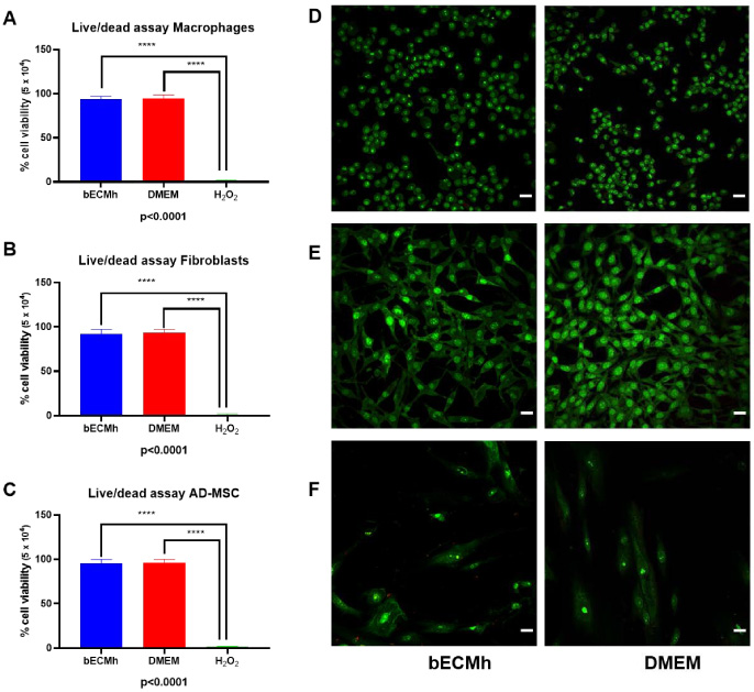
Cell viability of macrophages, fibroblasts and AD-MCSs in contact with bECMh extract was evaluated using LIFE/DEATH Cell Viability Assay by confocal microscopy. Cell viability did not show significant differences (bECMh versus DMEM) and differences were only found when compared to the positive control of dead (A, B and C) demonstrating that bECMh is biocompatible with multiple cell lines. Moreover, cellular morphology was not compromised after soluble extract exposure as was confirmed by confocal microscopy (D, E, and F).
Cytokines profile induction is important to get insight into bECMh impact on inflammation promotion. After hydrogel extract exposure on macrophages, we found an increase in pro-inflammatory cytokines IL-6 (79.18 ± 25.68 versus 11.10 ± 0.37 pg/mL) (Fig. 4A) and TNF-α (1157.99 ± 27.29 versus 247.37 ± 18.91 pg/mL) (Fig. 4B). For chemotactic cytokine MCP-1, we also found an increase (634.70 ± 16.38 versus 118.96 ± 6.40 pg/mL, p = 0.000) (Fig. 4D). On the other hand, IL-12 concentration (25.57 ± 5.26 versus 16.07 ± 7.39 pg/mL) and anti-inflammatory cytokine, IL-10 (43.22 ± 2.76 versus 40.39 ± 4.55 pg/mL) (Fig. 4C and E) concentration were not altered by bECM hydrogel extract exposure.
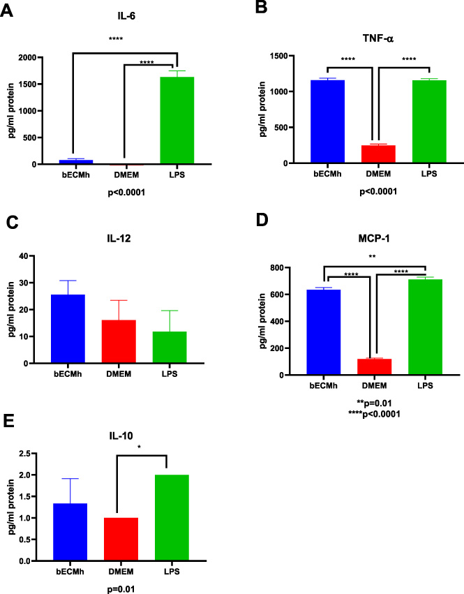
Proinflammatory cytokine production in macrophages exposed to bECMh extract. We found an increase in pro-inflammatory cytokines IL-6 (A) and TNF-α (B). For chemotactic cytokine MCP-1, we also found an increase (D). On the other hand, IL-12 concentration and anti-inflammatory cytokine, IL-10 (C and E) concentration were not altered by bECMh extract exposure.
In natural conditions, an injury would promote inflammation for increasing blood flow and vascular permeability, causing accumulation of fluid, leukocytes, and inflammatory mediators’ recruitment, as well as proinflammatory cytokine upregulation, especially IL-1β and TNF-α [38,39]. There is evidence that pro-inflammatory cytokines, such as TNF-α are required for hepatic tissue regeneration by increasing mitotic rates in a dose-dependent manner [40,41]. In this regard, IL-6 is also increased in hepatic tissue regeneration, it is known that TNF-α orchestrates IL-6 production [42] suggesting that bECMh hydrogel promotes both proinflammatory cytokines to initiate a reparative or regenerative process. Moreover, recent studies have demonstrated that the expression of the chemotactic cytokine MCP-1 is important in the promotion of angiogenesis and vasculogenesis, as well as, regeneration of dent pulp vascularization for tissue regeneration [43,44]. There is evidence that MCP-1 promotes angiogenesis by the regulation of VEGF and HIF-α by the activation of the ETS-1 transcription factor [45]. All this suggests that MCP-1 could be a promoter of angiogenesis by the activation of secondary angiogenic factors by the stimulation of bECMh. After 24 hours bECMh stimulation, the null detection of IL-12 pro-inflammatory cytokine, might be due to IL-12 rapid response in wound healing, suggesting its degradation before 24 h. Li and Cols found that IL-12 induces significantly more rapid onset and higher metabolic activity in the inflammatory response on wounded skin during the early time points of wound healing [46]. Besides, the lack of IL-12 and IL-23 exhibit enhanced bone formation by indirectly inhibiting bone marrow mesenchymal stem cells to osteoblasts differentiation in vivo [47], supporting that IL-12 is pivotal for inflammation initiation and metabolized in the early stages of regeneration. Nonetheless, further experiments are required to confirm these possible mechanisms.
After 24 h of bECMh inoculation into the dorsal sub-dermis, gross examination showed no damage in surrounded tissues (Fig. 5A) and that the hydrogel had good gelatinization, since remains stable in a well-defined area, which can help cells to occupy its space. Histological evaluation showed that, in fact, bECMh showed fibrillar bECMh structure and was populated by few host fibroblast-like cells and few inflammatory cells (Figs 5B and C), while, as expected, the inoculation of chloroform, resulted in severe tissues damage (Fig. 5D) with presence of a strong inflammatory infiltrate, mostly epithelioid-type macrophages, neutrophils and fibroblast-like cells (Figs 5E and F). These results agree with those shown by Výborný et al., given that their extracellular matrix hydrogel derived from the human umbilical cord also enhances the colonization of host cells in the implantation zone [48]. Future studies are needed to investigate the application of bECMh to repair bone defects in animal models.
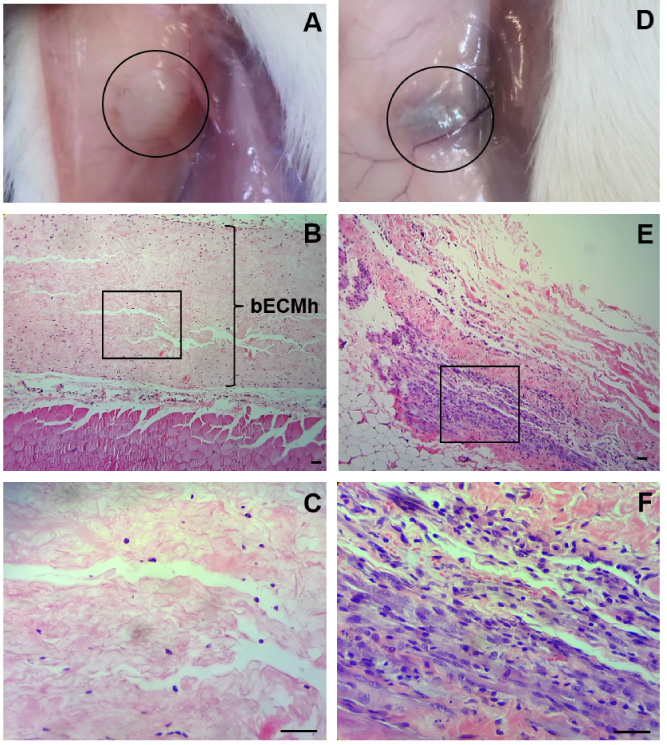
Biocompatibility in vivo of bECMh. 24 h after subcutaneous injection in the dorsal back region, of bECMh (A, B and C) and chloroform (D, E and F) as control of damage, reaction tissue was observed. bECMh remains stable in a well-defined area (black circle), without any abnormality of the dermis (A). At 10×, low cellularity was observed within the material (B). A higher magnification (40×) (black box) showed the fibrillar bECMh structure, with presence of few polymorphonuclear and fibroblast-like cells. In contrast, chloroform injection caused severe damage inducing an hematoma (D). At 10×, the inflammatory infiltrate can be seen (E) consisting in epithelioid-type macrophages interspersed with some fibroblasts, as well as small blood vessels in a dense connective tissue stroma, as showed at 40× (F). Scale bar 50 μm.
bECMh (ECM hydrogel derived from bovine bone) production methodology is reproducible and easy to implement. In vitro, this hydrogel behaves as a dynamic biomaterial that increases fibroblast proliferation induces the production of pro-inflammatory cytokines, among which IL-6, TNF-α, and MCP-1 stand out. In vivo, bECM allows the colonization of few host fibroblastic-like cells and polymorphonuclears, without tissue damage or inflammation. These results indicate that bECMh is a biocompatible material that could be used as a scaffold alone or in conjunction with cells, enhancing proliferation and allowing the filling of bone defects to further regenerate lost tissue.
Footnotes
Acknowledgements
None to report.
Author contributions
AAH, MAM, MB, JLG, GJG, MAF and ELV conducted laboratory tests. HCU, JGRQ and JLF collected the data and analyzed the results. RRP, MAM, JGRQ and CLC conceived and designed the study. RRP, MAM and CLC wrote the manuscript. All authors read and approved the final manuscript.
Conflict of interest
The authors declare no conflicts of interest.