
Abstract
BACKGROUND:
Hypertensive disorders during pregnancy pose significant risks to both maternal and fetal health, necessitating safe and effective therapeutic interventions.
OBJECTIVE:
This study aimed to investigate the potential of an extract derived from Falcaria vulgaris (FV), loaded with exosomes to form the Exo/FV complex, as a novel therapeutic agent for the management of hypertension in pregnant mice: antioxidants, antimicrobials, and phenolic compounds present in FV lower blood pressure.
METHODS:
The isolation of exosomes was done by ultracentrifugation methods and the FV was loaded into the exosomes by electroporation method.
RESULTS:
The Exo/FV was found to be spherical with diameter ranges from 20 to 30 nm and they were tested for biocompatibility in NHI 3T3 cell lines and found to be effective. This research investigated in vivo hypertension in mice induced by L-NAME and treated with FV and Exo/FV and found that AChE and MAO determine mice's redox state tends to reduce blood pressure. Increased non-protein thiol (NP-SH) and decreased lipid peroxidation were also found, and PDE-5, ACE, Arginase, and MDA activity has also been tested.
CONCLUSION:
This analysis showed that Exo/FV effectively treated hypertension during pregnancy.
Keywords
Introduction
Pregnancy hypertension (preeclampsia) is a common condition among pregnant women and was recorded as the second-highest cause of maternal mortality [1]. Preeclampsia is responsible for the deaths of more than 70,000 mothers and 500,000 infants and young children [2]. Worldwide, 5–10% were affected by hypertensive disorders of pregnancy (HDP) [3]. The types of hypertensions that may develop during pregnancy include preeclampsia, gestational hypertension, and preeclampsia on top of chronic hypertension. Consequently, the new criteria for defining stage 1 hypertension include a diastolic reading of 80–89 mmHg and a systolic reading of 130–139 mmHg. The most recent studies indicate that hypertension during pregnancy is characterized by blood pressure readings above 140 mm Hg systolic and 90 mm Hg diastolic. It is recommended that after giving birth, the blood pressure level should remain below 120/80 mm Hg to decrease the likelihood of adverse events [4]. The L-NAME was used to induce hypertension in mice models where the blood pressure was increased up to 130 mm Hg where the treatment of hypertension with angiotensin-converting enzyme (ACE) inhibitors has been associated with fewer side effects, shorter treatment times, and the development of novel, supposedly cost-effective anti-hypertensive drugs derived from plants [5]. This may result in significant risk factors for renal and cardiovascular disorders, hypertension is linked to oxidative stress and increased immunological responses [6]. To overcome these issues, this study found that extracts of Falcaria vulgaris (FV) were more effective than other plant extracts in treating hypertension, which is concerning given the global prevalence of the disease [7]. In traditional Chinese medicine, human immunostimulants made of herbs have been used for a long time to lead a healthier life [8]. The fast-growing plant FV, which is known as Ghazyaghi in Turkish and Paghazeh in western Iran, averages 30 cm in height and is a member of the Apiaceae (Umbelliferae) family which was used as a traditional medicine [9]. As a medicinal plant, FV has a long history of usage for various purposes, including wound healing, anti-inflammatory, antioxidant, antibacterial, antifungal, antiviral, reduces blood pressure, and is used as a bleeding inhibitor [10]. In addition to lowering blood pressure, scientific evidence suggests that FV, whether in the form of a cooked infusion or the leaves of the plant, can act as a carminative, febrifuge, vulnerary, stomachic, and hemostatic agent. Additionally, they are non-toxic when used therapeutically and can be taken as a supplement [11]. Thus, loading the FV extract into exosomes has improved its activity in treating hypertension. When cells undergo normal or abnormal processes, they release vesicular structures called exosomes, which are found to be nanosized and can range from 30 to 150 nm [12]. They serve as a tiny carrier for drug delivery and evade immune recognition, their inherent targeting ability and non-existent toxicity to cells and tissues make them the peak of achievement in optimal drug delivery. Their composition is rich in non-lamellar-forming lipids, which could lead to a bilayer curvature that facilitates drug delivery [13,14]. Human umbilical cord mesenchymal stem cells, or hUCMSCs, are a promising source of multipotent stem cells for clinical use due to their strong proliferation ability, great differentiation potential, low incidence of immunological rejection, and ease of acquisition with less ethical concerns. They have various biological roles, such as regenerating membranes, regulating biological processes through miRNA transfer, and delivering antigenic molecules to T-cells in immune responses [15]. Their physicochemical and pharmacodynamical properties, such as low solubility, poor cell penetration, hepatic disposition, and fast uptake by normal tissues limit their pharmacological use. However, these limitations are circumvented when exosomes are used as a vehicle for drug delivery [16].
In this study, the process of loading FV extract into exosomes was validated through characterization studies. The synthesized drug was tested for cytotoxicity and antioxidant properties. In vivo, studies were conducted with mice models to assess the efficacy of the extract in treating hypertension, which involved inducing hypertension in the mice and then treating them with the Exo/FV.
Materials and methods
Reagents required
Ethanol, PBS, BCA protein assay kit (P0012, Beyotime, China), Butylated hydroxytoluene, 2,2-diphenyl-1-picrylhydrazyl, potassium persulfate, DMEM, FBS, N-nitro-Larginine methyl ester (L-NAME), DMSO, APC Annexin-V, PI staining solution, Dithiobisnitrobenzoic acid, Semicarbazide, Benzylamine, Thiobarbituric acid, α-isonitrosopropiophenone, sodium dodecylsulfate were obtained from Sigma Aldrich, USA, was the source for all of the other analytically graded compounds that were utilized.
Plant sampling and preparation
The plants of FV were purified after being purchased from the store. After cleaning, the stems and leaves were dried in the shade for a full week. A powdery consistency was achieved by milling them. A mixture of 100 g of powder and 70% ethanol was prepared in the following proportions (1:4). They were submerged in water at 35°C for 48 hours and then the solution was filtered through Buchner funnel filter paper using a vacuum pump. The highly concentrated extract was obtained using the rotary device after separating the solution from its solvent. This process was repeated until the desired concentration was reached. The next step was to use 0.2 μm filter paper to filter the extract, after which they were sterilized [17].
Isolation of exosomes
The hUCMSCs were cultured in a medium devoid of exosomes for approximately 48 hours. A conventional ultracentrifugation procedure was used to separate the exosomes from the culture medium. Centrifugation was used for 20 minutes at 2000g to extract debris from the medium, followed by 30 minutes at 10000g at 4°C. The exosomes that had been purified were mixed with PBS and kept at a temperature of −80°C until the next experiments. The exosomes were isolated as per the literature [18]. The protein content of the exosomes that were collected was ascertained using a bicinchoninic acid (BCA) protein assay kit and the presence of exosomes was confirmed by TEM analysis.
Preparation of FV-loaded exosomes
The FV was loaded into exosomes derived from hUCMSCs using the electroporation method. This method delicately combined 100 μg of exosomes purified with 50 μg of FV. Next, 200 μL of electroporation buffer was added and kept at 4°C. The mixture was kept at 37°C for 30 minutes after electroporation in 0.4 cm cuvettes at 350 V and 150 μF to ensure that exosome plasma membranes were completely repaired (Bio-Rad, USA). Following this, the unreacted compounds were removed by ultracentrifugation after being washed twice with cold PBS [19]. To ensure that the exosomes contained FV they were analyzed by DLS, zeta potential, FT-IR, and TEM.
In vitro anti-oxidant activity
The 2,2-diphenyl-1-picrylhydrazyl (DPPH) assay was used to measure the antioxidant activity. When adding 20 to 100 μg/mL of FV and Exo/FV, followed by addition of 5 mL of a 0.004% (w/v) DPPH solution, the mixture was vortexed. After 30 minutes of incubation at 22°C in a dark condition, the absorbance was measured at 520 nm. Butylated hydroxytoluene (BHT) was used for comparison. The ABTS assay was carried out with 10 mL of potassium persulfate and 1 μg/mL of FV and Exo/FV (20 to 100 μg/mL) then the mixture was allowed to settle down and the absorbance was measured at 734 nm using UV-visible spectrophotometer with vitamin C used as a control measurement were taken in triplicate. Where t denotes the absorbance of the test sample, c represents the absorbance of the control.

The growth and maintenance of the NIH-3T3 cells were carried out in a controlled environment with 5% CO2 and 37°C. The recommended medium was DMEM, which contains D-glucose supplemented with 10% FBS, 100 U/mL of penicillin, and streptomycin [20]. The MTT assay was utilized to determine the effect of FV loaded into exosomes (Exo/FV) for cell cytocompatibility on NIH-3T3 cells. The cells were subjected to trypsinization and hemacytometer counting in this particular assay. A density of 1 × 10⁵ cells/well was used to seed the cells in 96-well plates with 200 μL of DMEM overnight. Drug concentrations ranging from 20 to 100 μg/mL were tested on them at intervals of 48 hours. Afterward, 20 μL of MTT reagents was added to each well and maintained at 37°C for 4 hours. After the MTT medium was removed, 200 μL of DMSO was added to dissolve the formazan crystals, and the absorbance was measured at 570 nm.
Morphological analysis of the cell
The morphology of NIH-3T3 cells was examined before and after the administration of the Exo/FV. We exposed cells to increasing concentrations of Exo/FV (20 to 100 μg/mL) for 24 hours after placing them in 12-well plates. The morphological alterations were observed through inverted phase microscopy (Zeiss Axiovert 100) at a magnification of 20×.
Flow cytometer analysis
NIH-3T3 cells were analyzed using flow cytometry after 24 hours of incubation at 37°C at a density of 5 × 10⁴. We detected cell death with Annexin-V, PI staining solution, and Annexin binding buffer (BD Biosciences, San Diego, CA, USA). Before collection and washing with 1× PBS, cells were treated with Exo/FV at various concentrations (20 to 100 μg/mL) for 48 hours. After suspension in binding buffer, the cells were subjected to a 15-minute dark incubation with Annexin V and PI. After that, cells were counted using a flow cytometer (Millipore Corporation, Billerica, MA, USA).
In vivo studies on mice model
The study protocol was reviewed and approved by the Ethics Committee of the First Affiliated Hospital of Guizhou University of Traditional Chinese Medicine (No. 20230968). During the experiment, pregnant female albino mice (3 to 9) weeks old with a weight of 30–50 g approximately were chosen. The mice were randomly assigned to treatment and control groups and maintained on a 12-h light/dark cycle. A healthy diet and access to clean water were made possible for them. The hypertension was induced in the mice by L-NAME (Nω-nitro-L-arginine methyl ester), which was given orally to the mice (10 mg/kg/day) and analyzed [19]. We observed changes between the normal conditions and conditions induced by hypertension in the mice, and we grouped the animals based on the treatment given, and the grouping is shown in Table 1.
Grouping of animals according to the treatment given
Grouping of animals according to the treatment given
The systolic (SBP) and diastolic (DBP) pressure readings were recorded on conscious mice using an automatic sphygmotonography instrument (BP-98A, Softron, Beijing, China) and the non-invasive tail-cuff plethysmography method. Systolic and diastolic blood pressure readings were recorded during the final week of treatment after the mice had been trained to use the apparatus.
Preparation of homogenates from tissues
The heart, lung, and kidney tissues were ground in cold saline (1/10 w/v) using a teflon glass homogenizer for around ten half-strokes at 1200 revolutions per minute. The pellet at the tube bottom was removed after 10 minutes of centrifugation at 3000g, and the supernatant was retained for additional biochemical analysis. A standard known as bovine serum albumin was used to quantify the protein concentration using Bradford's Coomassie blue technique.
Determination of acetylcholinesterase activity
In this method, hypertension was determined using the AChE enzyme assay. The reaction mixture included 100 mM K+-phosphate buffer with a pH of 7.5 and 1 mM 5,5'-dithiobisnitrobenzoic acid (DTNB). To determine the yellow anion, 5, 5'-dithio-bis-acid-nitrobenzoic forms, this method measures the absorbance at 412 nm while the mixture is incubated at 25°C. The acetylcholinesterase test was initiated by adding 0.8 mM acetylthiocholine iodide after the homogenate had been pre-incubated for 2 minutes. The units per milligram of protein were used to assess the enzyme activity.
The Monoamine oxidase (MAO) inhibition assay
A 2 mL fraction of the supernatant containing 50 μg of protein, FV ranging from 0 to 100 μg/mL, a 0.025 M phosphate buffer with a pH of 7, 0.0125 M semicarbazide, and 10 mM of benzylamine make up the reaction mixture (pH adjusted to 7). The mixture was centrifuged after boiling for 3 minutes. Then 1 mL of supernatant was mixed with 2.5 mL of benzene and 0.05% of 2, 4-DNPH. The benzene layer was separated and blended with 0.1 N NaOH in an equal ratio. An 80°C-heating operation was undertaken on the decanted alkaline layer for 10 minutes and the color change was measured at 450 nm.
Lipid peroxidation and thiobarbituric acid reactive species
A 0.1 M Tris-HCl buffer with a pH of 7.4 was combined with 30 μl of the supernatant to conduct the lipid peroxidation experiment. To make up the volume up to 300 μl with water, the mixture was incubated at 37°C for 1 hour. The reaction mixture was supplemented with a 300 μL solution of 8.1% SDS to generate the color reaction. Further, 500 μL of a combination of acetic acid and hydrochloric acid with 500 μl of 0.8% TBA (pH 3.4) and incubated for an hour at 100 and measured at 532 nm.
Non-protein thiols (NP-SH) assay
The homogenate from the heart was combined in a 1:1 ratio with 10% trichloroacetic acid. Carefully, the supernatant was collected after centrifuging the solution at 10,000g for 5 minutes at 4°C to determine the free SH groups the methodology was followed as per the literature [5].
Phosphodiesterase-5 assay (PDE-5)
For the preparation of a 5 mM working solution, 55.84 mg of substrate p-nitrophenyl phenylphosphate was dissolved in 20 mL of Tris buffer (pH 7.4). In a water bath temperature set at 37°C for 10 minutes, the mixture of cardiac homogenate, phosphodiesterase enzyme, and substrate was then allowed to rest. A water bath maintained at 37°C was used to incubate the mixture, after mixing the concentrated tissue with 100 μL of the enzyme. After 30 seconds, the reaction mixture was supplemented with 1 mL of the substrate, and the changes were observed at 400 nm and calculated.
Angiotensin-I-converting enzyme assay
This study was carried out with 125 mM Tris–HCl buffer along with 50 mL of lung homogenate and 150 μL of Hippurylhistidylleucine (Bz-Gly-His-Leu) (pH 8.3) was incubated at 37°C for 30 minutes. The reaction was then inhibited by adding 250 μL of 1M HCl. The reaction led to the dissociation of the Gly-His bond, and the hippuric acid was extracted using 1.5 mL of ethyl acetate. Centrifugation was carried out to separate the ethyl acetate layer from the remaining mixture. After the residues were dissolved in clean water, the absorbance was measured at 228 nm [21].
Arginase assay
The rate of urea generation using α-isonitrosopropiophenone was calculated after learning about the arginase activity in kidney homogenate. To activate the enzyme, a 30-minute pre-incubation at 37°C was conducted on a combination of 50 μl of the homogenate, 75 μL of Tris–HCl (50 mmol/l, pH 7.5), and 10 mmol/L of MnCl2 then the 50 mL of L-arginine (0.5 mol/l, pH 9.7) was combined with activated arginase a combination and left to incubate at 37°C for an hour after that the hydrolysis process was carried out. To end the reaction, 400 μL of an acid solution combination [H2SO4, H3PO4, and water in a ratio of 1:3:7 (volume/volume/volume)] was added. To carry out the calorimetric measurement of urea, the mixture was heated at 100°C for 45 minutes after adding α-isonitrosopropiophenone. After subjecting the sample to darkness for 10 minutes at 22°C, the urea concentration was determined by measuring the absorbance at 550 nm using a microplate reader.
Malondialdehyde (MDA) level determination
The levels of MDA were measured using the kidney homogenate. 200 μL of kidney homogenate, sodium dodecylsulfate (SDS), acetic acid solution (2.5 M HCl, pH 3.5), and 0.8% TBA were taken to get the reaction mixture, it took 90 minutes to bring the mixture up to 95°C. At 532 nm, the absorbance was measured to indicate the tissue MDA level in nmol MDA/mg protein.
Histopathology studies
To make the subcellular fraction, the hearts, lungs, and kidneys of pregnant mice were rinsed in a freezing solution containing 1.15% KCl. Before the histological examination, these organs were sliced and immersed in a 10% formaldehyde solution. A slice cut from the preserved tissue sample was about 1 cm thick. Alcohol was used by the tissue processing device to dry the tissues after they were sliced and placed into cassettes. Following treatment with 4% paraformaldehyde, biopsy samples of these organs were embedded in paraffin. We stained 5m thick embedded material slices with both hematoxylin and eosin (H&E). Images of the colored tissues were captured using a light microscope (B203TR, Optec, China) at 400× magnification.
Statistical analysis
For statistical analysis, we compiled the data using GraphPad Prism 8.02. All data were presented in mean SD format, and the statistical significance level was set at P 0.05. Bonferroni post hoc testing was performed after one-way or two-way analysis of variance (ANOVA) to compare the differences between the groups.
Results and discussion
Examinations of the characteristics of the FV loaded into exosomes
The exosomes were harvested from hUCMSCs and then electroporated with an extract from FV Here, an external electric pulse was used to activate the exosome membranes, resulting in the formation of holes that let the membrane flow through [22]. The DLS analysis validated the development of Exo/FV by revealing a small size distribution for the particles. The exosomes formed from hUCMSCs were spherical vesicles with a diameter ranging from 30 to 100 nm [23]. The particle size of the exosomes loaded with FV lies between 70–85 nm which was depicted in Fig. 1A. Where the DLS of FV was found to lie within 100 nm which confirms the formation of Exo/FV. The zeta potential of Exo/FV was found to be −20.12 mV observed in Fig. 1B. The zeta potential of the exosomes-mediated black bean was observed between 19.23 mV and −11.09 mV [15]. The FTIR spectra were used to confirm the formation of Exo/FV from Fig. 1C and observed the presence of the phenolic compound in FV which was responsible for reducing blood pressure. The figure shows a broad peak at around 3000–3500 cm−1 due to the presence of the hydroxyl group. In addition, the peak observed at around 2000 to 2800 cm−1 represents the C-H bond and the peak at 1000 to 700 cm−1 denotes the C-C and C-O. Whereas similar peaks were obtained when it loaded with exosomes and it confirms the presence of various groups [24]. The TEM was used for the morphological analysis, from Fig. 1D it was found to be spherical. The plant-derived exosomes also exhibited a spherical form with a diameter ranging from 100 to 150 nm. The particles encapsulated inside the exosomes were confirmed by the TEM analysis [25].

Determination of Exo/FV properties: This section presents the results of the DLS analysis of the Exo/FV (A), the zeta potential analysis (B), the FT-IR of the FV and Exo/FV (C), and the TEM image of the Exo/FV (D).
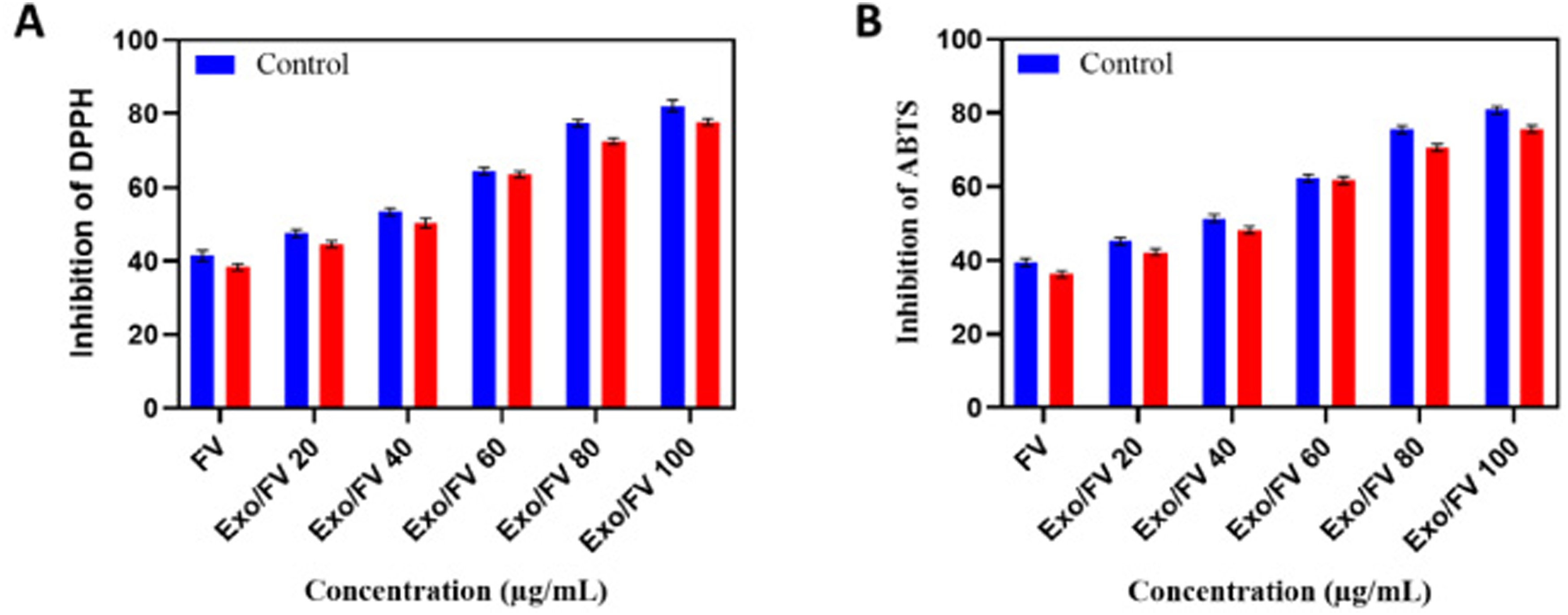
The antioxidant characteristics of the Exo/FV were evaluated in two ways: (A) using DPPH assay with varying concentrations of FV and Exo/FV while holding the BHT concentration constant, and (B) using ABTS assay with varying concentrations of FV and Exo/FV to analyze the antioxidant property with vitamin C as constant.
The antioxidant activity was measured using DPPH, and ABTS assay with FV and Exo/FV ranges from (20–100 μg/mL). These assays for determining the antioxidant activity results of the Exo/FV have a high free radical scavenging activity. Hence, Fig. 2A observed the DPPH free radical scavenging property of FV and Exo/FV and it was observed that Exo/FV has high scavenging activity similar to the BHT. From the literature, it was observed that the DPPH activity of FV was found to be 392 which was similar to the BHT [26]. ABTS was performed with Vitamin C as a control where it has strong activity making it an ideal control for studies that examined the efficacy of sugar-lowering compounds such as polyphenols, flavonoids, terpenoids, and others [27]. The FV and Exo/FV were analyzed and proved that Exo/FV has high radical scavenging activity as observed in Fig. 2B. The synthesized compound was found to have high antioxidant activity. Unlike the ABTS radical cation, DPPH does not need new preparation as it is a free stable radical. On the other hand, the ABTS test uses a higher wavelength, making it less susceptible to interference from colored material [28]. The DPPH and ABTS assay for Citrus reticulata Blanco tends to have high antioxidant properties [29].
MTT assay
The cell was treated with different concentrations of the FV and Exo/FV were analyzed by MTT assay for 48 hours to analyze the cytotoxicity effect on NHI 3T3 cell lines. These cells were used for an in vitro model to evaluate the toxicity of the synthesized compound was given in Fig. 3A and observed that the Exo/FV shows high cell viability when compared to the extract of FV. When the extract is combined with the exosomes it shows better activity and retains cell viability even at 100 μg/mL, and exhibits no toxicity. The absorbance rate was determined at 570 nm, which indicated extraordinary viability on the 3T3 cell line. A similar effect was shown on AgNPs by Thymus kotschyanus extract which is non-toxic and has great cell viability [30]. The best activity was observed for an extract of R. epapposum loaded with AgNPs was proved to be non-toxic [31]. Hence it was shown that the Exo/FV has high cell viability and is found to be non-toxic.
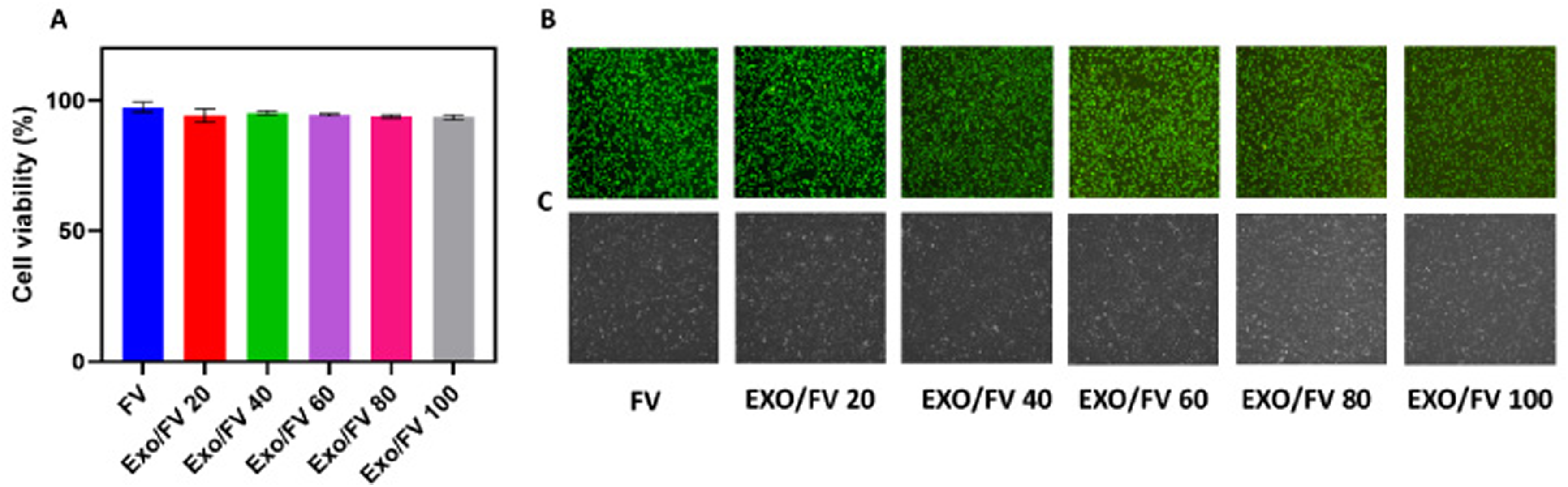
(A) The viability of cells In vitro was assessed using the MTT assay, with varying concentrations of Exo/FV, (B) denotes the fluorescence images of the 3T3 cell lines at various treatments with Exo/FV and (C) denotes the phase images of 3T3 cell line the images at 10× magnification.
Fluorescence and phase change analysis was performed, as shown in Fig. 3 (B and C), on cells treated with FV and Exo/FV at different concentrations. The results showed that the cells did not suffer any harm during treatment, confirming the synthesized molecule was non-toxic.
Flow cytometer analysis
The cell lines were exposed to FV and Exo/FV at concentrations ranging from 20 to 100 μg/mL, and modifications were noted. The analysis of Fig. 4A revealed that the cells were unharmed after the treatment. The drug was thus considered innocuous because maintained its viability at 100 μg/mL. Even though it had no impact on cell viability, pomegranate flower extract exhibited comparable effects when tested on normal 3T3 cell lines [32]. Figure 4B shows the % apoptosis observed that when treated with the drug there was no significant change in the cell apoptosis rate. It was clear that the plant extract is less toxic and has no impact on the cell lines throughout treatment since the cell viability has been considerably enhanced when treated with H. rhamnoides compared to the treatment with H2O2 [33].

(A) Flow cytometry analysis of the 3T3 cell line with different concentrations of FV, Exo/FV (20–100 μg/mL) and the apoptotic activity was determined. At 100 μg/mL, Exo/FV had a low apoptotic rate, revealing its non-toxicity, as seen in (B), which depicts the % level of apoptosis at different concentrations of Exo/FV.
The study was conducted using the cardiac tissues of pregnant mice. Research was conducted using FV and Exo/FV, and an analysis of conditional alterations has been completed. Mice with hypertension brought on by L-NAME showed signs of redox imbalance and dysfunctional biomolecules associated with heart function. Since elevated blood pressure is a symbol of hypertension, the fact FV and Exo/FV of concentration 100 μg/mL were used to analyze the effect of lowering L-NAME levels in mice with hypertension. Following these tests, their validity was established.
The systolic/diastolic blood pressure
In Fig. 5A, observed the systolic blood pressure (SBP) readings of the pregnant mice in the control group and the hypertensive group. Interestingly, when the drug was given to the normal mice, their SBP levels remained unchanged. However, when the hypertension-induced mice were given FV and Exo/FV of concentration 100 μg/mL, their SBP levels effectively decreased, with the best results observed which shows the stronger blood pressure-reducing ability. Whereas the literature shows that FV has a high effect in controlling heart pressure rate which also shows no toxic effect on heart tissue [34]. In a similar vein, the diastolic pressure rate was shown in Fig. 5B, and it turns out that normal mice DBP remains the same when they're given L-NAME the rate tends to be elevated, but when treated with FV and Exo/FV at a concentration of 100 μg/mL, their DBP reduced, proving that the treatment is effective. However, the antihypertensive effects may describe how rats given a diet rich in M. oleifera showed considerably lower systolic and diastolic blood pressure as observed from the literature [21].
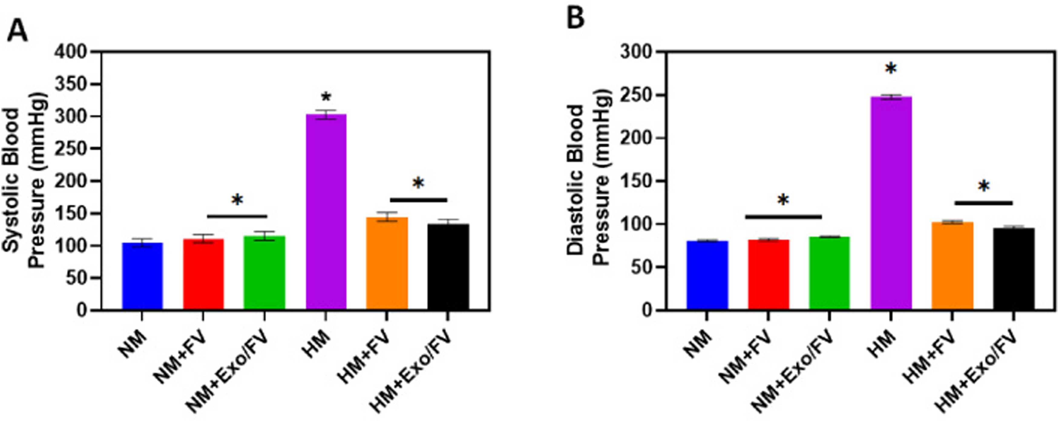
The pregnant mice's blood pressure, shown in (A), shows the difference between their systolic blood pressure before and after L-NAME caused hypertension. The blood pressure of animals given FV or Exo/FV changed, and Exo/FV tended to lower blood pressure, (B) displays changes in diastolic blood pressure as a result of FV and Exo/FV treatment, and the * denotes (p < 0.05) significantly different from control.
The homogenates of the heart tissue were processed and used for further investigation in the presence of AChE. No significant difference was seen between the control and normal groups, although there were notable increases in AChE activity in the groups with hypertension with a significance difference of p < 0.05 when treated with FV and Exo/FV at a concentration of 100 μg/mL as observed in Fig. 6A. According to the reviewed literature, cadmium administration causes problems due to an increase in AChE activity. The phenolic components of AChE enable them to give electrons and stop the chain reaction. Phenolics are a major family of phytochemicals [35,36]. The AChE activity has been linked with an increase in instances like anxiety, inflammation, heart rate recovery, and myocardial infarction.

(A) Heart homogenate acetylcholinesterase activity in normal and L-NAME induced hypertensive rats: an analysis of FV and Exo/FV, (B) the impact of monoamine oxidase function, (C) TBARS activity and (D) overall thiol level activity, and the * denotes that (p < 0.05) significantly different from control and hypertension-induced.
Figure 6B shows that the MAO activity has a small variance when treated for the normal mice when the L-NAME was given to the normal mice to induce hypertension the MAO activity tended to be increased. Further, when treated with the drug FV and Exo/FV of concentration 100 μg/mL the activity was shown to be reduced and this shows that treating with Exo/FV indicates better activity when compared with FV. Many studies have stated that the presence of phenolic compounds reduces the AChE and MAO activity in various organs. The levels of the neurotransmitter's serotonin, norepinephrine, and dopamine are reduced in hypertensive patients due to increased MAO activity in the heart, according to the literature [37].
Lipid peroxidation and thiobarbituric acid reactive species
The induced group's elevated lipid peroxidation was assessed using TBARS. From Fig. 6C it can be seen that when the induced group is treated with FV and Exo/FV with 100 μg/mL, the amount of lipid peroxidation tends to reduce and the level of TBARS was found to be decreased. The literature shows that an increase in TBARS level, a marker of radical-induced lipid peroxidation in cardiac tissue may trigger oxidative damage and other cardiovascular disorders [38]. An increase in coronary flow was observed after administering an FV extract, indicating that it would not cause myocardial damage. Because of this, it has been shown that the drugs FV and EXO/FV may protect against cardiovascular problems by decreasing TBARS levels [34].
Non-protein thiols (NP-SH) assay
The results of this test demonstrated that cardiac tissues are susceptible to free radical damage due to a low antioxidant content. It is common for CAT, SOD, and GSH levels to be low in heart tissue. Mice with hypertension had lower thiol levels, which is indicative of a redox imbalance and poor antioxidant status in cardiac tissue. From Fig. 6D, it can be seen that there was a notable decrease in the NP-SH level after treatment with the medicine FV and Exo/FV with 100 μg/mL, which increased the thiol level. In contrast, an elevated non-protein thiol level was seen in normally developing mice. Hence these tests showed that the level of hypertension decreased when treated with the drug and a similar effect was found in Moringa oleifera leaves and seed which show the same activity [21].
PDE-5 analysis
This assay was carried out in heart tissues as given in Fig. 7A it was observed that the level of PDE-5 was found to be elevated in the hypertension group when compared to the normal group. When treated with the drug FV and Exo/FV with the concentration of 100 μg/mL the level of PDE-5 was found to be decreased which proves that the drug was effective in treating hypertension. The herbal extracts that were rich in flavonoids were found to reduce the activity of PDE-5 and were reported on analyzed from the literature [39]. LC-MS/MS analysis revealed that the studied FV extracts contain several classes of phenolic compounds, mainly different types of flavonoids hence it was proven to be effective [40].
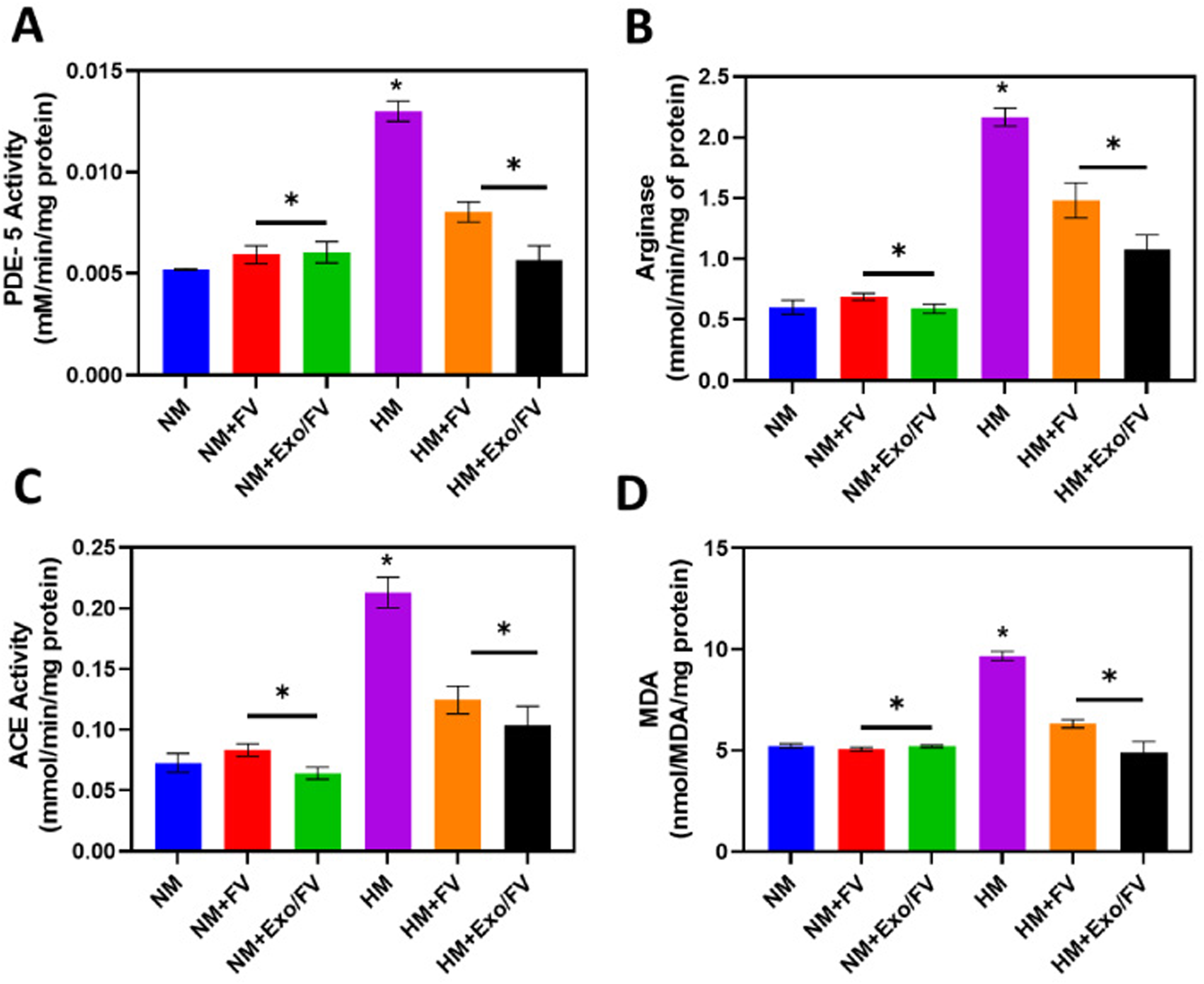
(A) Before and after hypertension, the effects of FV and Exo/FV were examined, and the impact of PDE-5 activity on cardiac homogenate was examined, (B) denotes the ACE activity on lung homogenate, (C) shows the arginase activity on kidney homogenate and (D) denotes the MDA activity, the * denotes (p < 0.05) significantly different from control and hypertension induced.
When the drug FV and Exo/FV with the concentration of 100 μg/mL were administered to the hypertension-induced group, the results showed that the drug had a greater impact in regulating hypertension, as the ACE activity was measured in lung homogenate the changes has been observed from Fig. 7B. Literature has shown that phenolic-rich plant extracts may reduce ACE activity. This inhibitory effect may be due to the formation of hydrogen bonds and hydrophobic interactions between the polyphenolic chemicals and the hydrophobic active site of the enzymes [36].
Arginase activity
This assay was conducted using kidney homogenate. From Fig. 7C it was observed that the hypertension-induced groups tended to have higher arginase activity, while the treated groups tended to have lower levels. The activity of arginase was also related to the activity of the phenolic compound found in the FV extract. After being loaded into exosomes, the activity of FV was significantly increased. The literature revealed that the polyphenolic substance decreased hypertension in mice models [41].
MDA analysis
We also performed an MDA analysis using the kidney homogenate depicted in Fig. 7D. We found that MDA levels were higher when hypertension was induced, while lower when FV and Exo/FV with a concentration of 100 μg/mL were used as treatments. There was a correlation between MDA and lipid peroxidation and found that hypertensive mice given FV had lower renal MDA levels. This might be because the phenolic components in FV created a compound that inhibits the catalysis of the first step of lipid peroxidation. That the plant may prevent cellular lipid peroxidation and lower blood pressure is supported by this study [42].

Histopathology images of various organs before and after the induction of hypertension. The changes were analyzed when treated with FV and Exo/FV: Heart, lungs, and kidney.
In the case of hypertension, the research was conducted on cardiac tissues. The effects of FV and Exo/FV with the concentration of 100 μg/mL were examined. The results showed that in normal conditions, the muscles in heart tissues were closer together, but in the hypertension-induced state, there was a noticeable expansion of the gap between them (Fig. 8A). Treatment with Exo/FV with the concentration of 100 μg/mL resulted in improved activity, the restoration of intermuscular space, and the prevention of muscle damage. Therefore, these results demonstrate that Exo/FV was a successful treatment for hypertension. A comparable finding was made in cardiac tissues that were exposed to pomegranate juice: at low concentrations, the tissue showed minimal damage, but at high concentrations, a small number of damaged cells were detected [43]. Where the effect of treatment of FV and Exo/FV has been tested in lungs and kidney tissue and the changes have been observed from Fig. 8B and 8C. The control tissue has no space in between whereas in hypertension conditions the tissues have a space in between then they were regained after the treatment with FV. The objective of the study was to reduce hypertension that occurs during pregnancy where the FV is rich in alkaloids, phenolic compounds, and volatile essential oils they were found to have a high antioxidant effect and prevent cells from damage when combined with Exo the activity has been enhanced [9]. Nowadays, people are paying more attention to extracts that are packed with Exosomes the rising prevalence of hypertension, together with the drug's possible side effects, made Exo an attractive alternative and complementary therapeutic option for the disease's prevention, treatment, and maintenance. Hypertension was effectively treated by the phenolic compound due to its structural resemblance to cholinesterase inhibitors [44]. These activities have been tested in mice models and proved that the synthesized drug was effective in treating hypertension.
Conclusion
This study highlights the potential of an extract derived from Falcaria vulgaris, loaded with exosomes, as a promising therapeutic strategy for the treatment of hypertension in pregnant mice. Both In vitro and In vivo investigations demonstrated significant anti-hypertensive effects, improved endothelial function, and reduced oxidative stress markers associated with hypertension. The formed Exo/Fv was found to be spherical with a diameter of 20–30 nm. They were found to be biocompatible. Hypertension in mice was induced with L-NAME; subsequent treatment with Exo/FV resulted in a decrease in blood pressure, an improvement in biomolecule activities (AChE, PDE-5, and MAO), and a resolution of the redox imbalance linked to cardiac dysfunction. These biological effects may be related to the presence of certain phenolic chemicals to validate the results, histopathology analyses were performed. This conclusion provides insights for further scientific exploration or potential therapeutic developments.
Footnotes
Conflict of interest
The authors declare that they have no known competing financial interests or personal relationships that could have appeared to influence the work reported in this paper.
Data availability
All data generated or analyzed during the study are included in this published article. Further queries can be directed to the corresponding author.