
Abstract
BACKGROUND:
Generation and utilization of the specific monoclonal antibodies against testis antigens is reported to identify the antigens that are important in reproductive field.
OBJECTIVE:
Current study aimed to introduce a hybridoma that producing a specific anti-testis monoclonal antibody to identify the testis antigens that can be important in the reproduction field.
METHODS:
To make hybridoma against testis antigens, mice were immunized with testis cell lysate. After cell fusion, resulted hybridomas were screened by indirect ELISA, then cloned by limiting dilution and finally the produced monoclonal antibody were characterized by some of the molecular laboratory techniques such as immunohistochemistry, immunocytochemistry and western blot.
RESULTS:
By using hybridoma technique, cell fusion was performed and ten (8A11, 8D6, 8D7, 9F6, 9G11, 10C3, 10B3, 10B2, 10C2 and 10H7) antibodies specific to the testis antigens were produced finally. Among the produced antibodies, 10C3 was found to cross-react with testis, but not detected in other tissues. mAb 10C3 recognized the sperm and testis antigens, specifically the intertestitial tissue of testis, spermatogonia, primary and secondary spermatocyte antigens, so they were most likely the target of generated mAb. Also our mAb could totally detect the mouse sperm antigens and the specific antigens of head and tail of human sperm. In western blotting analysis, mAb 10C3 could recognize the specific protein bands of sperm and testis extracts. Also in this study the testis specific genes that were target of generated mAb, were selected according to the mouse EST profile available at UniGene part of NCBI.
CONCLUSIONS:
So this stable anti-testis mAb has a potential for laboratory researches and also for diagnostic procedures in fertilization. Thus it could be exploited as a suitable tool for target-specific diagnosis and research in several diseases.
Introduction
The physiological process of spermatogenesis is responsible for the production of male germ cells but poorly understood generally. There are a pool of spermatogonial cells on the basement membrane of the seminiferous tubules that proliferate and give rise to a progeny of differentiating spermatogenic cells (primary spermatocytes, secondary spermatocytes, spermatids and spermatozoa). Spermatogenesis is regulated by a variety of factors and molecular signals [1, 2].
On the other hand, the antibodies that come from one type of cell, the hybridoma cell, are called monoclonal antibodies (mAbs). Hybridomas are cells that have been engineered to produce a specific antibody in large amounts. A hybridoma is produced by the injection of a specific antigen into a mouse and the subsequent fusion of the antigen-specific plasma cells (antibody-producing cell) from the mouse’s spleen with a myeloma cell (a B-cell cancer). The monoclonal antibodies have the qualities of the two different kinds of cells: first, the ability of myeloma cells to grow continuously; secondly, the ability of production of large amounts of antibody by immunized B-cells. The hybrid cell must be cloned to produce many identical daughter clones that can secrete the immune cell product. Monoclonal antibodies have the same isotype that produced by a single clone. The production of a single clone that has ability to secrete the specific antibody is one of the most important level of production monoclonal antibodies by hybridoma technology [3, 4]. The monoclonal antibodies are the specific antibodies that are the essential tools of many biomedical investigators and have the great avail [5]. Also humanized mAbs are a rapidly rising group of antibodies in clinical trials and can be used for the detection of the substance of interest for the produced drugs [6]. In fact, about 30 mAbs currently approved by FDA for using in the treatment of various human diseases including cancer, transplantation, cardiovascular diseases and infections. Nowadays, antibodies have become vital research tools in several laboratory techniques such as western blotting, immunocytochemistry, immunohistochemistry, immunoprecipitation and enzyme-linked immunosorbant assay (ELISA) [7]. So, the global value of these specific antibodies is approximately $20 billion per year [8, 9]. The three advantages of monoclonal antibodies are described below:
Diagnosis (for instance: assaying of hormones by monoclonal antibodies in pregnancy, classification and diagnosis of viral diseases such as influenza virus). Immunopurification (for instance: separation of one substance from a complex of similar mole- cules). Therapy.
Also, the monoclonal antibodies can recognize the antigens present in different cell types such as in the various stages of testicular development and could be a useful tool for the characterization of the specific molecules which may participate in germ cell differentiation and/or interactions during testis development. Numerous studies have been reported to generate monoclonal antibodies that are specific to the markers of these germ cells. Antisperm antibodies are the invaluable tools for the identification of sperm proteins that are involved in fertilization [10]. However, very few monoclonal antibodies generated are considered to be testis-specific and the candidate genes that are probably the target of these monoclonal antibodies rarely were investigated in these researches. Therefore, the purpose of this study was first to generate a stable hybridoma secreting monoclonal antibody against testis specific antigens and then characterize the produced monoclonal antibody as a good candidate for using in prognostic and even treatment of infertile patients in future studies. So, in this project we used the testis tissue lysate instead of pure synthetis antigens to immunize and generation of anti-testis mAb. Mice were immunized with the testis cells and best mice with highest antibody titers were selected for generation of hybridoma. Finally, the clone with highest absorbance in ELISA test was chosen for screening and single cell cloning. Afterwards three different molecular techniques were specifically performed to characterize the generated mAb. Also, we attempt to search in some biological databases for finding the most probable target genes which are recognized by these specific monoclonal antibodies.
Antigen preparation and Immunization
To produce monoclonal antibodies by hybridoma technology, the B-cells should be isolated from the spleen of animals, usually mouse, already immunized with the desired antigen. Our antigen was prepared by extracting testes of mice. Groups of testes were mixed with phosphate buffered saline 1X and homogenized by Ultrasound Technology (Hielscher UP100H Lab Homogenizer). The homogenates were centrifuged at 500 g for 10 min, then the supernatants were collected and held at
Notably, The process of working with laboratory animals is approved by the Ethics Committee and the animals were treated to procedures consistent with international regulations relating to experimental animals.
Evaluation of immune responses by indirect enzyme-linked immunosorbent assay
One week after each injection and also 3 days after final boost, all mice were bled and sera were tested by indirect ELISA. For this purpose, flat-bottomed 96-well ELISA plates were coated with 100
After 3 times washing, three-fold serial dilutions (1/100–1/500–1/1000) of serum were added to the wells. Then, the plates were incubated for 1–1.5 hour at 37
The antibody-binding was visualized by adding 100
Cell fusion and hybridoma technology
The SP2/0 cell line, myeloma cells as fusion partners were cultured (in logarithmic phase) at 37
After 5-min incubation at 37
Screening of antibody titers by ELISA
Hybridoma clones with the antibody-producing ability were selected by indirect ELISA, as described above, and the positive clones were counted to perform limiting dilution. The cells were cloned four times by limiting dilution to obtain the stable hybridomas.
Screening of the produced monoclonal antibody
The desired antibody in the supernatants of hybridoma clones were characterized by the following methods.
Immunocytochemical method
Semen samples from healthy donors were allowed to liquefy at room temperature and separated from seminal plasma by centrifugation (1000 g) for 20 min at room temperature. After washing in PBS, the pelleted spermatozoa were frozen at
Immunohistochemical method
All procedures related to the care and treatment of animals were in accordance with the institutional and National Institutes of Health guidelines. The testis as well as kidney, skin and spleen of Balb/C mouse were fixed in PBS containing 4% (w/v) paraformaldehyde for 18–24 h depending on the size, and then embedded in paraffin wax. All samples were cut into 2–5
SDS-PAGE and western blot
Cell extracts were mixed with a concentrated SDS sample buffer and boiled for 5 min at 95
A healthy monoclone hybridoma growing 10 days after limiting dilution (Inverted Phase Contrast; Magnification 20X).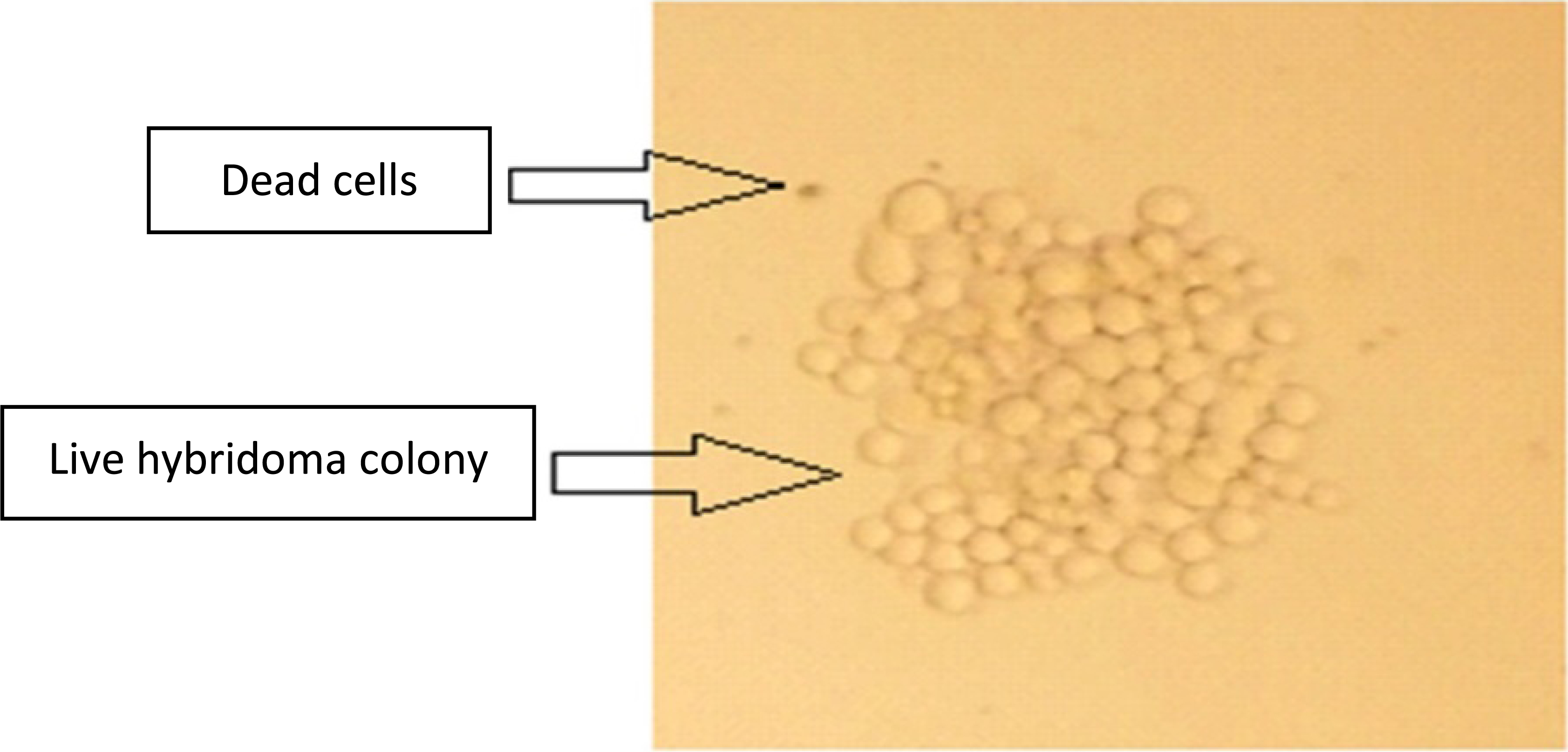
ELISA OD values obtained by the supernatants of produced hyridoma. All of the results are in comparison with the control group (serum-free DMEM).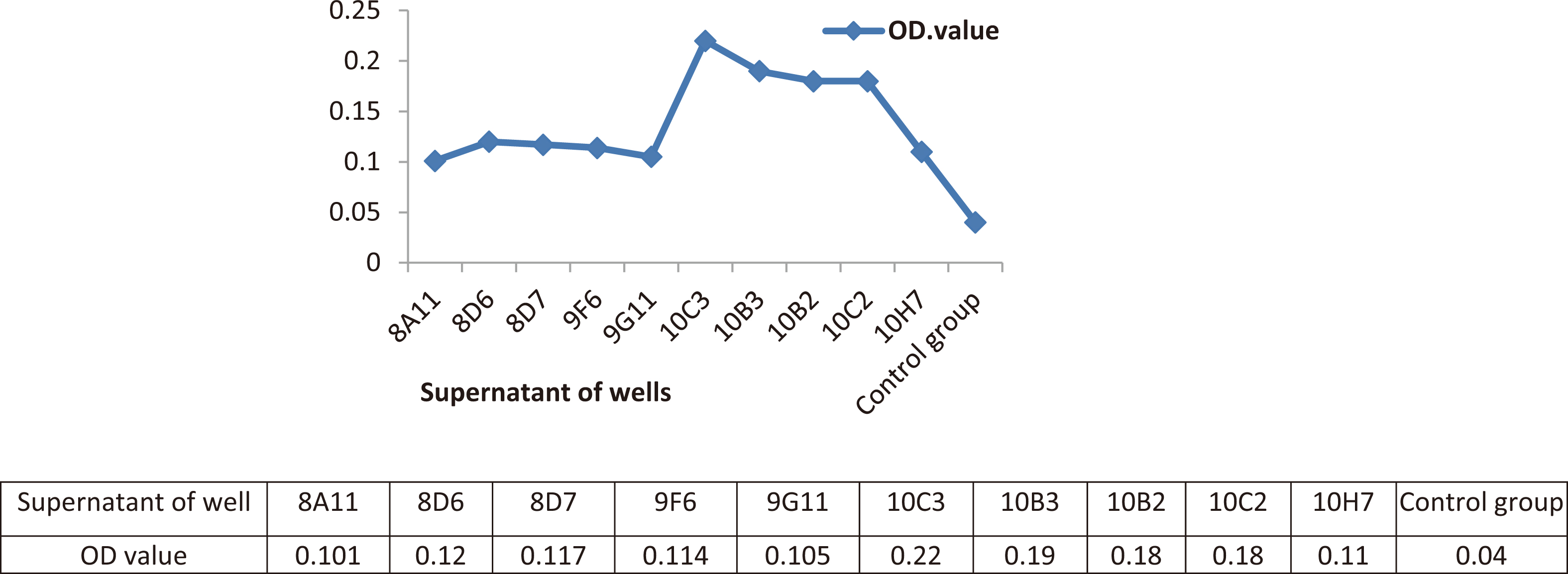
In order to select testis specific genes that are probably the target of our monoclonal antibody, the mouse EST profile available at UniGene part of NCBI was analyzed and the corresponding protein size and function were retrieved from UniProt. According to the results given in Uniprot, the testis specific genes were selected as the target of desired mAb [20].
Results
To obtain monoclonal antibody against mouse testis antigens by hybridoma technique, mice were immunized by the immunogen, the extract of testes. Then hybridoma cells were generated by fusion SP2/0 cell line with the spleen B-cells of the mouse immunized. Two important post fusion techniques are necessary for visualizing the stable hybridoma in culture: (a) sub-cloning and limiting dilution, (b) screening of wells for antibody activity. So the wells containing more than one hybridoma clone were screened and sub-cloned and the limiting dilution was done for such wells. A monoclone hybridoma colony is demonstrated in Fig. 1. The screening of hybridoma clonies was performed by ELISA test.
The ELISA test is one of the best strategy that could be used successfully to identify hybridomas that are secreting the appropriate monoclonal antibody. For this purpose, the strength of the immune response (titer) used as a guideline to predict the frequency of positive clones. The clone with the highest titer correlates well with the number of B cells that are producing circulating antibody and which also correlates with the greatest number of the positive clone resulting from successful fusion. In our research, we generated 110 clones and finally selected 10 clones (8A11, 8D6, 8D7, 9F6, 9G11, 10C3, 10B3, 10B2, 10C2 and 10H7) with the highest OD in ELISA method. ELISA data demonstrated that all clones exhibit a concentration-dependent reactivity to testes antigens, but not to an equivalent amount. So the clone with the highest OD in 450 nm, 10C3, was selected as the appropriate hybridoma cell for expansion (Fig. 2). The results were reported in comparison with the control group (serum-free DMEM).
(I) Immunofluorescent localization of mAb 10C3 by (FITC)-conjugated goat polyclonal anti-mouse IgG antibody in the mouse sperm totally (A), sperm head and tail of human (B); (II) control group, Immunofluorescent localization of 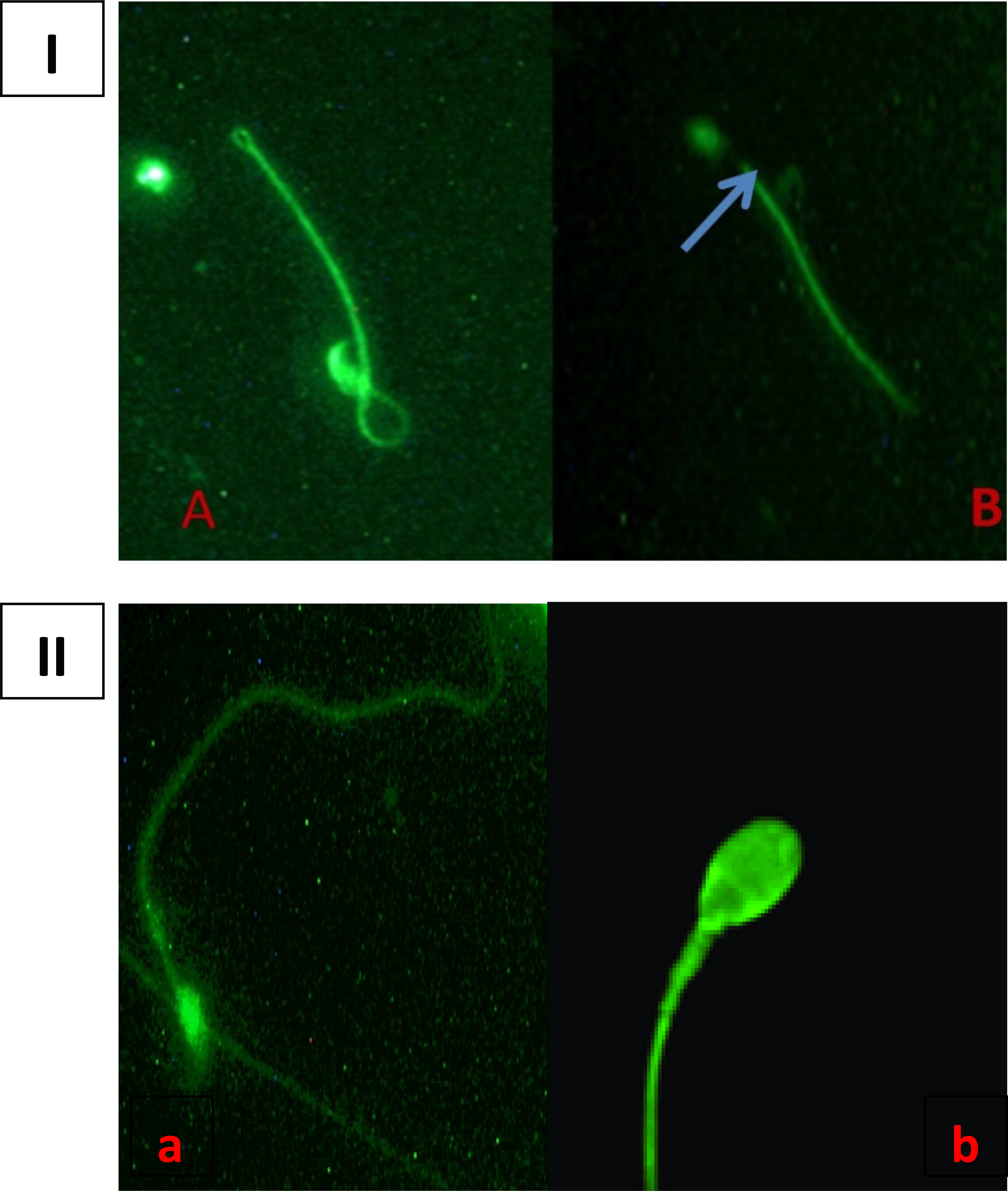
Detection of the mAb 10C3 antigens in mouse tissues. Immunohistochemistry was performed for (A) testis sections ((a) FITC staining, (b) DAPI staining, (c) merged image), (B) Detecting of the mouse testis antigens with UB (P
On the other hand, the characterization of generated monoclonal antibody was performed by several methods. To detection the specificity of monoclonal antibody on the sperm, the media of 10C3 clone, the clone with the highest titer of desired antibody, was collected and screened by Immunocytochemistry. The results of immunocytochemistry staining on the mouse and human sperm have compared with the control group, (immunofluorescent localization of
Western blot analysis of various mouse adult tissues by mAb 10C3. (A) Lysates from human sperms. (B) Lysates from mouse sperms. (C) Lysates from mouse testes. (D) Lysates from mouse liver. (E) Lysates from mouse kidney. (F) Lysates from mouse muscle (blue arrow shows the prestained protein ladder and Black arrows on the left side of image show the significant bands and the arrows on the right side of image show the protein sizes of prestained protein ladder).
Selected testis specific genes with protein size between 66 to 75 kDa (these testis specific genes were selected according to the mouse EST profile available at UniGene part of NCBI)
To elucidate the specificity of produced monoclonal antibody on the mouse tissues, especially on the testis, we carried out immunohistochemistry staining on the mouse testis and also on the several tissues of mouse such as liver, kidney, muscle, skin, as the control of tissue. The results were indicated that the localization of the supernatant of 10C3 clone, as the specific monoclonal antibody, was only on the testis when compared with control group (usage of UB (P4D1) primary antibody, as the control of antibody), and showed the significant detection on the spermatogonia and spermatocytes membranes specifically; whereas other tissues didn’t show any significant detection by our mAb (Fig. 4). In this regard, we suggest that the obtained monoclonal antibody could detect the spermatogonia and spermatocytes antigens and could react with the antigens that are distributed on the basement membrane of each seminiferous tubule. The logical reason for this observation might be that the monoclonal antibody was designated for the proteins that are located on the membrane of spermatogonia and spermatocytes.
To determine the size of protein(s) that can specifically detect by obtained mAb, western blot analysis was performed from several tissue extracts. As shown in Fig. 5, two protein bands recognized by mAb 10C3 from the lane of human sperm that were about 70 and 40 kDa, but as you see, specifically obtained mAb has show a significant binding to the mouse sperm protein that are about 53 kDa and also one specific protein band with about 73 kDa recognized by prepared mAb in the mouse testis lysate. As shown in Fig. 5, mAb 10C3 does not show an efficient binding to kidney and spleen proteins, as the control group. The results suggest that antigens recognized by mAb 10C3 appear in a testis and sperm-dependent manner. One possibility is that testis and sperm-derived molecules are a target of generated mAb 10C3. However, mAb 10C3 didn’t recognize any protein from other tissues, so we could confirm the specificity of produced mAb against the testis antigens.
Finally the most probable target genes which were recognized by produced monoclonal antibody were investigated by some biological databases such as UniGene part of NCBI. The results reported in Table 1 show the compatible genes with our experimental results that could be target of our mAb.
In this study with the purpose of investigating the testis antigens involved in fertilization, we have reported the characterization of the produced monoclonal antibody that are specific to the antigens of testis cells and for this aim the tissue-specificity studies were performed to select the antibodies that have the minimum cross-reactivity with other mouse tissues. In order to maximize the recovery of antibody-positive hybrid clones, the ELISA assay was utilized to select the best clone with the highest titer of desired antibody. The positive ones were then screened by Immunohistochemistry, Immunocytochemistry and western blot analysis. Also by using bioinformatics data, we were investigated some biological databases to select the candidate genes that could be the target of these produced monoclonal antibodies.
For over a century, many papers published by researchers in the monoclonal antibody field have addressed in great findings on the anti-sperm antibodies and methodologies for their detection [21]. Also in recent years, the effect of these anti-sperm antibodies upon diagnosis and treatment of infertility have investigated by many experts, because the production of anti-sperm antibodies can cause destroying of antigen molecules and infertility. A unique scheme of antibody preparation, termed as hybridoma technology, has been employed in these studies to obtain the monoclonal antibodies. The process of hybridoma technology relies on animal immunization to yield many antigen-specific cells that can be fused with an immortal myeloma cell line for producing the hybrid cells [22, 23].
As the means to produce the specific monoclonal antibodies, hybridoma technique has been used successfully in several laboratories. It has been applied to produce several kind of antibodies against different antigens and diseases, such as: antibodies against the human sperm coating antigens, cancer/testis tumor-associated antigens, human spermatozoal surface antigens, producing human-human hybridomas from lymphocytes of patients with various autoimmune diseases and also producing the diagnostic monoclonal antibodies against the rare genetic disorders such as Congenital Adrenal Hyperplasia (CAH) [24, 25, 26, 27]. Todays, the useful advantages of mAbs in biotechnology and genetic engineering have been proved for development of modern vaccines and gene therapy [28]. The mAbs can be used for immunotherapy for instance in the post-transplantation patients as a vaccine that can be prevent the recurrence of HCV infection and in diagnosis of acute myeloid leukemia [29, 30]. Also, one of the most important of mAbs benefits is their specificity and flexibility as the molecular drug targets against the common diseases such as multiple myeloma and for detection and treatment of different biomarkers of cancers, for example breast, ovarian and stomach cancers [31, 32, 33, 34].
In all cases, donor mice were immunized first with the desired antigens which mixed with an equal volume of “Complete Freund’s Adjuvant”. Then the same mice were immunized with the complex of desired antigens and “Incomplete Freund’s Adjuvant”. According to this scheme, we have utilized mice testis lysate as the desired antigens. Then the hybridoma protocol was performed by the spleen B-cells of immunized mice. For this purpose, the B-cells were fused with the SP2/0 cell line and several hybridoma clones were obtained after culturing the best hybridoma cells. Then hybridoma clones have been selected for the ability to produce a significant amount of desired antibody. In the ELISA analysis, all of them showed the high OD in comparison with the control group (Fig. 2). However, the best clone (10C3) with the highest OD were selected for characterization and localization by several methods. At this point, there is a concern that this clone may fail to produce specific antibody to the desired antigen. However, further analysis by western blotting or immunohistochemistry and immunocytochemistry methods have demonstrated that 10C3 clone out of 10 clones really produce specific antibody to testis antigens of mouse.
Immunocytochemistry analysis of mouse and human sperm has demonstrated that the mAb 10C3 antigens are present in all parts of the mouse sperm, but not in the middle segment of human sperm. This is consistent with our scheme of the specification of produced antibody.
Immunohistochemistry of several tissues has showed
that the localization of produced mAb is specifically in the spermatogonia and spermatocytes antigens of mouse testis, when compared with the positive control group. It is interesting to note that the antigens of several other tissues such as kidney, liver, skin and muscle, as the control group, are not recognized by the mAb 10C3 (Fig. 4).
As discussed earlier, Western blotting of testis and sperm lysates of mice with the produced mAb 10C3 has also demonstrated that the target antigens are recognized as a single protein band by western blot, indicating that the target molecule of mAb 10C3 is proteinous substance with the molecular weight of 73 KDa for mouse testis protein and 53 KDa for mouse sperm protein. These suggests that the specific proteins of testis and sperm are the target for generated antibody. But the target antigens are recognized as multiple protein bands with 40 and 70 KDa of human sperm by western blotting. In general, a mAb can only recognize one epitope. However, as shown in Fig. 5, 10C3 mAb reacted with two protein bands of human sperm (70 and 40 kDa) on the western blot test. As we know, one protein might be processed by alternative splicing [35]. So our desired 10C3 mAb may be detect the protein that appears to be processed and finally two bands of the human sperm proteins detect on the western blotting by the produced mAb 10C3.
It is also possible that mAb 10C3 recognizes other specific protein targets of human sperm in comparison with kidney and spleen of mouse, as the control group. Because of the difficulty in purifying the target molecules for mAb 10C3, we do not know the molecular structure of the protein targets now and then it should be clarified by further investigation.
This idea is potentially important that the mamma- lian spermatogenesis is a complicated process which consists of three events that three kind of testis cells are involved in that: mitosis (spermatogonia), meiosis (spermatocyte) and post-meiotic (development of spermatid to spermatozoa) [36, 37]. The results of present study suggest that a sperm dependent event is the target of obtained antibody, so we suggest that the specificity of our monoclonal antibody is just for sperm and testis of mouse. In conclusion, we described the analysis of the specific monoclonal antibody that produced by hybridoma technique on the several tissues of mouse and found that the generated antibody could not detect the proteins of other tissues such as kidney and spleen, however in testis and exclusively on the sperm have shown the significant detection. Sperm-derived molecules by itself could be a candidate for the target of mAb 10C3 and we believe this is possible because western blotting analysis of lysates prepared from up to 10000 sperm demonstrated visible band. Also some of the human sperm proteins could be detected by produced mAb. The localization of the mAb 10C3 antigens to the spermatogonia and spermatocyes and spermatozoa allows us to suggest the biological functions of produced mAb. However further experiments will be necessary to determine the functional role of mAb 10C3 antigens in the mouse embryogenesis, especially in the early development of testis.
Despite all these efforts, the identification of genes that are involved in the biological relevances of male fertilization are necessary to develop reliable diagnostic methods for clinically usage of anti-sperm and anti-testis antibodies. So in order to find the most probable target genes which are recognized by the prepared monoclonal antibodies, some biological databases were investigated and compatible genes with our experimental results reported in Table 1. In this regard, the expression profile of more than 78000 mouse EST clusters was evaluated and the clusters with specific gene names and high testis specificity were selected. Then, according to western blotting results, only proteins with molecular weight between 66 to 75 kD were selected. This implies that they were introduced as the probabilistic specific targets of our produced mAb.
In summary, this is the first report regarding the preparation of the monoclonal antibodies based on the mouse testis antigens content and selection the target genes which show the dedicated functional of these antibodies on the mouse testis proteins, which is absent in the other tissues tested in this study. On the other hand, we have demonstrated that the monoclonal antibodies prepared by hybridoma technique against testis antigens can be used to analyze the functional display of mouse testicular proteins. Our present study not only provide a reference for further researches on the production of monoclonal antibody against various tissues of mouse, but also can facilitate the studies in the reproductive immunology fields that are going to investigate the function of genes that are involved in mammalian fertilization. Moreover, further studies are necessary to verify safety and applicability of our generated antibody in clinical trials before any therapeutic application.