
Abstract
BACKGROUND:
Previous studies have shown that the FABP9/PERF15 gene is expressed in mice and in some other mammals in the testicles and in the spermatozoa, and its possible effect on the ability of the sperm to form and maintain the nucleus until fertilization.
OBJECTIVE:
Since the FABP9 homologue gene exists in humans, and so far no research has been done to indicate the exact location of this gene in the organism, it is necessary to find a better interpretation of its possible performance by its localization in the testis.
MATERIAL AND METHODS:
Biopsied testicular tissue samples after sectioning and embedding on class slide were subjected to IHC with specific monoclonal antibody and underwent final staining with hematoxylin and eventually evaluated by light microscope.
RESULTS:
The antibody could successfully bind and detect its related protein, FABP9, in Leydig cells rather than spermatogenic cells.
CONCLUSION:
The expression of FABP9 in a different cell type rather than spermatogenic cells in other mammals, reports of a plausible different function for the gene product like its involvement in fertility potential in homo sapiens.
Introduction
Interactions of FABP9 with its neighboring proteins. LIPE: Lipase, CYP2U1: Cytochrome P450; FABP4: Fatty acid binding protein 4; CYP2J2: Cytochrome P450 family 2, subfamily J, polypeptide 2; CYP2R1: Cytochrome P450, family 2, subfamily R, polypeptide 1; MBNL1: Muscleblind-like splicing regulator 1; CEL: Carboxyl ester lipase (bile salt-stimulated lipase); LAMC2: Laminin, gamma 2; CA2: Carbonic anhydrase II; RBM4: RNA binding motif protein 4. 
FABP9 (Fatty Acid Binding Protein 9) encoding gene (ENSG00000205186) is located on chromosome 8 and codes for a 132 amino acids protein with molecular weight of 15093 Dalton (Fig. 1) (
The overall gene structure of FABPs is highly conserved, consisting of four short exons and three variables in size introns [4, 5] while their positions are similar for all FABPs, but the intron length is variable between isoforms [6, 7]. This gene shares high homology with myelin P2, A-FABP, and H-FABP, thus it is proposed that it has a binding ability to membranes. FABP9 gene expresses in salivary and mammary glands in addition to testis [2]. In rodents testis, it is expressed in spermatogenesis cells [8] and play a probable role in the structural arrangement and stability of the organelle [9]. It is also, proposed that FABP9 plays a protective role for sperm from oxidation [10] but did not show any enrolment in sperm function for oocyte fertility [11].
The protein-protein interactions of FABP9 were searched in STRING database. The results obtained from this website showed that FABP9 plays possibly in fatty acid signaling processes in various organs (Fig. 1). Recently, a carcinogenic involvement has been reported for FABP9 when its higher levels were in malignant prostatic cell lines compared to their levels in the benign cells with a prognostic significance [12].
Sampling
The data consisted of observations from testis biopsy received from a man who died of stroke, immediately was put into fresh bouin solution, and transferred to 4
The present study was approved by the ethics committee of Shahid Beheshti University of Medical Sciences (IR.sbmu.msp.REC.1393.13/1376).
IHC method
Based on routine immunohistochemistry (IHC) techniques, paraffin blocks were prepared for the tissue samples, cut with a thickness of 4 microns and placed on an IHC adhesive slide (Silane-coated slides) which were then incubated for 24 hours. After dehydration by xylene and a serial dilutions of ethanol (20 to 100%), wash and dilution with methanol, and peroxidase solution, they were incubated with 100
Three sets of slides were prepared as followings: Without antibody negative controls; with secondary only antibody negative controls to confirm the observed staining is true and reliable; and test slides incubated with both primary and secondary antibodies, respectively.
Results
The immunohistochemical assay using specific human FABP9 monoclonal antibody and the goat- secondary antibody showed that the protein is specifically expressed in Leydig cells from the interstitial tissue of normal testis instead of in dividing cells of the gonad including spermatogonia, spermatids, and elongating spermatozoa (Fig. 2). There was no sign of reactivity in the negative controls except that the nucleus was colored blue in all tubal cells (Fig. 3).
FABP9 expression in leydig cells. As is observed the brown color in the connective tissue cells shows the location of the gene product after staining with DAB. Staining with hematoxylin provides blue color which indicates the spermatogenesis cells’ nucleus residing in seminiferous tubules.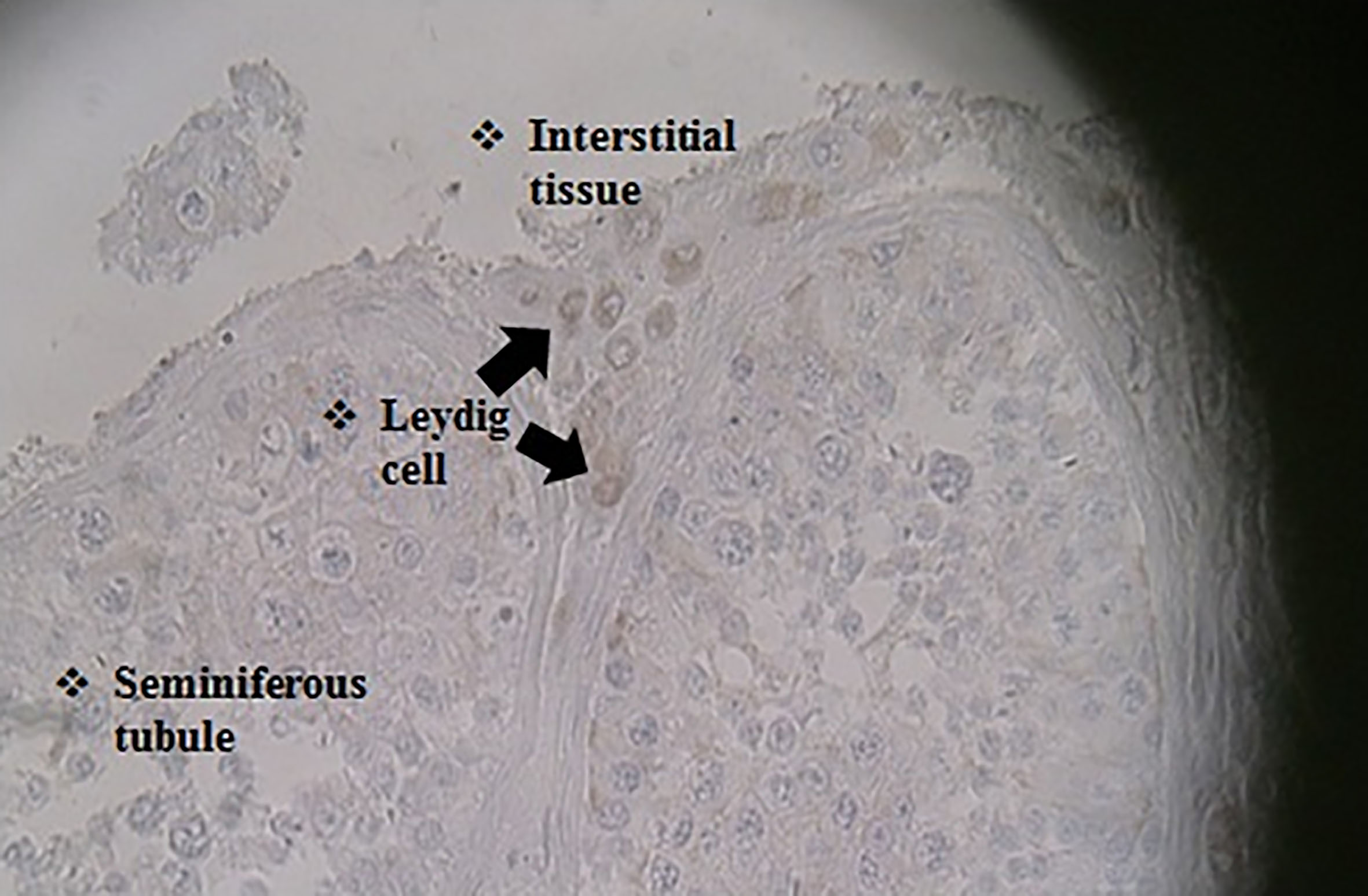
Secondary antibody only control. A weak color was observed, which could be due to the adherent nature of the tissue.
Previous studies have shown that FABP9 plays an important role in morphology and sperm function [13]. This protein is a member of the FABP gene family that exists only in the mammalian genome [9, 14]. In addition, the protein product of this gene seems to affect the programmed cell death of spermatocytes and their growth, as well as a possible protective role for oxidation against sperm fatty acids, which in turn increases fertility potential [10]. Previous studies have shown that the removal of the protein has slightly effect on the morphology of sperm and its motility [11].
Experimental experiments with recombinant FABP9 suggest new functions contributed to the gene product including binding activity, growth retardation, and cellular agglutination against bacteria and suggests that FABP9 participates in response to bacterial infections and lipopolysaccharides through detection the cell surface antigens of bacteria and hemocytes when defense [15]. The protein levels elevation with age and type of germ cells appearance has been associated with its likely active role in differentiating the mass of the cells in the last stages of spermatogenesis [16]. There was no mutation in the structure of FABP9 when sequenced in 100 infertile male specimens bearing dysmorphic sperms as the primary cause of infertility [17] which in term rejects the protein involvement in germ cells functionality. Moreover, the report could rebut the previous hypothesis of expressing this gene during human testicle meiosis and its effect on the structure and ultimate performance of the sperm.
The results of the present study prove the presence of FABP9 in Leydig cells (not in spermatogenesis cells). The presence of this protein in the Leydig cells may affect survival and growth of the germinal tissue in humans. Grete Lottruph and colleagues have shown that in patients with reproductive disorders such as infertility, Leydig cells have abnormal performance [18]. There is still no evidence of expression of FABP9 in the heterozygous subpopulations of Leydig cells with various gene expression profiles [18]. However, due to the morphological and functional changes of Leydig cells in reproductive patients, the discussion of the expression of structural genes seems more important than gene regulation. Therefore, investigation around the gene expression profile using sequential tissue sections is likely to prime future reports of FABP9 gene expression with different patterns in reproductive impairment patients.
Footnotes
Acknowledgments
Thanks to the SBMU research council head that facilitated this investigation performance and special thanks to the Ethics Committee of Iran’s Forensic Medicine in Isfahan who permitted the use of the tissue samples. This study was funded by SBMU Research Council (grant number 13/1376).
Conflict of interest
The authors report no conflict of interest.