
Abstract
At present, cancer is a major health issue and the second leading cause of mortality worldwide. Researchers have been working hard on investigating not only improved therapeutics but also on early detection methods, both critical to increasing treatment efficacy and developing methods for disease prevention. Diagnosis of cancers at an early stage can promote timely medical intervention and effective treatment and will result in inhibiting tumor growth and development. Several advances have been made in the diagnostics and imagining technologies for early tumor detection and deciding an effective therapy these include radiomics, nanobodies, and aptamers. Here in this review, we summarize the main applications of radiomics, aptamers, and the use of nanobody-based probes for molecular imaging applications in diagnosis, treatment planning, and evaluations in the field of oncology to develop quantitative and personalized medicine. The preclinical data reported to date are quite promising, and it is predicted that nanobody-based molecular imaging agents will play an important role in the diagnosis and management of different cancer types in near future.
Introduction
Cancer is emerging as a global health problem throughout the world and is the second leading cause of death worldwide exceeded only by cardiovascular diseases [1]. Tumors develop due to mutations in genes, or due to activation of oncogenes resulting in loss of cell cycle control system, leading to uncontrolled mitosis of cells [2]. At present, radiotherapy, chemotherapy, and immunotherapy are the most common methods used for the treatment of cancer and have significantly improved the prognosis of cancer patients [3, 4, 5]. However, the treatment of malignant tumors in particular late-stage tumors is difficult as the seeding of tumor cells at distant metastatic sites. Once metastasis has occurred, the conventional treatment methods fail to inhibit tumor growth, leading to decreased prognosis [6]. In most cases of tumor development, tumors go undetected at early stages, therefore advances in the detection and diagnosis of early-stage tumors will significantly help in improving patient survival [7]. Tumor detection methods have significantly advanced and molecular diagnosis has become an essential part of cancer prognosis. The evolution of molecular diagnostics has driven several technological developments, which have a clinical relevance of incorporating the novel investigations in diagnostic practices [7]. Presently molecular characterization of cancer has become a very fundamental consequence in cancer treatment both for prognostic testing as well as classification of different tumors [8].
The development of robust, high-quality diagnostic tests for technical as well as clinical support will significantly improve the overall survival (OS) and relapse-free survival (RFS) of cancer patients [9]. Several advances in the diagnostics methods including radiomics, nanobody-based diagnosis, and aptamers in diagnosis and detection of tumors show promising results. Radiomics equipped with computational power, machine learning, as well as artificial intelligence technology have been found to significantly quantify diagnosis and prognostic biomarkers and proving ancillary guidance in the field of oncology and are proving promising in developing quantitative and personalized medicine [10].
Besides, recent advances point in radiomics point that the concept of encryption of different tumors and their microenvironment based on phenotypes will significantly improve early detection and diagnosis [10]. Likewise, aptamers, short non-coding, single-stranded oligonucleotides (RNA or DNA) show promising results in the detection and diagnosis of tumors [11]. Antibodies are currently widely used in the detection and diagnostics of tumors, however high immunogenicity, low stability, and product cost limit the use of antibodies [12]. In contrast, aptamers possess a high ability to bind to specific targets with high affinity, with low immunogenicity, production costs, and high stability [11]. Also, diverse aptamer-nanobody conjugates have been developed and widely investigated for diagnostics and targeted therapy in different cancers and point to promising outcomes [13].
Nanobodies are heavy chain only antibodies (HcAbs) and were serendipitously discovered in camelids [14]. The HcAbs comprises of just two heavy chains, with a single variable domain and possess significantly low molecular weight compared to normal antibodies [15]. These nanoscale VHHs were defined by the name “nanobodies” and could retain full antigen-binding potential upon isolation, establishing them as the smallest, naturally-derived antigen-binding fragment [16]. Several studies report that nanobodies due to their high specificity can be utilized in specific diagnostics of tumors and as molecular imaging probes [15]. Also, the high stability of nanobodies in harsh conditions such as high temperature, pH, or ionic strength further points towards their application [17]. At present several nanobodies are in clinical trials for the detection and imaging of diverse tumors [15].
Here in this review, we will shed light on the recent advances in radiomics, aptamers, and nanobodies in the detection, diagnosis, and imaging of tumors.
Radiomics in tumor diagnosis and prognosis
The concept of radiomics was first proposed in 2012 and has subsequently attracted the responsiveness of researchers all over the world [18]. It involves the extraction of quantitative, and subvisual features of radiological images to develop mineable databases [19]. Interestingly, the features of the radiomics have been demonstrated to correlate with disease pathologies in particular tumors [10]. Thus, a lot of interest has been taken to utilize radiomics in tumor screening to increase sensitivity and specificity. In addition to diagnosis, radiomics can also be utilized to predict the outcome of therapies in cancer patients [20]. Radiomics typically initiates using data acquisition, where it depends on a huge tome of medical images, thus conforming clinical data in revealing the association that exists between them Fig. 1 [21]. It was first of all proposed using CT images and then in the analysis of MR images, and later PET images were also utilizing the related strategy [21, 22, 23, 105]. Several studies have been done demonstrating the value of radiomics in improving the diagnosis and treatment of cancer [20]. In brief, the aim of radiomics is to translate the tumor-inherited phenotypes using noninvasive imaging [24]. Presently radiomics analysis involves images that are mostly digital imaging which includes; CT, magnetic resonance imaging (MRI), and positron emission tomography (PET) [25]. The innovations in high-throughput computing and machine learning algorithms have led to the advent of the radiomics concept which refers to a collective categorization as well as quantification of biological information example; metabolomics, proteomics and genomics. In simple radiomics refers to a programmed intellection of either 2-dimentional or 3-dimentional medical images and their subsequent application in data mining and analysis of techniques describing shape, histogram as well as texture features extracted from several imaging modalities such as; CT, MRI or FDG-PET (18-fludeoxyglucose positron emission tomography) [26].
Work flow of radiomic approach. (A) Workflow of extracting radiomic features: (I) A lung tumor is scanned in multiple slices. (II) Next, the tumor is delineated in every slice and validated by an experienced physician. This allows creation of a 3D representation of the tumor outlining phenotypic differences of tumors. (III) Radiomic features are extracted from this 3D mask, and (IV) integrated with genomic and clinical data. (B) Representative examples of lung cancer tumors. Visual and nonvisual differences in tumor shape and texture between patients can be objectively defined by radiomics features, such as entropy of voxel intensity values.
Ever since the radiomics has come into the medical field, several studies have been done improving the diagnosis and treatment of several types of cancers including; lung cancer, head-and-neck cancer, liver cancer, prostate cancer, gastric cancer, colorectal cancer, brain cancer, and breast cancer [27]. The standard brain imaging although grades tumors, however, improve was needed for high precision [28]. Bai et al. first exploited the diffusion kurtosis MRI, categorizing different gliomas, and reported promising results in gliomas grading [28]. Besides, radiomics attracted the attention for predicting molecular subtyping of brain tumors [29]. The precision diagnosis of gene expression patterns utilizing could pave and promote targeted therapies thereby augmenting prognosis. Additionally, in the prediction of molecular subtyping of brain tumors, mutations in Isocitrate dehydrogenase (IDH), O6-methylguanine-DNA methyltransferase (MGMT), 1p/19q co-deletion, EGFR expression level, Ki-67 expression level, p53 status, and ATRX have been the main focus [27]. Besides, utilizing radiomic analysis in multi-parametric MRI helped in the prediction of molecular characteristics. More importantly, imaging phenotypes were shown to be associated with molecular pathway activities that may determine the type of targeted therapy [30].
Moreover, in glioblastoma patients, identifying the imaging phenotypes helped in the prediction of treatment evaluation and prognosis in patients. For example; utilizing radiomics different responses were predicted for the bevacizumab treated recurrent glioblastoma patients [29, 31]. Besides, studies demonstrate that characteristics detected by MRI and PET images predict the survival of glioma patients [32].
Radiomics has been widely explored in head and neck cancer [25]. An MRI-based signature that can be used in radiomics was built by Ren et al. aiding in predicting the tumor stage in head and neck cancer. The study reported that MRI-based radiomic signature significantly decimated different stages of tumor [33]. Additionally, a CT radiomic signature reported by Leijenaar et al. was found to predict the status of HPV (p16) in oropharyngeal squamous cell carcinoma [34].
Breast cancer is one of the most common malignancy among women and the leading cause of death [35, 36]. The high heterogeneity of breast tumors demands precise diagnosis and early prediction of response to the treatment. Presently, radiomics, in combination with data from multi-modality imaging and clinical information, is now used widely in breast cancer research. Breast cancer is detected by several imaging techniques, including the US, PET/CT, mammography, and MRI [37, 38, 39, 40, 41]. Consequently, the imaging data can be utilized in radiomics to predict tumor heterogeneity, diagnosis, and prognosis [27]. Many studies have employed radiomics in predicting subtypes of breast cancer or the expression of hormone receptors/markers etc. Also, Antropova et al., proposed the combination of US, MRI, and mammography data fusion to improve diagnosis and detection of breast tumors [37]. Moreover, in breast cancer radiomics was employed to study the response of patients to therapies [42]. Using a pretreatment MRI scan, Chan et al. predicted the treatment failure in early breast cancer patients [42]. Also, Braman et al. reported that intra- and peri-tumoral features of DCE-MRI could help in pretreatment prediction of pathological complete response (pCR) [43].
Additionally, radiomics has been utilized in diagnosis, treatment evaluation, and prognosis in lung cancer [44, 45]. Lung cancer at present is the most lethal type of cancer and its incidence continues to rise worldwide due to smoking, pollution, and other factors [1]. Using LIDC-IDRI datasets, Kumar et al. was able to classify the malignant and benign liver tumors and achieved a sensitivity of 79.06% and a specificity of 76.11% [46]. A deep learning model based on CT image data predicted better results for malignant lung nodule in contrast to previous methods [47]. Evaluation of response to treatment is important in lung cancer due to its significance in the treatment decision. Studies revealed that radiomic signatures could aid in predicting the recurrence after Stereotactic Ablative Radiation Therapy (SART) in Lung Cancer [48, 49]. Also utilizing delta-radiomic features predicted the outcomes in Stage III NSCLC patients during the radiation therapy [50]. Their results suggest the change of radiomic features due to radiation therapy would be indicators of tumor response. Also, Coroller et al. reported that analyzing the pretreated CT radiomic features could aid in predicting the pCR in patients with advanced NSCLC after neoadjuvant chemoradiation [51]. Furthermore, Song et al. concluded that radiomic signatures based on CT are associated with prognosis and thus could help to predict progression-free survival after therapy with tyrosine kinase inhibitors [52]. In addition to therapeutic outcomes, many radiomic studies have focused on prognosis. For example, Aerts et al. demonstrated that the clusters of radiomic features correlated with the prognosis of lung cancer [53]. Also, the relationship between CT radiomic characteristics and overall survival in patients with also reported in NSCLC [54]. Furthermore, Balagurunathan et al. analyzed the reproducibility and prognosis of quantitative radiomic features from CT images, and many features were associated with the prognosis of lung cancer [55].
Together these results paint the picture of applications of radiomics in oncology. However, radiomics is still a naive and developing field and several advances on reproducibility and interpretation are needed.
Vaccines and monoclonal antibodies have been explored at length for diagnostics and as therapeutics in many diseases including cancer [56, 57, 58]. Monoclonal antibodies possess a large size that limits their function in solid tumors, in large part due to unique characteristics of tumor microenvironment such as high pressure of tumor interstitial fluid [59]. Smaller formats of antibodies have been developed to tackle such limits, which include antigen-binding fragments, single-chain variable fragments, single variable domain of camelid antibody (so-called nanobody (Nb) or VHH) [60, 61]. Three decades ago, it was serendipitously discovered that camelids (dromedaries, camels, llamas, alpacas, guanacos, and vicuñas) possess unique efficient heavy-chain-only antibodies [62]. These heavy chain-only antibodies (HCAbs) display alike affinity to their cognate antigen, even though these have only one single variable domain (VHH) for antigen recognition [15]. Studies have revealed that these autonomous VHH are the smallest natural intact antigen-binding fragment and retain their full antigen-binding potential [63, 64]. Nanobodies have a very small size (15 kDa) and dimensions in the nanometer range (
Therapeutic and diagnostic potential of nanobodies in cancer. Nanobodies due their high size, low immunogenicity, and very small size demonstrate potential in treatment as well as in diagnostics and imaging tumors.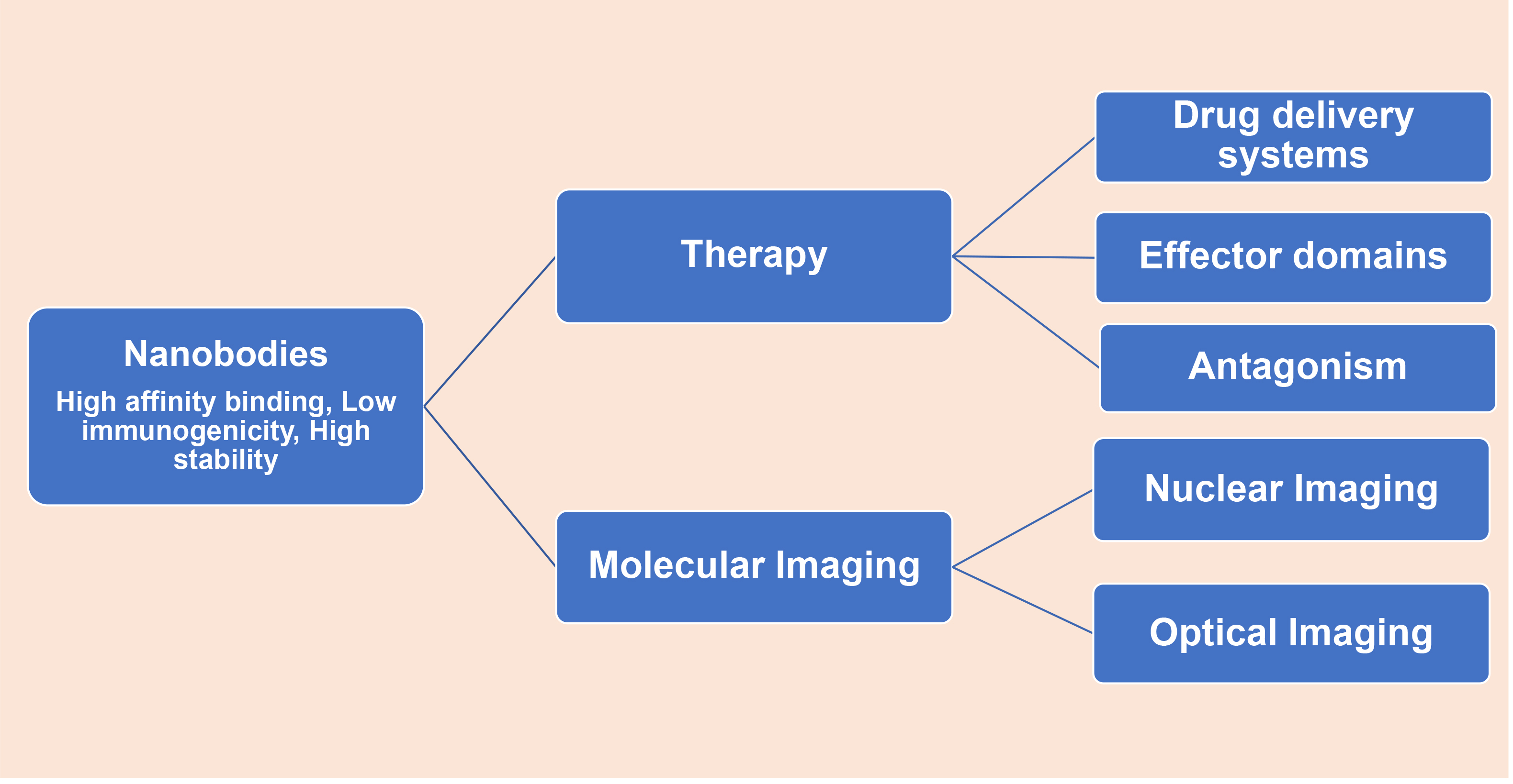
Nanobodies due their high size, low immunogenicity, and very small size demonstrate potential in treatment as well as in diagnostics and imaging tumors. Nanobodies in addition to their use as therapeutic agents can make highly effective as well as non-invasive molecular imaging reagents Fig. 2. Also due to their high penetration, quick elimination, low immunogenicity, and easy production and selection, nanobodies can be used to produce diagnostic tools [67, 68, 69]. To increase the chances of survival, it’s essential to diagnose tumors very early. Due to their small size, high stability, high target specificity, and affinity nanobodies have been engineered into Nanobody-detective constructs for non-invasive in vivo molecular imaging [70, 71]. Such nanobody constructs developed can sharply reach a maximal contrast between the signal in the pathological tissues and that in healthy tissues [68, 71]. For optimal in vivo molecular imaging, sharpness in reaching maximal contrast is vital. Furthermore, nanobodies have a very short half-life and the excess of non-targeting Nanobodies are rapidly cleared from the bloodstream via the kidney and bladder. Their rapid clearance from blood guarantees a high tumor to background ratio early after administering the nanobody probe [72]. Several imaging techniques have been developed to date for clinical application and manifestation, for instance; single-photon emission computed tomography (SPECT), magnetic resonance imaging (MRI), optical, ultrasound, and photoacoustic imaging positron emission tomography (PET), computed tomography (CT) [73].
Molecular imaging of cancer using nanobodies
In comparison to the conventional monoclonal antibodies, nanobodies have small size and very low molecular weight, are exceedingly strategic especially in the field of molecular imaging because it facilitates rapid tumor accumulation, homogenous distribution as well as efficient blood clearance, contributing to high tumor-to-background ratios [74]. Besides, due to their high specificity, nanobodies in conjugation with several kinds of imaging agents provide their use as safe [74, 75]. Various imaging techniques developed that use nanobodies for imaging tumors include; SPECT, PET, etc. Single-photon emission computed tomography (SPECT) is a
List of nanobody probes used in cancer imaging
List of nanobody probes used in cancer imaging
Positron emission tomography (PET) and single photon emission computed tomography (SPECT) images obtained using nanobodies that are labelled with distinct radionuclides in preclinical and clinical studies. I. Uptake of 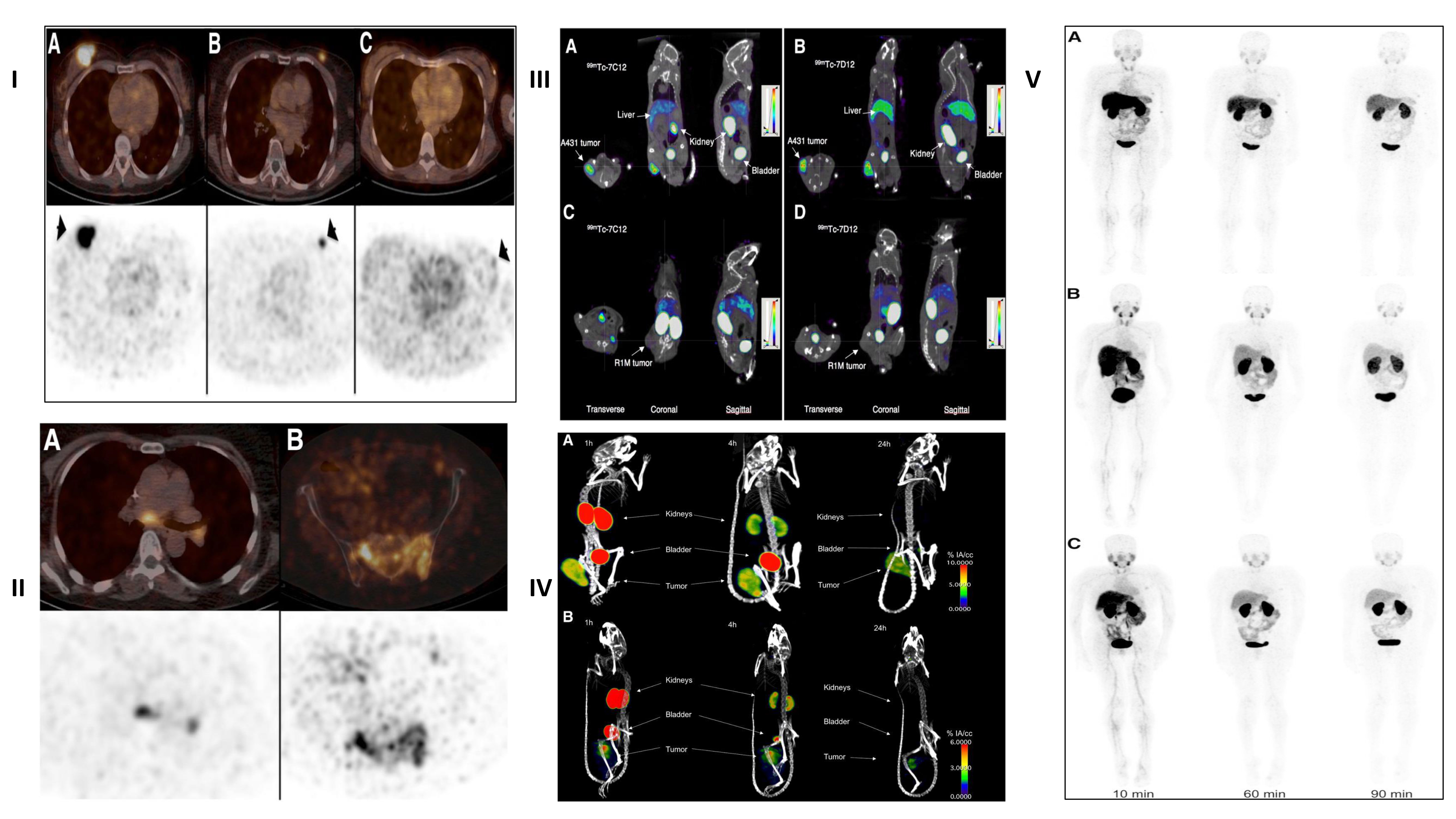
Schematic representation of different aptamers in tumor diagnostics.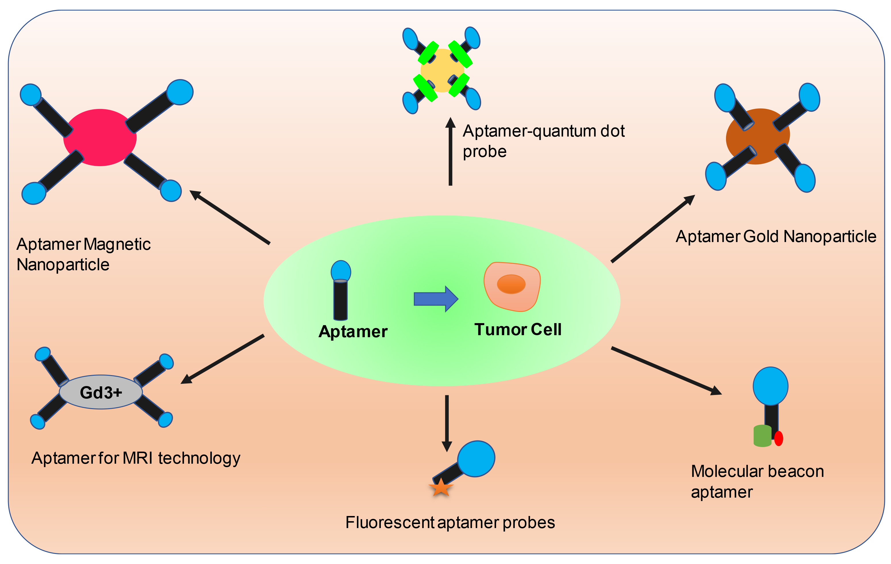
Among the numerous cancer-specific receptors, EGFR and HER2 represent two interesting targets for tumor visualization [79, 88, 76, 89, 90, 91, 92]. In the radio-immunodetection of EGFR overexpressing tumors, 8B6 a
Recently,
Apart from this, no adverse effects or immunogenicity against the administrated nanobody were detected, representing this construct to be appropriate to enter phase II clinical trials [89]. Using a nanobody NJB2, specific for an alternatively spliced domain of fibronectin, recently have been exploited for extracellular matrix (ECM) targeting for non-invasive imaging of tumor progression, metastasis, and fibrosis. This study has revealed that in in vivo immuno-PET/CT imaging, NJB2 helps in the detection of primary tumors and metastatic sites with excellent specificity in multiple models of breast cancer, including human and mouse triple-negative breast cancer, as well as in melanoma. Additionally, NJB2 could detect not only PDAC tumors but also early pancreatic lesions known as; pancreatic intraepithelial neoplasias, representing a very challenging innovation in comparison to being detected by any current imaging modalities, with excellent clarity and signal-to-noise ratios that outperformed conventional 2-fluorodeoxyglucose PET/CT imaging [95]. Also, OA-cb6 nanobody with
List of aptamers developed against specific markers expressed by tumor cells
The term aptamer was coined by Ellington et al. in 1990 and has been derived from a Latin word ‘aptus’ which means fit [97]. Aptamers are single-stranded DNA, RNA, or altered nucleic acid sequences that have a high affinity for target-specific binding developed by SELEX i.e. systemic evolution of ligands by exponential enrichment technology [98, 99]. Because of their high specificity, stability, and non-immunogenicity, aptamers are well-thought-out to be the most promising therapeutic agents which can specifically bind to their targets. Because of their unique three-dimensional structure, aptamers can bind to the target with high specificity and affinity. Few aptamers have a unique function as regulatory proteins and can regulate the function of target proteins via binding to such proteins. There are diverse targets for aptamers some of which include; proteins, ions, cells etc. [100, 101]. Aptamers are termed as chemical antibodies that can be produced at low cost with easy modifications. In comparison to antibodies, aptamers have several applications: high specificity, stronger affinity with the target proteins, can be easily prepared and modified, high stability, and stress-free storage [102]. Until now, a range of aptamers have been screened in diagnostics and therapeutics [103]. Due to the limitations in traditional cancer treatments and diagnostics, it is vital to discover effective methods for cancer diagnosis, early detection, and treatment strategies [104, 105] and the distinctive properties of aptamers have contributed to the rapid analysis, diagnosis as well as treatment of cancer Fig. 4 [106]. Different modifications in aptamers are being applied regularly to make them more versatile, such as with biotin, fluorescent dyes, or even radionuclides [107]. Aptamers are capable to deliver drugs, proteins, or nucleic acids more efficiently into specific structures in the cells via conjugation to siRNAs, nanobodies, or drug molecules reducing the toxic side effects [108, 109, 110]. With the help of aptamers that can recognize specific antigens or biomarkers on the tumor cells in combination with nanoparticles, actively targeted aggregation of nanoparticles can be achieved in the tumor cells with the reduction in toxicity to normal surrounding cells [111].
Presently, aptamer-nanoparticle conjugates have proved significant applications in cancer diagnosis and therapy. Such conjugates have been widely applied for providing the early diagnosis as well as targeted therapy with high affinity biomarkers in the detection of circulating tumor cells (CTCs), introducing a novel outlook for personalized medicine of cancer [112, 113, 114]. The development of cell-SELEX aptamer screening technology is used to categorize cancer cells on the basis of cell surface markers that are overexpressed or altered due to multiple oncogenic mutations Table 2 [115, 116, 117, 118, 119, 120, 121, 122, 123, 124, 125].
The use of aptamers represents a class of ligands that are highly specific as well as having a high affinity towards their target [126, 98]. Presently, a range of aptamers have been screened that can target tumor cells; for example; A10, PMSA aptamer (anti-prostate-specific membrane antigen) [127], AS1411, anti-nucleolin aptamer [128, 129], EpCAM, anti-epithelial cell adhesion molecule aptamer [130, 131], MUC1, anti-mucin1 aptamer [132], Sgc8, anti-protein tyrosine kinase 7 aptamer (PTK7) [133, 134] etc.
Cancer being the second leading cause of death throughout the world [135, 1], therefore an accurate diagnostic technology will show a positive clinical application, which will help clinicians to suggest early treatment strategies, evaluation of treatment effects, monitoring tumor relapse and metastasis as well as assessment of prognosis. Hence aptamers due to their high affinity and specificity to targets hold an apparent advantage in stability, modification and cost production. There is a vast application of aptamers in the field of cancer diagnosis. Circulating tumor cells (CTCs) represent specific tumor markers that are secreted by tumor cells into the blood circulation act as a good target for liquid biopsy [136]. Thus, the delicate recognition of cancer cells plays an important role in diagnosis as well as prognosis [137]. The detection of CTCs in the early stages of cancer in an accurate and efficient concentration will help to monitor the condition and progression of cancer among patients [138]. Recently several methods have been applied for CTCs detection in conjugation with different signal reporting technologies like; fluorescence, magnetic, calorimetric, and electrochemical technology [139, 140, 142, 142]. For example, using a DNA network comprising repeating adhesive aptamer domains, Karnik et al. had developed a podium to isolate and capture CTCs. Similarly, Kashefi-kheyrabadi et al. had established an electrochemical technique for CTCs detection of liver cancer using a liver cancer HepG2 cell-specific aptamer conjugated to a gold plane electrode [140]. For isolation of cancer cells from blood, Fan et al. had developed a podium that links multivalent DNA aptamer nanoparticles with microfluidic devices [143]. Likewise, a dual-aptamer-targeted high-sensitivity CTCs detection podium have been established by Wang et al. conjugating VEGF aptamers with magnetic beads for capturing and concentrating CTCs, using MUC1aptamer-conjugated Pt-Au nanoparticles. Aptamer-functionalized nanostructures have also been developed for cell hook and isolation.
Since recent times, molecular imaging is of great importance in disease detection, observation and prognosis [144]. Aptamers in combination with imaging molecules, like; radionuclides, fluorescent molecules etc. are fetching a prominent tool in cancer diagnosis [145, 146, 147]. A number of imaging technologies used in tumor diagnosis include; computed tomography (CT), luminescence imaging, radionuclide-based positron emission tomography (PET), magnetic resonance imaging (MRI), and single photon emission computed tomography (SPECT). IRD800CW labeled CD30 aptamer have been used for in vivo imaging of lymphoma [148], Cy5 labeled pancreatic cancer specific aptamer have been used for in vivo imaging of pancreatic cancer [149], AS1411 aptamer coupled with blood-brain barrier targeting peptide for in vivo imaging of gliomas [150], etc. In addition to imaging with fluorescent molecules, aptamers in conjugation to magnetic beads can enhance MRI [147]. For example, vascular endothelial growth factor receptor 2 (VEGFR2) aptamer conjugated with magnetic nanomaterials for glioma MRI imaging [151]. Also, Epidermal adhesion molecule (EpCAM) aptamer in conjugation with magnetic nanomaterials is used for MRI imaging of gastric cancer [152], etc. All of these conjugated aptamers significantly improved the tumor-targeted imaging capability, sensitivity as well as biocompatibility, and ultimately reduced the cytotoxicity, advancing the clinical application potential of MRI. In current years, aptamers have shown application features superior to antibodies and are being widely used in immunohistochemical analysis (IHC) analysis of tumor cells. The ICH analysis of formalin-fixed-paraffin-embedded lymphoma was performed by Zu’s group, who found that CD30 aptamer showed almost the same staining pattern as CD30 antibody. Unlike antibodies, aptamers do not cause non-specific staining of necrotic areas in tissue samples when applied to ICH [153]. Moreover, Li et al. had screened a group of aptamers that can specifically identify metastatic lymph node tissues in colon cancer. Using aptamers as probes, IHC analysis could specifically identify colon cancer tissues showing lymph node tissues with colon metastasis. no such signal was stated in non-metastatic colon cancer tissues, or other control tissues, which suggest that aptamers recognize the target related to the process of metastasis. This can be used in the early diagnosis of cancer [154]. Apart from this, a number of chemical drugs are being used to kill cancerous cells, the only drawback is they destroy normal tissues causing severe adverse reactions. As a result, targeted drug delivery is the key approach for current cancer treatment. And aptamers have grown into a novel path in targeted drug therapeutics because of their unique biological and chemical properties [155] viz. aptamer-drug conjugates (AptDC), aptamer-functionalized nanoparticles, therapeutic aptamers, and aptamer mediated immunotherapy [156].
Conclusion
Globally, the high mortality rate due to cancer is still challenging irrespective of advances in treatment including radiotherapy, chemotherapy, and immunotherapy. The use of imaging biomarkers in cancer diagnosis has paved the way for personalized medicine with the accuracy and predictive diagnosis and treatment alternatives. Advances in radiomics, nanobodies, and aptamers in diagnosis and tumor imaging will open new doors for precision treatment and increase the overall survival of cancer patients. Irrespective of their vast applications in cancer biology, they still need to improve target efficacy as well as affinity. Besides their modifications with other drugs, such technologies can be conjugated with several imaging agents which could help in efficient detection of cancer type as well as monitor drug efficacy and disease progression. We believe that with the continuous advancements in such innovative technologies, these will play an important role in future oncological applications.
Footnotes
Acknowledgments
The author acknowledges support and cooperation of Department of Radiological Sciences and Medical Imaging, College of Applied Medical Sciences, Majmaah University, Almajmaah-11953, Kingdom of Saudi Arabia.
Conflict of interest
The author declares that has no conflict of interest.
Funding
No funding was received for the said project.