
Abstract
Migraine is a common disorder which is placed among the top ten reasons of years lived with disability. Cytokines are among the molecules that contribute in the pathophysiology of migraine. In the current study, we evaluated expression levels of IL-6 coding gene in the peripheral blood of 120 migraine patients (54 migraine without aura and 66 migraine with aura patients) and 40 healthy subjects. No significant difference was detected in expression of IL-6 between total migraine patients and healthy controls (Posterior beta
Keywords
Relative expression of IL-6 in migraine patients and controls based on their gender. Red cross indicates outliers.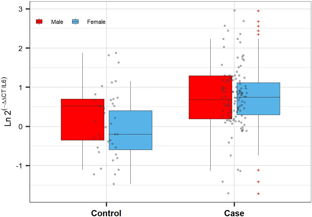
Migraine is a frequent disorder with reported annual prevalence of up to 9% among men and up to 25% among women [1]. It is among the top ten reasons of years lived with disability, worldwide [2]. Several molecular mechanisms have been identified that explain some aspects of migraine pathophysiology [3]. Plasma protein extravasation, glutamatergic transmission and electrophysiological alterations are among the possible mechanisms in this regard [4]. Moreover, substantial evidence implies the role of cytokines in the induction of pain and activation of neurovascular inflammatory responses which are linked with migraine episodes [5, 6, 7]. Interleukin-6 (IL-6) is among cytokines with appreciated roles in the pathogenesis of migraine [5]. Moreover, this cytokine is among molecules whose concentrations are elevated in inflammatory circumstances and upsurges in IL-6 are associated with pain severity during time [8, 9]. Elevated levels of this cytokine in the serum of migraine patients imply tits role in the initiation of pain or immune cascades. In spite of these observations, the serum concentrations of this cytokine has not been decreased in migraine patients taking topiramate [5]. Therefore, high IL-6 levels throughout pain-free intervals may make patients susceptible to pain episodes [5]. Moreover, animal studies have shown that IL-6 increases the excitability of dural afferent neurons which are involved in the pathogenesis of migraine. This effect has been exerted probably through ERK-mediated alterations in a kind of Na+ channel [10]. Based on the possible role of IL-6 in the pathobiology of migraine and incompleteness of data regarding its expression pattern in migraine patients, we designed the current study to measure its expression levels in migraine patients and healthy subjects.
Relative expression of IL-6 in migraine patients compared with controls
Relative expression of IL-6 in migraine patients compared with controls
*Group: Case/control; Gender: Female/male; **estimated from frequentist methods. The Bonferroni correction and 95% CrI were used simultaneously for subgroup analysis.
The difference in IL-6 expression between study subgroups according to the Bayesian quantile regression model
*Reference: Control group. **Adjusted for age and gender.
Enrolled persons
A total of 120 migraine patients (54 migraine without aura and 66 migraine with aura patients) were enrolled in the current project. Cases include 20 males and 100 females with mean age (
Expression assays
Peripheral blood samples were obtained from migraine patients and healthy subjects in EDTA tubes. RNA was extracted from blood specimen using the RNA extraction Kit from the GeneAll Company (Seoul, South Korea). The OneStep RT-PCR Series Kit (BioFact™, Seoul, South Korea) was used for conversion of RNA to cDNA. Expression of IL-6 coding gene was measured in all samples using the HPRT1 gene as the normalizer. Reactions were prepared using the RealQ Plus 2x Master Mix (Amplicon, Denmark). The nucleotide sequences of primers and probe are as follow: F: ATGCAATAACCACCCCTGACC, R: CCATGCTACATTTGCCGAAGAG and FAMACCACAAATGCCAGCCTGCTGACGTAMRA.
Statistical methods
Relative expression of IL-6 gene was compared between cases and controls using the multiple Bayesian quantile regression model to reduce the uncertainty that may occur when an outcome has a non-normal distribution. This model allowed for controlling the effects of gender, age, and interaction effects. Subgroup analysis was conducted within gender group if the interaction effect between gender and group was significant. The Bonferroni correction was used along with 95% credible interval (CrI). We used asymmetric Laplace family prior with 2000 Burn-Outs as well as 5000 iterations. The adequacy of model was examined using Rhat, Posterior predictive plots, and Loo. We used the Stan, ‘ggplot2’, ‘brms’, and pROC packages to conduct statistical analysis in the R v.4 environment.
Results
Expression levels of IL-6 in study groups
Figure 1 depicts the overview of IL-6 expression levels in migraine patients and healthy controls.
No significant difference was detected in expression of IL-6 between total migraine patients and healthy controls (Posterior beta
Then, we compared expression of this cytokine coding gene between healthy controls and patients with and without aura. Expression of IL-6 was significantly higher in patients with aura compared with controls (Posterior beta
Finally, we appraised the diagnostic power of IL-6 transcript levels in differentiation of patients with migraine from healthy subjects. Expression levels of this gene could differentiate these two sets of samples with sensitivity of 91.67% and specificity of 42.5%. The area under the receiver operating characteristic (ROC) curve was 0.668. Figure 2 shows the details of ROC curve analysis.
ROC curve showing the diagnostic power of IL-6 transcript levels in differentiating migraine cases from controls.
In the present project, we examined peripheral expression of IL-6 coding gene in a cohort of Iranian patients with migraine and healthy controls. IL-6 has a prominent in the process of perception of the harmful stimulus by sensory nervous. Expression of this cytokine, its receptor and molecules involved in the related signaling are increased in peripheral nerves throughout experimental induction of pain [12]. Moreover, this cytokine alters expression of numerous pain mediators. Finally, IL-6 antagonism affects the perception of pain, thus IL-6 has been suggested as a remarkable target in modulation of nociception [12].
We detected no significant difference in the expression of IL-6 between total migraine patients and healthy controls. Yet, expression of this gene was significantly higher in female patients compared with female controls. The differences in the pathobiology of migraine between males and females have been noted previously [13]. Moreover, IL-6 has been shown to be a sex-specific susceptibility factor for immune disorders [14, 15] which is suppressed by estrogens [16]. Therefore, it is possible that over-expression of this cytokine exerts more detrimental effects in female subjects making them more susceptible for migraine attacks.
Consistent with our results, Sarchielli et al. have assessed IL-6 levels in the consecutive blood specimens obtained from the internal jugular venous of migraine patients without aura during headache episodes. They detected a temporary elevation in IL-6 levels in the first 2 hours following initiation of migraine attack compared with the time of catheter insertion [17]. However, Oliveira et al. have shown decreased levels of IL-6 and a Th1-dominant cytokine profile in female patients with migraine as being demonstrated by elevated plasma levels of TNF-
Moreover, we detected higher expression of IL-6 in patients with aura compared with controls in spite of similar levels of this cytokine between patients without aura and healthy subjects. Therefore, this cytokine might participate in the observed extra-axial immune responses in migraineurs with aura [19].
Finally, we demonstrated that IL-6 transcript levels have sensitivity of 91.67% and specificity of 42.5% in differentiation of patients with migraine from healthy subjects. However, the diagnostic power of these transcripts were less than being regarded as appropriate biomarker in this regard.
Taken together, our data suggest that IL-6 might be involved in the pathophysiology of migraine among females and migraine with aura among both sexes.
Footnotes
Acknowledgments
The current study was supported by a grant from Hamadan University of Medical Sciences (Grant number 9809126715).
Conflict of interest
The authors declare they have no conflict of interest.