
Abstract
Superresolution is a concept to increase the resolution. The main objective of this paper is the study of iterative curvature based method for super-resolving low resolution of a leaf diseased images. The domain specific prior is incorporated into superresolution by the means of iterative curvature SR based estimation of missing high frequency details from infected leaf images. The model is composed of two step pixel filling approach. Through this proposed work, fine edges of SR images are preserved without applying complex mathematical algorithms based on wavelet, fast curvelet, etc. In this paper, we have validated proposed scheme over 9 infected leaf images of various crops like soybean, cotton, rose, citrus family etc. shows better result in visual as well as subjective quality as that of complex multi frame SR algorithms like reconstruction and registration along with less computational time. This concept is most useful for agricultural expert for helping our farmers for exact leaf disease detection and accurate remedial actions. The experimental result shows the best visible SR result of an infected leaf along with MSE and PSNR i.e. Statistical results. It also shows the comparison of proposed method with the existing techniques successfully.
Introduction
In agriculture, the analysis of infected leaf area is of great importance for the application of techniques such as pruning, fertilization and planting density [2]. A feature that can be extracted by analyzing the leaf area is the quantification of damage caused by pests and diseases. Such damage can be detected through the study of damaged leaf area by pests [2]. Detecting the precise amount of damaged leaf area is essential to determine control actions such as application of pesticides, since a small damaged leaf area may dispense control measures. In this paper, we have analyzed infected leaf image using adaptive based image superresolution techniques in order to recover the high frequency details such edges, various features, etc.
Obtaining a high-resolution (HR) image from single or multiple low-resolution (LR) images, known as “super-resolution” has been a classic problem. High resolution means high pixel density, also refered to as high-definition (HD). An HR image brings out details that would be blocked out in an LR image. In this paper, using artificial intelligance, infected HR of a diseased image is exactly detected and diagonsed.
Super resolution problem is an ill-posed inverse problem. Estimating details is an inverse problem since low resolution observation is the result of a smoothing and downsampling process [3]. Basically, SR technique is broadly categorized in two parts.
First is traditional non-adaptive image reconstruction and registration technique [5, 6] in these methods attempt to solve the problem by employing and fusing a number of low resolution images. The images are of an underlying scene are positioned into a common coordinate frame by sub-pixel shifts of images. Most of the literature available on super-resolution is for multi-frame and majority of them are based on the motion as cue. The super-resolution idea was introduced by Tsai and Hung, where a pure translation motion has been considered [1]. In such methods the quality of reconstructed SR image obtained from a set of LR images depends upon the registration accuracy of the LR images and some prior knowledge of imaging system [5, 6]. Nearly all SR reconstruction algorithms are based on the fundamental constraints that provide less useful information as the magnification factor increases also less computationally efficient to get more accuracy. Baker and Kanade found these limitations and developed a SR algorithm by modifying the prior term in cost to include the result of a set of recognition called as recognition based super-resolution or hallucination [11].
And second is single image adaptive learning based SR methods [7] which is more powerful and useful, when only a single observation image is available and several other high resolution images are present in the data set. All high resolution images from data set will act as training images. This method is classified under the motion free superresolution scheme as the new information required for predicating the HR image is obtained from a set of training images rather the subpixel shifts among low resolution observations.
In this paper, we have proposed an adaptive iterative curvature based spatial SR method for accurate analysis of infected leaf diseased images.
Existing superresolution techniques
As per the superresolution imaging analysis is concerned, there are two main domain i.e. Frequency domain and Spatial domain approach for image registration [3].
In our case, the results of the spatial domain are very much better in visual quality as well as in analytical parameter also than the frequency domain, which typically failed to adequately register images.
By the nature of frequency domain, Fourier transform methods are limited to only global motion models. In the early stages, most of the research work is carried out under frequency domain approach but as more general degradation models were considered; later research has tended to concentrate almost exclusively on spatial domain formulations.
Basically, image superresolution can be obtained in two categories – Non-adaptive SR and Adaptive SR in spatial domain approach.
Non-adaptive image SR
Non-adaptive image SR techniques are based on direct manipulation on pixels instead of considering any feature or content of an image. These techniques follow the same pattern for all pixels and are easy to perform and have less calculation cost. Various non-adaptive techniques are nearest neighbor, bilinear and bicubic [5, 25]. But these techniques having some drawbacks such as problems of blurring of edges or artifacts around edges. It stores only low frequency component of an original leaf image also produces blurry images quality. Mainly it misses the required information from superresolved infected leaf image. To overcome these, we approached for Adaptive Image SR for our agricultural information for more accuracy.
Adaptive image SR
This technique considers image features like intensity value, edges as well as texture informations. It also provides better visual image quality result as it preserves high frequency components from an original infected leaf image, so it is much easier for detection and classification accuracy. Various adaptive SR techniques are NEDI, DDT, FCBI, Learning based approach [17, 18]. Only main drawback is it requires much more computational time. So, here, we have worked over this problem while maintaining the SR quality of an infected leaf image. So, as far as infected leaf image problems are concerned, adaptive image SR approach is much better in practice and advantageous.
Machine learning based detection and recognition of plant diseases can provide clues to identify and treat the diseases in its early stages [11, 12]. Comparatively, visually identifying plant diseases is expensive, inefficient, and difficult. Also, it requires the expertise of trained botanist. There are several methods for measuring leaf area, however, in practice, it is used mainly three: the human evaluation, the method of leaf dimensions and the methods which use devices such as planimeter and area integrator [26]. Nevertheless, these methods require extensive work and are time-consuming. Moreover they have some degree of inaccuracy. And, the measurement techniques are not performed in the most cases by a farmer, but by an expert (agronomist), which delays the diagnosis. With the advances in computing, especially in the graphics processing, it is possible to develop alternative methods for determining the damaged leaf area. Plant diseases have turned into a dilemma as it can cause significant reduction in both quality and quantity of agricultural products.
Proposed methodology – adaptive iterative curvature based image super-resolution
Overall block diagram of step by step implementation.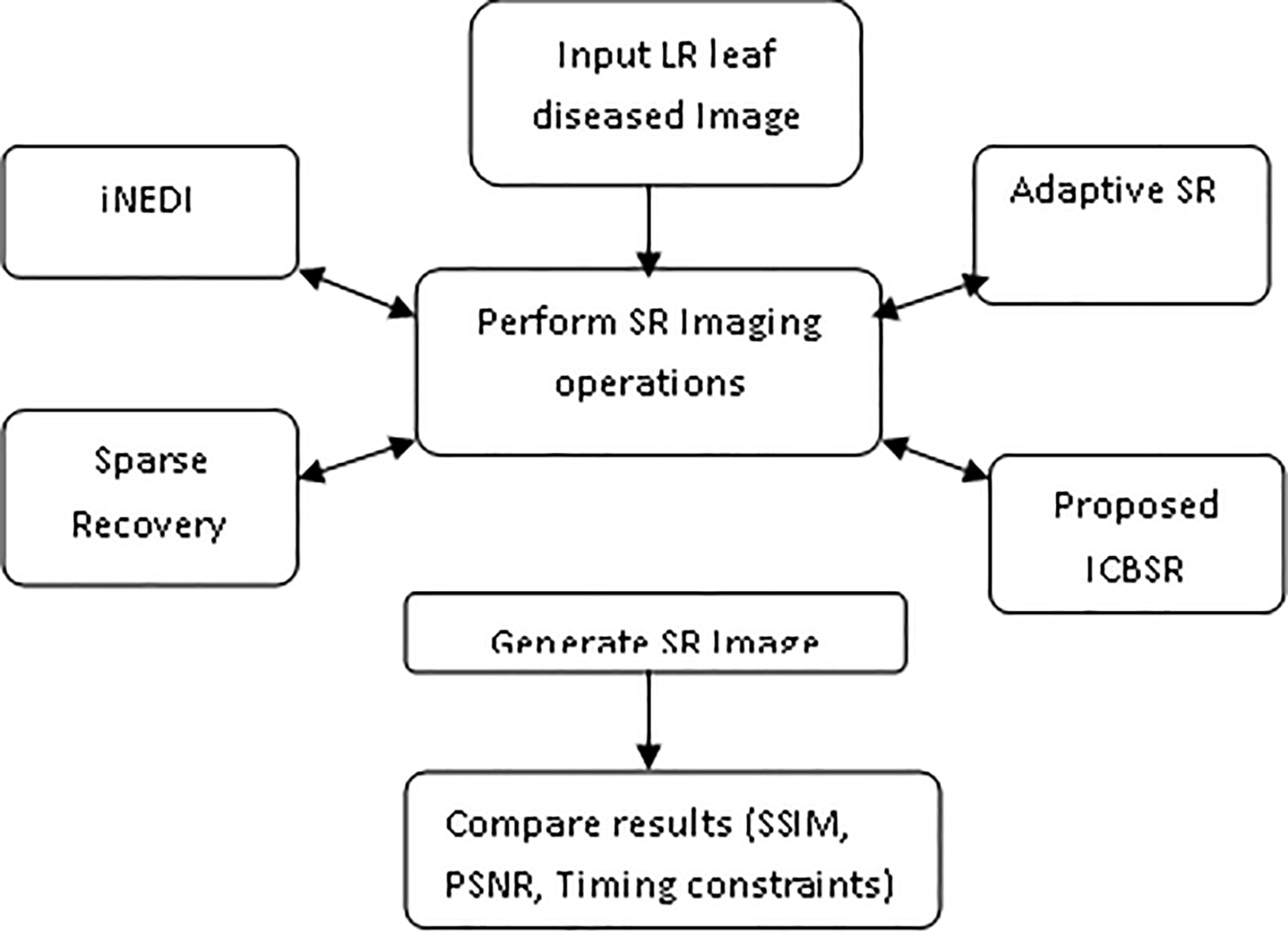
Figure 1 illustrates the overall step by step implementation of a proposed system. In block diagram, all the existing three techniques are compared with the proposed ICSBSR method and obtained analytical parameters are compared so as to validate a successful implementation of our proposed system.
Iterative curvature-based interpolation technique focuses on estimation of direction and based on second order derivatives. Main purpose of introducing ICBSR technique to minimize the artifacts presented in image compare to other technique like patch based learning and other adaptive and nonadaptive SR techniques. ICBSR technique has lower computational cost then other non-adaptive techniques. Image magnification generally results in loss of image quality. Therefore image magnification requires interpolation to read between the pixels. Generally the enlarged images suffer from imperfect reconstructions, pixelization and jagged contours. The proposed system provides error-free high resolution for real time infected leaf images. The basic idea behind the system comprises two basic steps: ICBSR technique is a combination of two techniques. In first technique, the new pixels are computed by interpolating along the direction (FCBI, Fast Curvature Based Interpolation). In second technique, we modified the interpolated pixels using iterative method with energy term for edge preservation purpose [14].
First technique, FCBI is same the Data Dependent Triangulation interpolation technique, but instead of taking the average value of two opposite neighbor pixels, we consider second order derivatives in two diagonal direction
In detail, as shown in Fig. 2, if we consider the first interpolation step, we compute the local approximation of the second order derivative
Equation (3) represents second order derivative
Edge-based interpolation based on a12-Pixels neighborhood: the largest second order diagonal derivative determines the interpolation direction.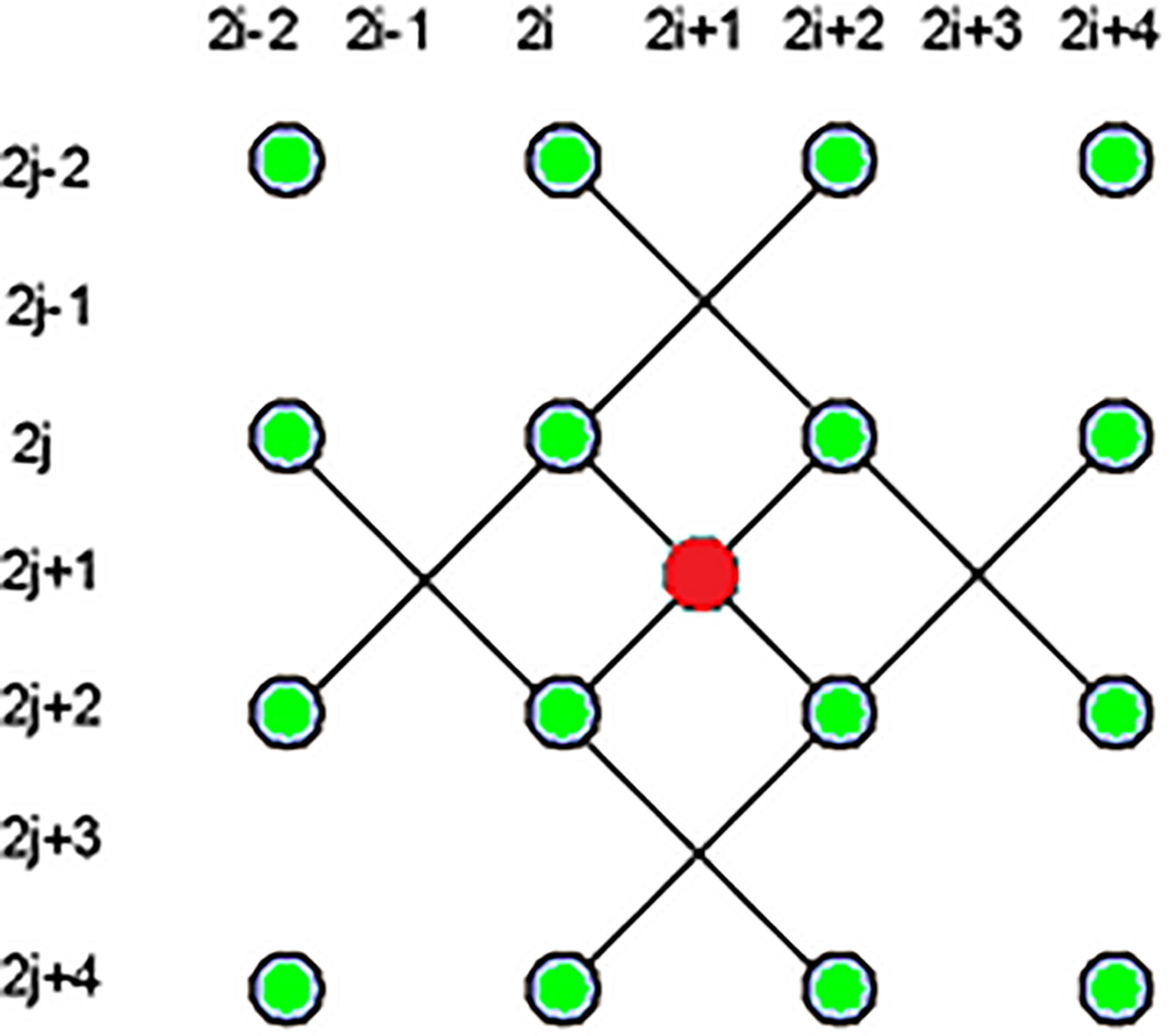
Modified formulas for the second step filling the remaining gaps are straightforward.
First we introduce the term as
From Eq. (3), where,
This energy term sums local directional changes of second order derivatives. Weights wi are set to 1 when the first order derivative in the corresponding direction is not larger than a fixed threshold and to 0 otherwise. In this way, smoothing is avoided when there is a strong discontinuity. The minimization of this term roughly corresponds to force continuity in the local curvatures of the continuous surface ideally describing the image.
The complete energy function for each pixel location
From Eq. (3), where,
Overall procedure for ICBSR technique is as follow:
The average of the two neighbors in the direction of lowest second order derivative (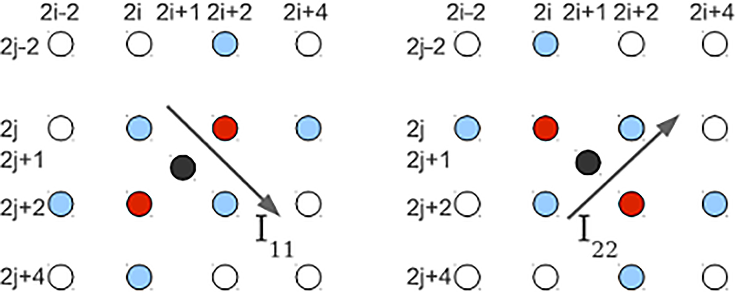
Figure 3 gives the detailing of proposed algorithm approach for pixel filling concept.
Mathematical analysis of SR is basically based on accurate MAP estimation [4]. According to the MAP estimator, the additive noise, the measurements, and the ideal image are all assumed stochastic signals. The MAP estimation of the unknown image
Bayesian approach provides a flexible and convenient way to model a priori knowledge concerning solution
The mathematical operation shows the final result as:
Where,
From Eqs (4) and (6), if we assume that the measurements additive noise is zero mean Gaussian random process with auto-correlation matrix
By considering the stochastic least mean square filtering operation in order to minimize the error function as
The solution can be achieved through following expression
In Eq. (4), the ratio
The analytical parameter such as MSE and PSNR can be calculated as, let,
PSNR avoids many problem of measuring image quality by scaling the MSE according to the image range. It is defined in Eq. (10) as
The MSE (Mean Square Error) and PSNR (Peak Signal to Noise Ratio) shows the better analytical result as that of conventional SR interpolation methods.
Consider, an original image size [
For Sparse Recovery and Example based SR technique,
For iNEDi and ICBSR technique,
Where,
The SSIM index is a full reference metric; in other words, the measuring of image quality based on an initial uncompressed or distortion-free image as reference. SSIM is designed to improve on traditional methods like peak signal-to-noise ratio (PSNR) and mean squared error (MSE), which have proven to be inconsistent with human eye perception.
SSIM considers image degradation as perceived change in structural information. Structural information is the idea that the pixels have strong inter-dependencies especially when they are spatially close. These dependencies carry important information about the structure of the objects in the visual scene.
At a high level, SSIM attempts to measure the change in luminance, contrast, and structure in an image.
The generated form of SSIM can be given as:
Where
Adaptive Neural Fuzzy Inference system is designed to allow IF-THEN rules and a membership function (fuzzy logic) to be constructed based on the historical data and also includes the adaptive nature for automatic tuning of the membership functions [24]. ANFIS refers to an inference system that integrates the best features of neural network and fuzzy logic. It is a system that predicts input/output relationship of given set of data. It consists of nodes and directional links through which the nodes are connected. Part or all of the nodes are adaptive, which means that their outputs depend on the parameter(s) pertaining to these nodes and the learning rule specifies how these parameters should be changed to minimize the error measure [24].
The ANFIS structure is broadly classified in five classes as below:
a rule base containing a number of fuzzy IF-THEN rules. a database which defines the membership functions of the fuzzy sets used in the fuzzy rules. a decision-making unit which performs the inference operations on the rules. a fuzzification interface which transforms the crisp inputs in to degrees of match with linguistic values. a defuzzification interface which transform the fuzzy results of the inference in to a crisp output.
Usually, the rule base and the database are jointly referred to as the knowledge base. Figure 4 shows the ANFIS flow architecture level-wise.
ANFIS architecture.
From Fig. 4,
Layer 1 is Fuzzifying Layer; Layer 2 is Implication Layer; Layer 3 is Normalizing Layer; Layer 4 is Defuzzifying Layer and Layer 5 is Output Summation Layer.
In the extreme case, we can even shrink the whole network into a single adaptive node with the same parameter set.
Thus, we have constructed an adaptive network that is functionally equivalent to a Sugeno Fuzzy Model. The flexibility is that we can combine layer 3 and layer 4 to obtain an equivalent network with only four layers. By the same token, we can perform the weight normalization at the last layer. Following Fig. 4 illustrates an ANFIS of this type. In the extreme case, we can even shrink the whole network into a single adaptive node with the same parameter set. Table 1 shows the ANFIS parameter specification.
Specifications of ANFIS model
In actual practice, first order Sugeno model is preferred because of its transparency and efficiency property. Figure 5 shows the ANFIS Sugeno fuzzy model, where weight normalization at last layer.
ANFIS Sugeno fuzzy model, where weight normalization at last layer.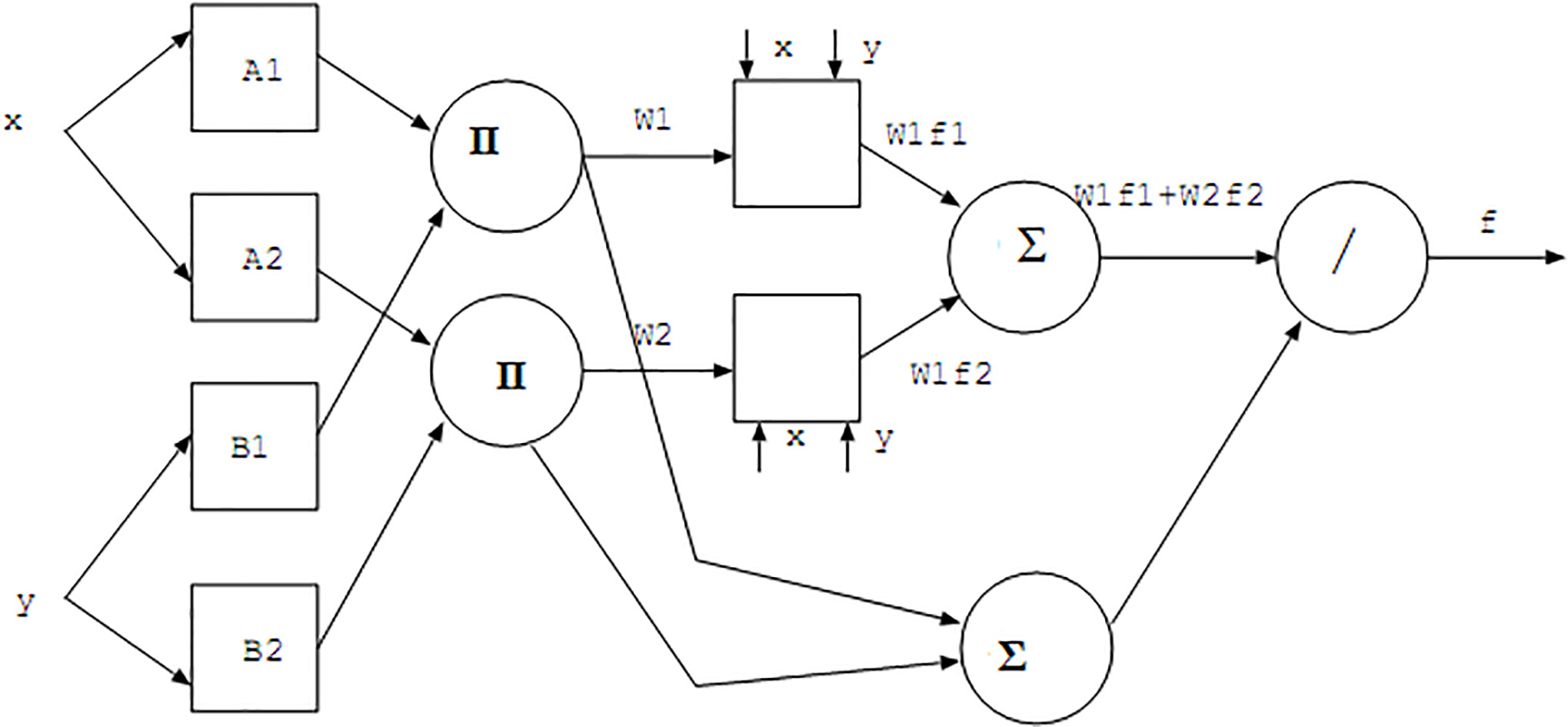
ANFIS training network also show the RMS error response against each 3000 epochs for different images. Figure 6 shows the ANFIS training response in terms of epochs Vs. RMS error performance where, epochs are limited 3000 against which we have got avg. RMS error of 0.170287 for different 10 leaf images. As epochs increases RMS error reduces exponentially. Reconstruction error is very less becoming one of the important criteria of inverse image superresolution problem. Fine edge as well as fine details can be detected by using this technique which is one of the most important parts of feature extraction and image restoration and image enhancement.
Comparison of output size, PSNR (dB), Timing constraints (sec) and memory space requirement with respect to increments of SR factor
Parameters of SR image quality: Structural Similarity Index Matching (SSIM)
ANFIS training response.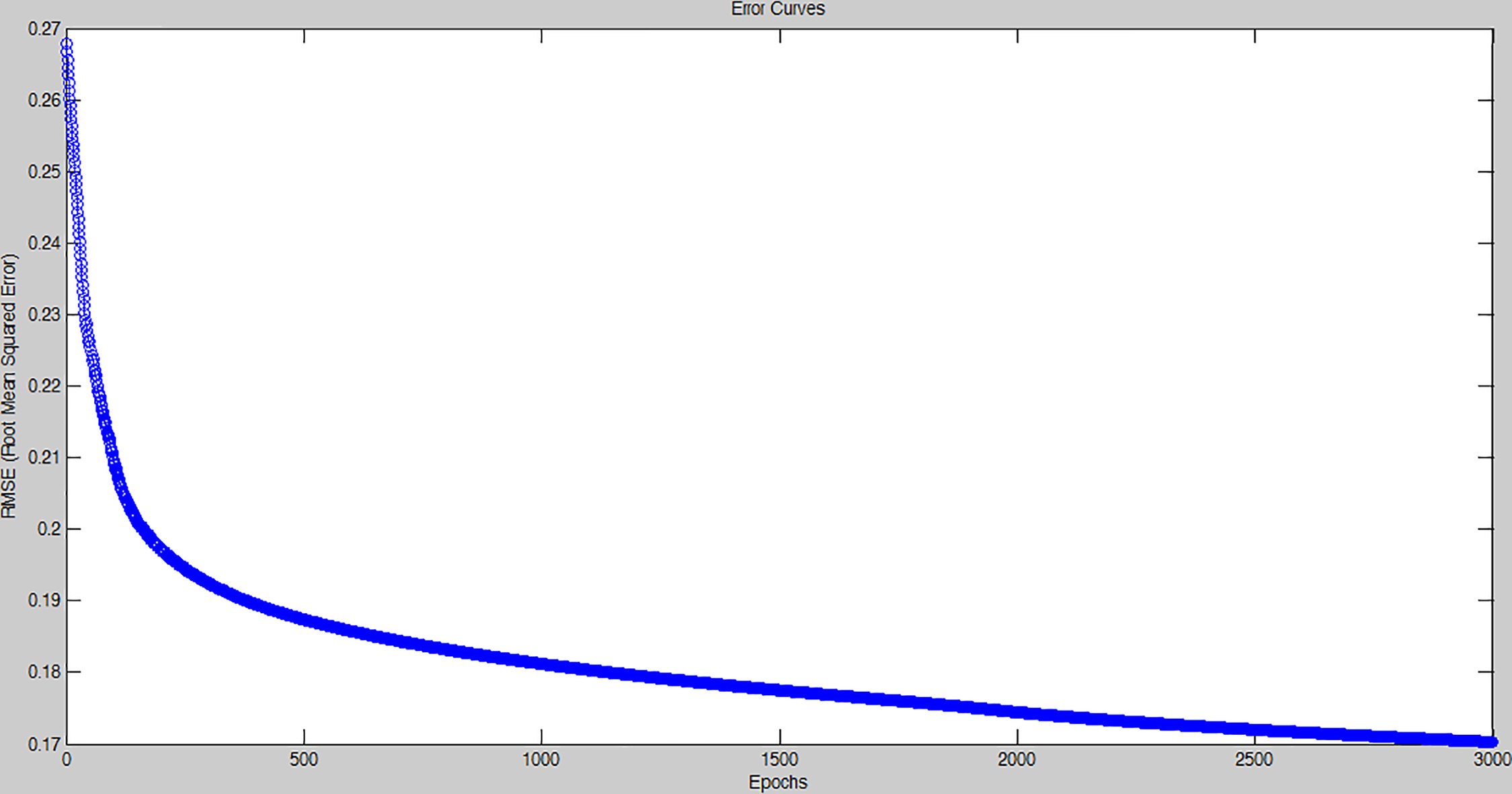
(a1), (b1), (c1), (d1), (e1), (f1), (g1), (h1): Original low resolution leaf diseased images. (a2), (b2), (c2), (d2), (e2), (f2), (g2), (h2): Super resolution leaf images by factor 4.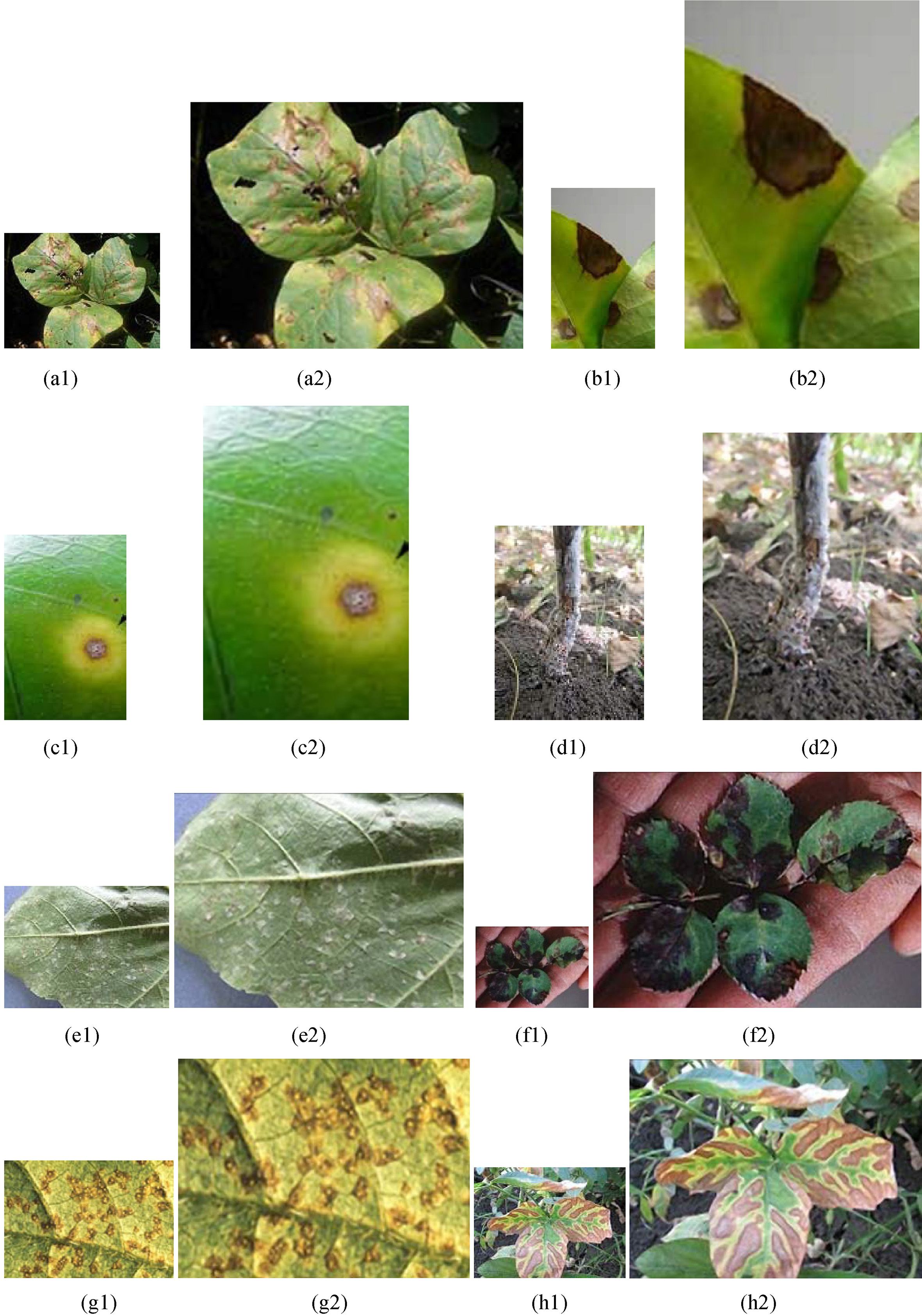
In this paper, we have tested and implemented proposed superresolution technique for agrobased application. Plant disease is one of the essential causes that reduces quantity and degrades quality of the agricultural merchandises. In agriculture, the analysis of infected leaf area is of great on great demand and important for the application of techniques such as pruning, fertilization and planting density.
We have tested randomly 10 sample images of some of the 10 diseased and insect’s LR images to test our robustness of our proposed adaptive learning based iterative curvature based SR techniques.
The overall testing work has been done over original images without any SR technique against example based patch learning SR algorithms, Sparse Recovery SR algorithm and our proposed SR algorithm.
The experiments were executed on an Intel Core TM 2 duo CPU @ 2.5 GHz with 3 GB RAM and results are obtained using MATLAB tool. Images are captured by using a low cost low resolution mobile camera with resolution which is pre-setted. Initially, we had captured all possible high resolution of infected leaf images from surveying various farm fields in order to prepare a huge database i.e. dictionary. And they are preprocessed to convert in low resolution for validation. Thus, we have implemented single image superresolution technique in this paper.
Again minimizes the error function requires for image superresolution operation. Implementation is also very simple and easy.
Comparison between PSNR of various SR techniques
Comparison between PSNR of various SR techniques
Comparison between timing constraints for various SR techniques
Statistical comparison result: Timing constraints.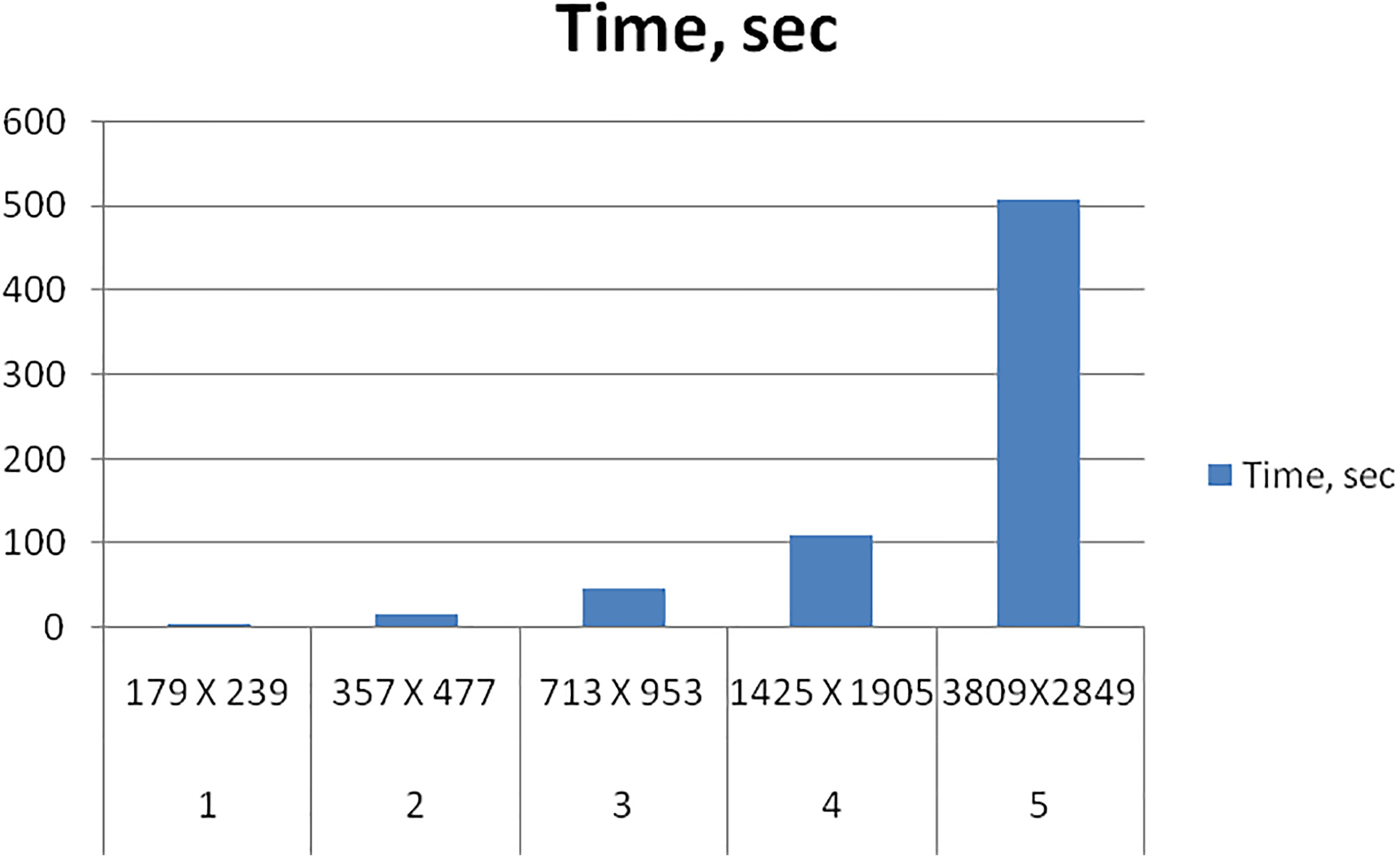
Statistical analysis of SSIM.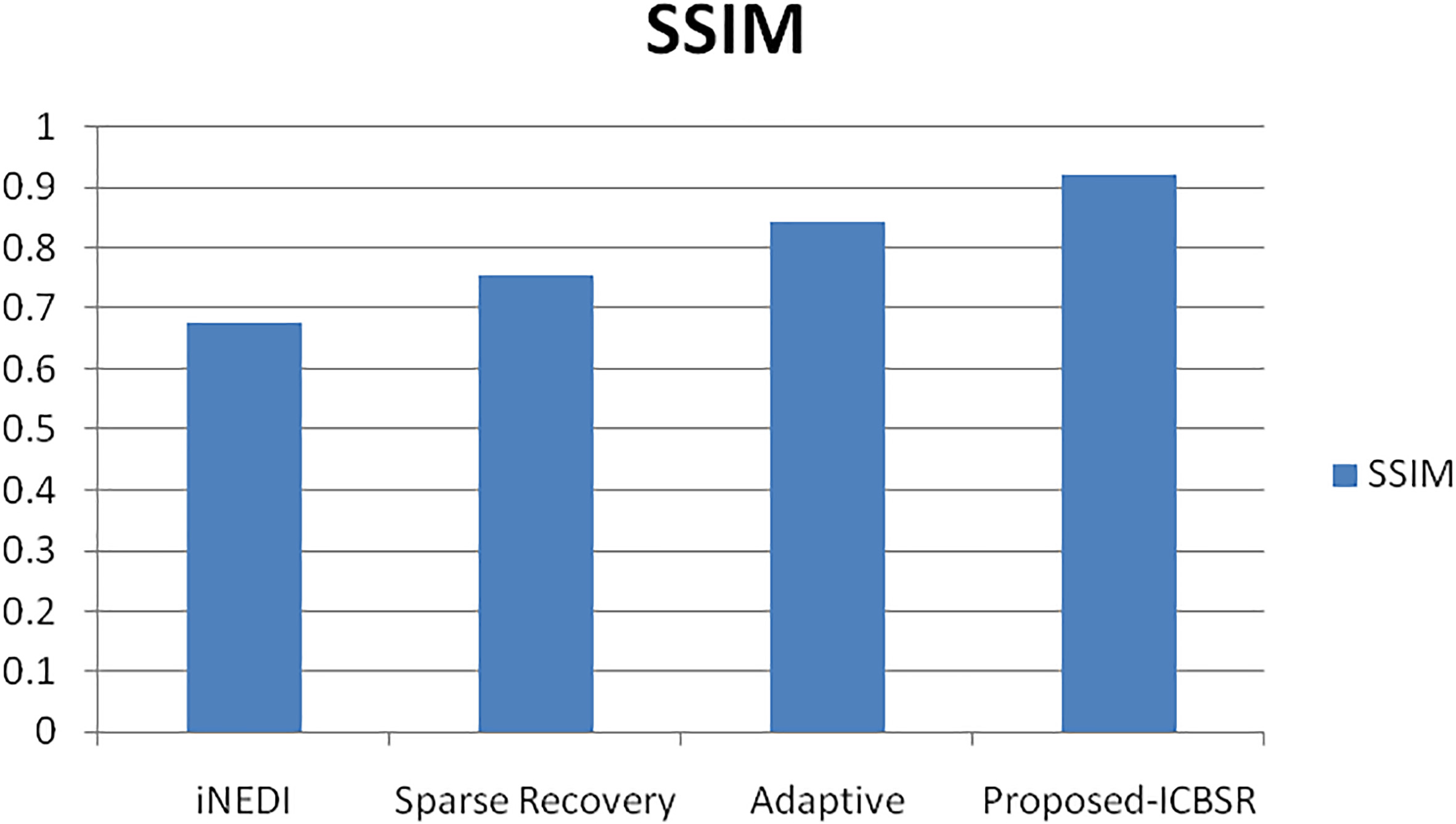
Avg. statistical PSNR results.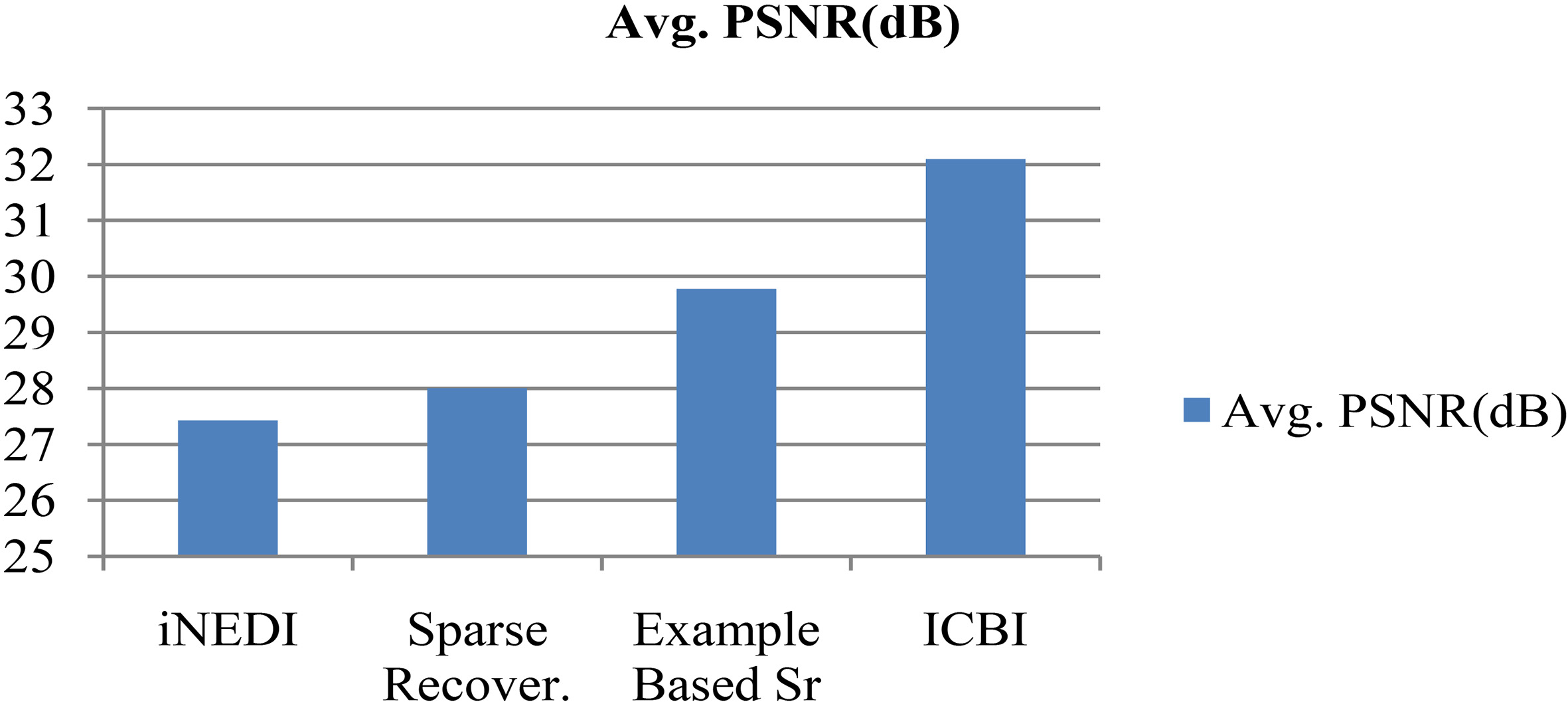
Avg. statistical timing constraints results in sec.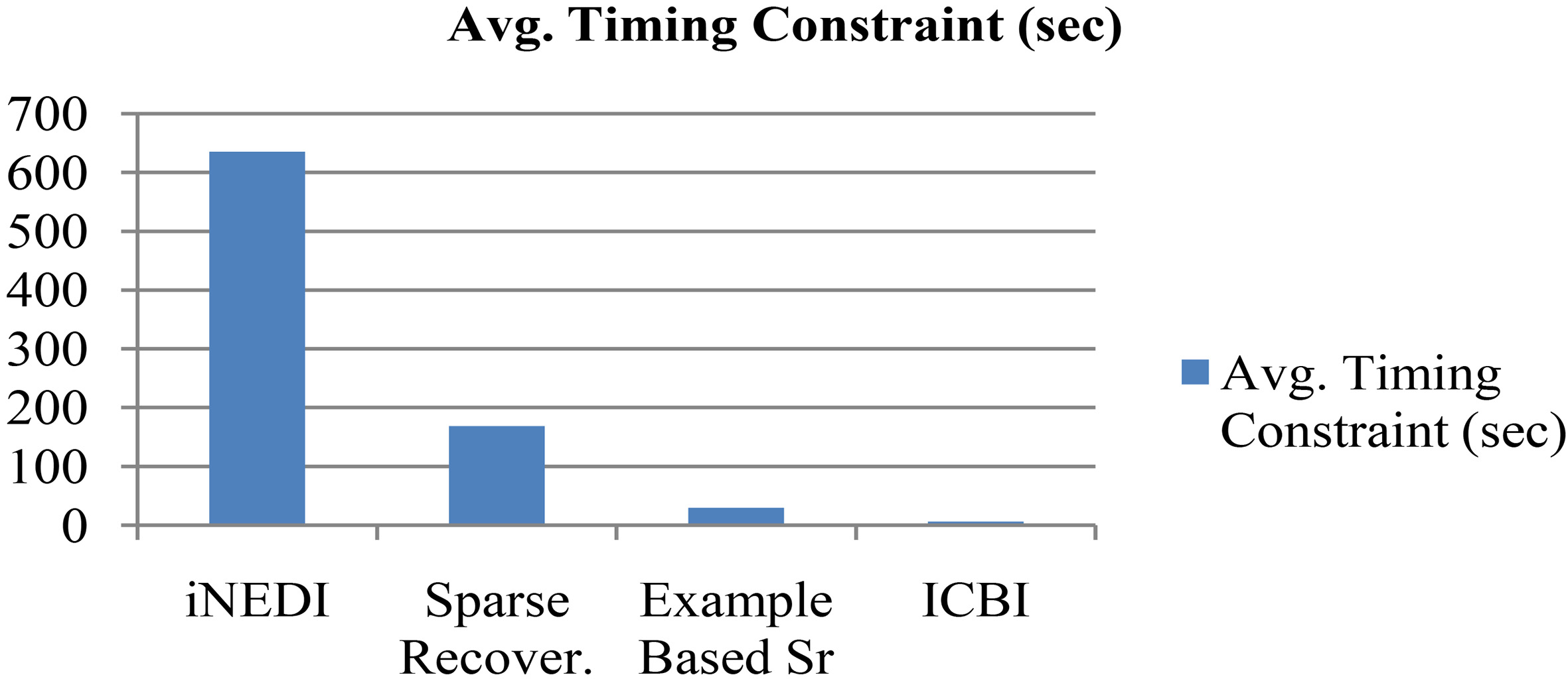
In this experimentation, we have considered various performance matrics such as Structural Similarity Index Matching (SSIM), Peak Signal to Noise Ratio (PSNR), timing constraints, and memory space requirements in order to validate the best result of our proposed methodology among various existing techniques. Figure 6 shows the original quality of images. Figure 7 outputs SR images with SR factor of 4. Table 2 shows Comparison of output size, PSNR (dB), Timing constraints (sec) and memory space requirement with respect to increments of SR factor (serially, 1, 2, 3, 4, 5) on a single image by our proposed ICSBR technique. While Table 3 shows the comparison results of Structural Similarity Index Matching (SSIM) parameter among existing SR techniques with respect to proposed technique respectively. Table 4 shows the comparison results of Peak Signal to Noise Ratio (PSNR) parameter in dB among existing SR techniques with respect to proposed technique respectively. Table 5 shows the comparison results of require timing constraints for all the techniques in second among existing SR techniques with respect to proposed technique respectively. Figure 8 shows Statistical Comparison Result in timing constraints as SR factor increases serially from 1 to 5. Figure 9 shows the average statistical comparison results of Structural Similarity Index Matching (SSIM). Figure 10 shows the average statistical comparison results of Peak Signal to Noise Ratio (PSNR) parameter in dB. Figure 11 shows the average statistical comparison results of require timing constraints for all the techniques in second.
In this paper, we have compared and analyzed obtained results among INEDI, Sparse Recovery, and Adaptive SR technique with the proposed superresolution technique.
Conclusion and future scope
From the observational result, it is verified that the disease infected single LR image with low cost camera is only sufficient to improve its resolution with better visual quality. Information from leaf edges are recovered successfully. The proposed algorithm is very much fast with reduced size of database due to
Properly analyzed infected leaf images are mostly useful for plant pathologist for the following purposes:
Identification of diseased leaf, stem, fruit; Identification and quantification of affected area by disease; Identification of intensity of diseases and their effect on productivity.
Our proposed methodology is the best option for costly and complex hyper spectral satellite imagery system.
This paper will definitely bring some smile on farmer’s face for improvement is crop production and agricultural development through agricultural experts.
In future, this concept can be extended to different plant pathologist for solve various agricultural engineering problems. There is a great scope for doing further research on the creation of self-learning database for any kinds of single image SR. Also, the work should be independent from the interpolating factor.
Footnotes
Acknowledgments
The proposed research work is carried out under the guidance of Department of Plant Pathology, College of Agriculture, Nagpur affiliated under Dr. Panjabrao Deshmukh Krishi Vidyapeeth, Akola. I kindly express my gratitude towards time to time support and making availability of the resources for the collection leaf images for the development of this project.