
Abstract
BACKGROUND:
The effects of a long-term static stretching program on physical performance parameters have not been elucidated completely, although the effects on muscle flexibility have a consensus.
OBJECTIVE:
This study aimed to investigate the effect of a long-term static stretching program on physical performance and muscle properties.
METHODS:
Participants performed a 2-min static stretching for the ankle joint 5 times per week for 4 weeks. Physical performance and muscle properties was measured before and after the static stretching program.
RESULTS:
Results showed that range of motion (ROM), dynamic postural stability, and muscle hardness were positively changed, whereas other variables i.e. maximal isometric plantar flexion moment, jump heights, muscle-tendon junction displacement and its angle, were not.
CONCLUSIONS:
Four-week of SS program may improve ROM, dynamic postural stability, and muscle hardness without decreasing physical performance.
Introduction
Stretching is speculated to improve skeletal muscle flexibility and considered useful for preparation for sports activity [1] and injury prevention [2, 3]. Static stretching (SS) is widely performed during warm-up and cool-down sessions at athletic practices or competitions. However, SS was found to decrease performance in the acute phase in some systematic reviews [4, 5]. In addition, dynamic movements rather than SS were reported to be important for post-exercise recovery [6]. According to previous studies, SS should not be performed before and after athletic activity.
Athletes also perform SS for daily conditioning. Muscle flexibility is decreased after training or sports activity [7]. A less flexible muscle can lead to muscle injury [8] and SS is one of the easiest methods to maintain flexible muscles. Therefore, understanding the effect of long-term SS as daily conditioning is very important. Some studies investigated the effect of a 5-week SS program and found that muscle hardness was decreased [9, 10, 11]. These findings have proved that SS is an effective method for maintaining muscle condition. With regard to physical performance, some studies reported improvement in muscle strength [12] and sprint times [13] after an SS program. However, other studies found no effects of SS on range of motion (ROM), running speed, or vertical jump height in healthy subjects [14]. Thus, the effects of an SS program on physical performance parameters have not been comprehensively elucidated. More evidence is warranted regarding the effect of, in particular, a long-term SS program in this context.
The purpose of this study was to investigate the effect of a long-term SS program targeting the plantar flexors on physical performance and muscle properties. We hypothesized that physical performance and muscle properties would improve positively after the SS program.
Materials and methods
Participants
Eighteen healthy men participated in this study. They were randomly assigned to either the SS group (
Intervention protocol
The training sessions, which included a warm-up and a cool-down, were supervised and lasted 10–20 min. The participants performed a 2-min SS five times per week for 4 weeks. All sessions were supervised and assessed by a physical therapist. The participants were familiarized with the exercise before the study began, and all participants were free to withdraw from the study at any time.
Intervention using a stretching device. Participants stood on a stretching device (a,c) in which the footplate was displaced into a dorsiflexed position (b) and a neutral position (c).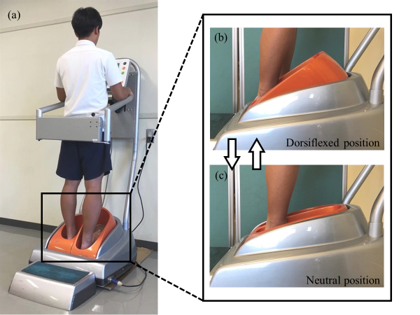
Participants assigned to the SS group performed the intervention using a stretching device (Rakkun walk-R-1; Maruzen Industrial Co., Ltd. and Hiroshima University, Japan; Fig. 1). This device can control the dorsiflexion angle. Participants were instructed to stand on the footplate of the stretching device and to set the maximum dorsiflexion angle themselves without suffering discomfort or pain. All sessions were performed on the non-dominant leg, with the participants’ hips and knees in full extension while standing on the footplates of the stretching machine. The participants grasped the arm support to prevent loss of balance. Participants assigned to the control group stood on the same device for the same length of time as those in the SS group, without changing the dorsiflexion angle settings from the neutral position.
Performance data in each group
SS: static stretching; ROM: range of motion; MIPM; maximum isometric plantar flexion moment; SJ: squat jump; CMJ: counter movement jump; DPSI: dynamic postural stability index; MLSI: medial-lateral stability index; APSI: anterior-posterior stability index; VSI: vertical stability index.
Before and after each training session during the 4-week study period, passive ankle dorsiflexion ROM, muscle hardness, muscle-tendon junction displacement (
Measurement of physical performance
Range of motion(degrees): Participants were instructed to lie supine on the reclining seat of an isokinetic dynamometer (BIODEX system 3, Biodex medical systems Inc., Shirley, NY, USA) with their hip and knee fully extended during ROM measurement. Their foot was placed on the footplate and secured with two Velcro straps and a rubber heel cup. To determine the ROM of dorsiflexion, the footplate was manually moved to dorsiflexion from 0 degrees (neutral position). The dorsiflexion angle was determined at the maximal angle at which the participant suffered no discomfort or pain. This measurement was performed three times and the average value was used for statistical analysis.
Maximal isometric plantar flexion moment (Nm/kg): Measurements of MIPM were performed while the participants lay in a prone position on the bed of the dynamometer with their hip and knee joints fully extended, and their non-dominant foot attached to a footplate at a fixed angle of 0 degrees with two non-elastic straps. Maximal plantar flexion moment was recorded during 3-s of isometric muscle contraction, and the value normalized to body weight was used for statistical analysis. The MIPM was measured three times and the average of the three measurements was used for subsequent analyses. All participants rested for 1-min after each MIPM test to avoid fatigue.
Squat jump and counter-movement jump (cm): Each participant performed two different maximal voluntary vertical jumps on the floor. The first jump was a SJ which consisted of a vertical jump from a semi-squatted position with no preparatory counter-movement. The second jump was a CMJ in which the participant started in an upright standing position and made a downward movement before jumping upward and, through flexion at the knees and hips, made a preparatory downward movement before jumping upward. Both jumps were performed on a jump gauge (Myotest SA, Sion, Switzerland) and participants were instructed to jump vertically to avoid horizontal or lateral displacement, and to land in their takeoff position. Participants maintained an upright upper body position with their upper limbs on their hips while jumping to maximize the utilization of the lower limb muscles [15]. The participants were allowed to recover for as long as they needed between jumps. Each jump was performed three times and the average jump height was used in subsequent analyses.
Dynamic postural stability index: Dynamic postural stability was evaluated using force plate analysis to assess the participants’ ability to land stably on a single leg after jumping with both legs in the forward direction. This approach has good intersession reliability with an intraclass correlation coefficient (ICC) of 0.86 (3, k) [16]. The jump height was standardized at 30 cm, and the jump distance was normalized to the participants’ body height. The participants were located at a distance of 40% of their height from the edge of the plate, and a 30 cm hurdle was placed at the midpoint between the starting position and the force plate [16]. The participants were instructed to jump forward over the obstacle using both legs and to land on the force plate with their non-dominant limb. They were instructed to stabilize themselves on their one leg as quickly as possible and to place their hands on their waist and hold still for 10-s while looking forward. Each participant performed the jump three times with a 1-min break between efforts. The task was repeated if the subject failed to jump or contacted the hurdle during the jump, or if the non-dominant limb touched the ground around the force plate. All subjects were able to perform three successful jumps in the pre- and post-training testing. The sampling rate used to collect ground reaction force data was set at 200 Hz. The global reference system was set so that the anterior-posterior axis was the y-axis, the medial-lateral axis was the x-axis, and the vertical axis was the z-axis. The DPSI was calculated using a Microsoft Excel macro to process ground reaction force data. These data were passed through a zero-lag fourth-order low pass Butterworth filter with a frequency cutoff of 20 Hz. Table 1 shows that the dependent variable is DPSI, which is a composite of the stability indices for the anterior-posterior (APSI), medial-lateral (MLSI), and vertical (VSI) directions. The DPSI was calculated using the ground reaction forces generated in the 3-s immediately following initial contact, which was identified as the instant when the vertical ground reaction force exceeded 5% of body weight. The force plate data were normalized to body weight. The average of three successful trials was used for further analysis [17].
Measurement of muscle properties
Ultrasound probe position for measuring muscle hardness of medial gastrocnemius muscle (MG). Elastography images were manually obtained by performing a light repetitive compression with the probe.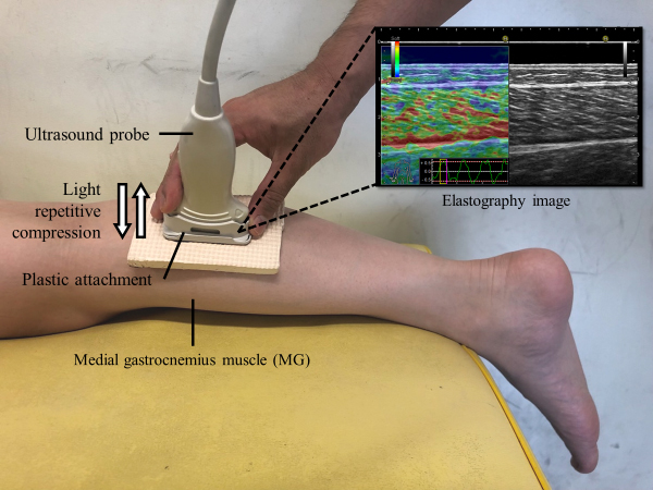
Muscle hardness (muscle/coupler): We measured muscle hardness of the medial gastrocnemius muscle (MG) as the flexibility index of the plantar flexors (Fig. 2). For the assessment of muscle hardness, we used ultrasound real-time tissue elastography (Noblus; Hitachi Aloka Medical Japan, Tokyo, Japan), which can evaluate the strain of tissues, as described by Yanagisawa et al. [18]. The ultrasound probe was placed over the center of the MG at 30% of the distance between the medial point of the popliteal crease and the center of the lateral malleolus to obtain the longitudinal image. The probe location was marked with ink on the skin surface and redrawn when it became faded. This was performed at exactly the same location before and after the 4-week study period for the measurement of muscle hardness. A reference material named acoustic coupler (EZU-TECPL1; Hitachi Aloka Medical Japan) was set between the probe and skin surface using a plastic attachment (EZU-TEATC1; Hitachi Aloka Medical Japan). The elasticity of the acoustic coupler, according to material testing performed by the manufacturer, was 22.6
Muscle property data in each group
SS: static stretching; MTJ: muscle-tendon junction.
Muscle-tendon junction displacement (mm) and muscle-tendon junction angle (degrees): these variables were evaluated using Ultrasound B-mode (Noblus; Hitachi Aloka Medical Japan, Tokyo, Japan) and an 8 MHz linear array probe to visualize a continuous longitudinal ultrasound image. The probe was placed within a manual fixation frame before securing it to the leg, which was attached to the skin over the target muscle with a tape. After obtaining the image, open-source digital measurement software (ImageJ, National Institutes of Health, Bethesda, MD, USA) was used to quantify
The effects of the intervention on all outcome measures were determined using a two-way repeated measures analysis of variance with group (SS, control) as a between-participant factor, and time (pre- and post-training) as a within-participant factor. In addition, a paired
Results
Change in physical performance
The results of the change in muscle properties are shown in Table 1. No interaction effect between the SS and control groups was found for ROM (F(1, 36)
Change in muscle properties
The results of the change in muscle properties are shown in Table 2. Interaction effects for muscle hardness at 0, 10, and 20 degrees (F(1, 36)
Discussion
This study aimed to investigate the effect of a 4-week static stretching program on physical performance and muscle properties. Significant interactions were found between group and time for MLSI and muscle hardness at all dorsiflexion angles. Our results indicated that the 2-min SS session 5 times per week for 4 weeks significantly increased ROM, improved dynamic postural stability, and decreased muscle hardness.
The significant increase in ROM as part of our results supported the findings of previous studies [9, 21, 22]. The increase after SS is attributed to the increase in the capacity to tolerate loading prior to stretch tolerance [23]. The changes in muscle mechanical properties have also been reported as a factor to increase ROM [24]. The SS program in the current study could increase the capacity to tolerate loading and improve muscle mechanical properties, which was indicated by decreased muscle hardness.
The index of dynamic postural stability significantly improved after the SS program. A previous study reported that ankle dorsiflexion ROM, ankle inversion/ eversion strength, and knee flexion/extension strength were predictors of dynamic postural stability [24]. Maeda et al. found that dynamic postural stability improved after cyclic stretching, enhancing ankle dorsiflexion ROM in the acute phase [17]. However, the dynamic postural stability did not improved after SS in the same study. The discrepancy of the results between the previous and current studies was attributed to the difference in effect on ankle dorsiflexion ROM and the intervention period. In the previous study, SS did not improve ankle dorsiflexion ROM in the acute phase, which could be caused by SS duration. In addition, a previous study investigated acute effects. Some reviews concluded that SS reduced muscle strength [4, 5] and central drive [4] in the acute phase. However, the current study investigated the long-term effect of SS program and found no reduction in isometric muscle strength.
Muscle strength, SJ, and CMJ did not decline after the SS program in the current study. Some studies reported decreasing strength as the effect of SS in the acute phase [25, 26, 27]. Muscle strength reduction immediately after SS is caused by neuro-muscular factors such as reduced muscle activity and sensitivity of the muscular spindles [28, 29]. Another cause of reduction in muscle strength could be altered contractile component (intramuscular length and velocity conditions) by SS [26]. Stiff musculo-tendinous units have been reported to allow more effective force transmission between the muscle and the skeletal system [30]. According to previous studies, muscle strength is likely to decrease after a long-term SS program. However, our study found no difference in muscle strength. The mechanism underlying these conflicting results was not clarified in this study. A systematic review of stretching suggested structural, neurological, cellular, and hormonal adaptation in the acute phase, but it did not include chronic adaptation. Therefore, elucidating the chronic adaptation is necessary in the future. With regard to SJ and CMJ, a previous study found a significant relationship between jump performance and ankle plantar strength [13]. In our study, muscle strength (ankle plantar flexion) was not significantly different after a long-term SS program. Therefore, due to muscle strength not being significantly different after long-term SS in our study, it may have led to SJ and CMJ also not being significantly different after the SS program. No reduction in muscle strength, CMJ, and SJ after a long-term SS program could be considered positive because several systematic reviews concluded that SS decreased physical performance in the acute phase [4, 5].
The finding of decreased muscle hardness after a long-term SS program in this study support previous reports [9, 10, 11]. A previous study reported factors related to muscle hardness; the cytoskeleton of the sarcomere and the intramuscular connective tissue consist of parallel elastic components (e.g., the endomysium, perimysium, and epimysium) that influence muscle flexibility [31]. However, these elastic components can readily adapt to imposed load and length demands [31]. In the investigation of acute and prolonged effects of SS, although muscle stiffness decreased 36.5% in the acute phase compared with pre-stretching, it had returned to 24.7% after 10-mins [32]. These previous studies suggested that muscle flexibility changes and readily returns to its original condition after SS. In the current study, muscle hardness decreased significantly (improved muscle flexibility). This result suggests that muscle flexibility could be maintained in the long term by following an SS program.
A few limitations of the study need to be considered. First, the study had a small sample size. Second, despite the fact that MG and lateral gastrocnemius muscle (LG) are the two-joint muscles that intersect in the knee and ankle joints, passive elongation and stretching are performed only in the knee extension position. Thus, whether the differences in passive muscle stiffness between the MG and LG in the knee-extended position can be observed in the knee-flexed position remains unclear.
Conclusions
A 4-week SS program may improve muscle flexibility, ROM, and dynamic postural stability. Furthermore, it does not decrease physical performance (muscle strength, SJ, and CMJ) and thus may be suitable for daily conditioning to maintain muscle flexibility.
Author contributions
CONCEPTION: Junpei Sasadai and Noriaki Maeda.
PERFORMANCE OF WORK: Junpei Sasadai, Noriaki Maeda and Shogo Sakai.
INTERPRETATION OR ANALYSIS OF DATA: Junpei Sasadai and Shogo Sakai.
PREPARATION OF THE MANUSCRIPT: Junpei Sasadai and Shogo Sakai.
REVISION FOR IMPORTANT INTELLECTUAL CONTENT: Noriaki Maeda and Yukio Urabe.
SUPERVISION: Yukio Urabe.
Ethical considerations
The Hiroshima University Center for Integrated Medical Research approved the study (protocol ID number: E-341), and written informed consent was obtained from all participants.
Funding
The authors report no funding.
Footnotes
Acknowledgments
The authors would like to thank the graduate students of Hiroshima University for technical assistance with the experiments.
Conflict of interest
The authors declare that they have no competing interest. Given his role as an Editorial Board Member, Noriaki Maeda had no involvement nor access to information regarding the peer review of this article.