



Abstract
We recently developed the National Dementia Biobank in México (BioBanco Nacional de Demencias, BND) as a unit for diagnosis, research, and tissue transfer for research purposes. BND is associated with the Facultad de Estudios Superiores Cuautitlán, Universidad Nacional Autónoma de Mexico (UNAM), Mexico. The donation of fluids, brain, and other organs of deceased donors is crucial for understanding the underlying mechanisms of neurodegenerative diseases and for the development of successful treatment. Our laboratory research focuses on 1) analysis of the molecular processing of the proteins involved in those neurodegenerative diseases termed tauopathies and 2) the search for biomarkers for the non-invasive and early diagnosis of Alzheimer’s disease.
Keywords
INTRODUCTION
In recent years, Mexico has experienced a reduction in infant natality and an increase in life expectancy as a consequence of the different social, technological, scientific, and health policies. Society must now be ready to make important changes due to the growing population of older adults and the chronic degenerative diseases related to aging. Currently in Mexico there are more than 13 million adults over 60 years of age [1]. Longevity is a significant risk factor for quality of life and autonomy of individuals. The incidence of neurodegenerative diseases, such as dementia, increases over the age of 65 years [2]. Dementia is a progressive and irreversible neurological disorder characterized by cognitive and behavioral impairment that interferes with the social and occupational functioning of people who suffer from it [2, 3]. There are approximately 860,000 adult Mexicans over 60 years of age affected by some type of dementia, and it is estimated that by 2050, this population will increase to more than 3.5 million people [1, 4]. Alzheimer’s disease (AD) is responsible for 50 to 75% of dementia cases [5]. It is characterized by progressive and irreversible alterations that include loss of memory and impairment of cognitive functions, language, judgment, and behavior [6]. AD can occur in two forms: a hereditary or familial form typically affecting people at a younger age (approximately 5% of cases) and a sporadic or late onset form (from 65 years of age). The familial form is autosomal dominant, associated with genetic mutations on chromosomes 21, 14, and 1; encoding the amyloid precursor protein (APP), presenilin 1 (PS1), and presenilin 2 (PS2), respectively [7]. On the other hand, the sporadic type has been related to various risk factors, including type 2 diabetes, hypertension, sedentary lifestyle, obesity, and head trauma, as well as exposure to metals [8]. Clinically, AD can be diagnosed with up to 90% certainty [9, 10] through various cognitive tests and imaging studies that allow the exclusion of those pathologies that share dementia as a characteristic [9, 11–13]. However, the definitive diagnosis can only be made on the basis of postmortem study of the brains of those who have suffered from the disease [14–17]. Macroscopically, a brain affected by AD presents symmetric atrophy with all lobes affected, increased depth of the grooves (Fig. 1, arrow), dilated ventricles (Fig. 1A, V), decreased weight and cerebral volume (Fig. 1A) [18]. At the microscopic level, AD is characterized by the presence of two types of fibrillar lesions, called neuritic plaques (Fig. 2A, large arrow; 2B, Aβ) and neurofibrillary tangles (Fig. 2A, short arrows; 2C, arrows). The neuritic plaques consist of extracellular deposits of the amyloid-β peptide (Fig. 2B, Aβ) [16, 19–22], associated with a large number of dystrophic neurites (Fig. 2A, B, arrows). Intracellular and extracellular neurofibrillary tangles (Fig. 2C, arrows) are composed of paired helical filaments, whose major constituent is tau protein [22–25]. So far, the origin of this disease is unknown and unfortunately the currently approved pharmacological drugs are limited to the treatment of symptoms in the early and moderate stages of this disorder [26]. In recent years, different cell and animal models have been developed [27–31]. However, since this neurodegenerative disorder is unique to humans, it is important to study this disease in human biomaterial.
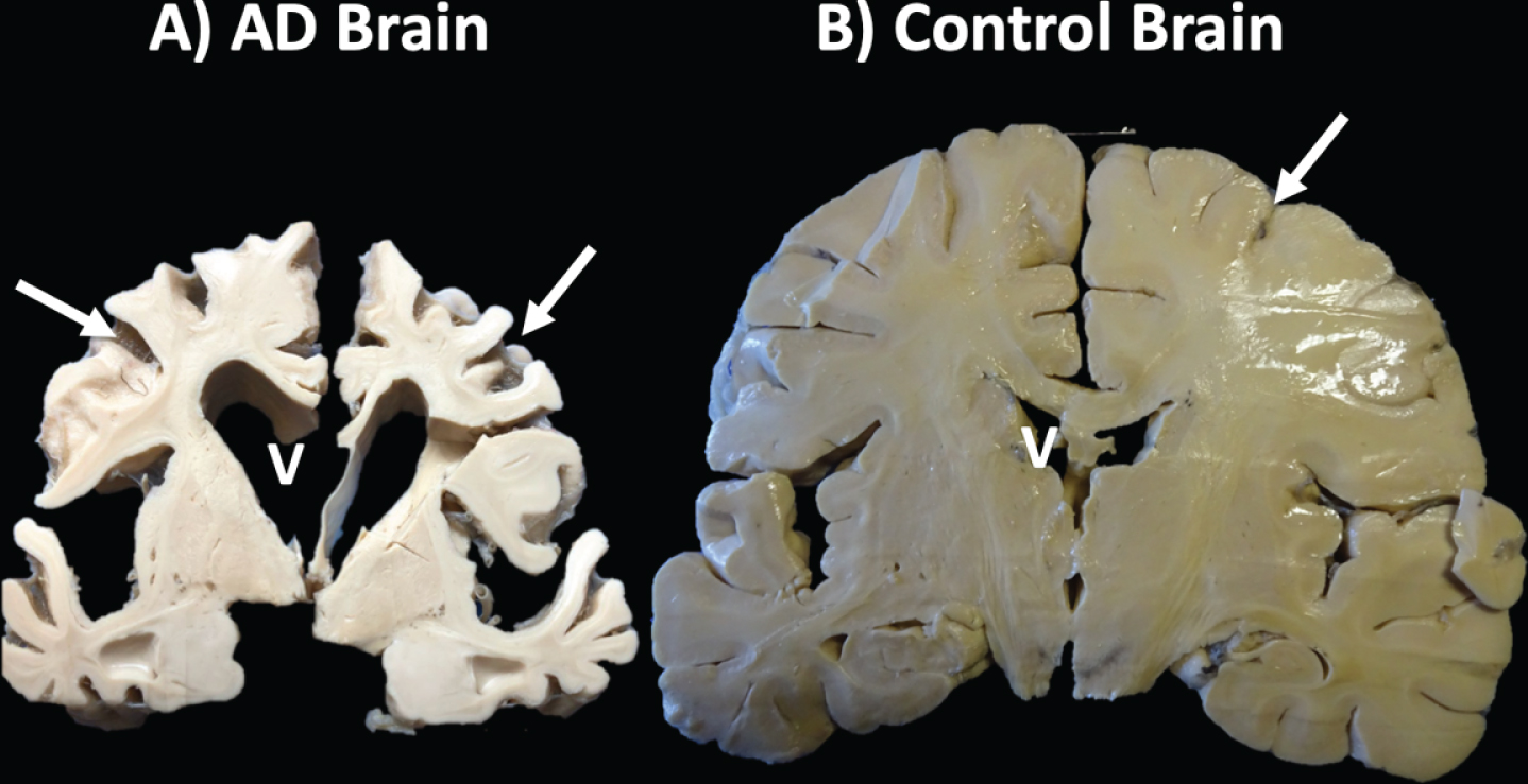
Coronal sections of an Alzheimer’s disease (AD) and control brain. A) A reduction in brain size, an increase in the ventricles (V), and the grooves of the convolutions in the AD brain are observed with respect to the B) healthy brain.
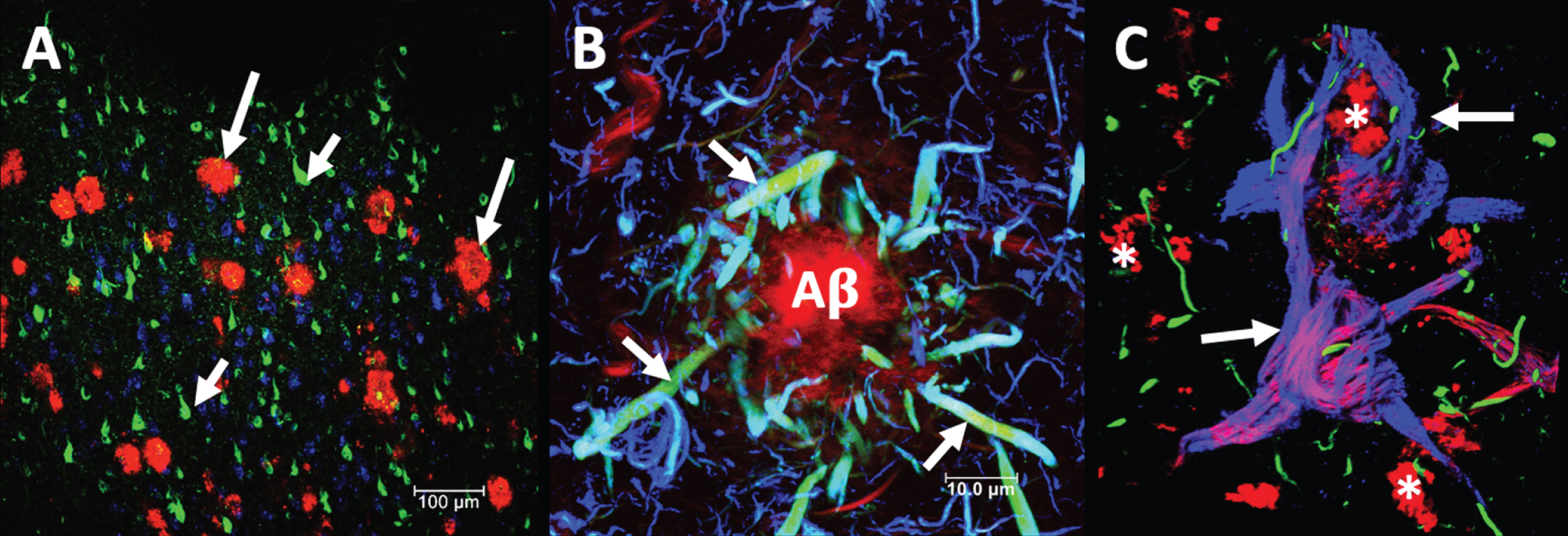
Neuropathology of a case with Alzheimer’s disease. Double immunostaining with two antibodies directed against the phosphorylated tau protein (green and blue channel), counter-stained with the dye thiazine red. A) Temporal cortex of a late case of Alzheimer’s disease at low magnification. Neuritic plaques are seen in the red channel (long arrows) and neurofibrillary tangles (short arrows). B) Neuritic plaque in which the amyloid (Aβ) fibrillar deposit is recognized by thiazine red. Associated with this deposit, a large number of dystrophic neurites (arrows) are observed in turquoise green color, where the two tau markers colocalize. At the periphery, blue dystrophic neurites are observed. C) Extracellular neurofibrillary tangles (arrows) stained for both phosphorylated tau protein (green channel) that shows dystrophic neurites and truncated tau protein with an antibody that recognizes the truncation in Glutamic 391 (blue channel), and which colocalizes with the thiazine red (red channel). Lipofuscin granules are autofluorescent in the red channel (*). The images were obtained using a Leica SP8 confocal microscope.
NATIONAL DEMENTIA BIOBANK
The National Dementia BioBank (BioBanco Nacional de Demencias, BND) is a diagnostic and research unit, where brain, other organs, and fluid (blood, saliva, and cerebrospinal fluid (CSF)) are collected and stored for an indefinite period of time in optimal conservation conditions [32].
BND has the trademark registration in Mexican Institute of Industrial Property (IMPI) with registration number 2027216, 2085417. The logo that identifies the BND (Fig. 3) shows a human brain icon frame corresponding to a lateral view, in a purple color emblematic of AD. Currently the BND is located within the facilities of the Facultad de Estudios Superiores Cuautitlán, Campus 1, UNAM. The developer and director of BND is Dr. José Luna-Muñoz. The national collaborators are Sandra Martínez and Erik Ballesteros from UNAM. The national scientific advisors are Dr. Oralia Barbosa and Bárbara Saénz Ibarra from Hospital Universitario, Dr. José E. González de la UANL, Nuevo León, México. The international scientific advisors are Dr. Mar Pacheco-Herrero from Pontificia Universidad Catolica Madre y Maestra, Dominican Republic and Dr. George Perry from the Department of Biology, University of Texas at San Antonio, San Antonio, TX, USA.
Logo representing the National Dementia BioBank (BND): a unit focused on the diagnosis and research of neurodegenerative diseases. In the logo we wanted to represent a brain in profile and also the hippocampus cut. The purple color is representative of Alzheimer’s disease.
Our vision is that the BND provides a facility for the conservation and storage of tissue and fluids. Each donation will be accompanied by a confirmatory diagnostic molecular test for neurodegenerative diseases and the corresponding medical history. The confirmatory test will be carried out in the BND center through immunohistopathological characterization. Remarkably, the donated biological material will serve to support research studies that elucidate the underlying changes that lead to neurodegenerative disease [33–37]. This tissue is made available for research projects undertaken by other national and international researchers [38, 39].
Specifically, BND has focused on the molecular processing of tau and associated proteins in order to find an early diagnostic method for AD.
The BND develops very specific activities for brain donation for research [40], including: Donation programs for neural tissue, organs, and fluids. Establishment of a multidisciplinary collaboration network with basic science and clinical researchers. Access to the autopsy service. Obtaining and maintaining human tissue for research. Molecular analysis and diagnosis through immunostaining techniques of proteins involved in neurodegenerative diseases by highly specialized staff. Postmortem confirmatory histopathological diagnosis of prion-encephalopathy (unique in the country). Support for students from other institutions to carry out the undergraduate or graduate research.
ORGAN DONATION PROTOCOL
Informed consent for tissue donation
BND collects fluids, brains, and other organs from deceased patients with or without diagnosed neurodegenerative disease. The donation may come from local hospitals (Mexico City) or from other cities, in cooperation with BND. The donation is voluntary, and donated tissue is used exclusively for research. Every donation is accompanied by clinical information, clinical diagnoses, and three copies of informed consent letter (according to health laws of every country). Consent for donation can be given by the patient or by the nearest relatives, if the patient does not have the capacity to consent. On this late case, when the patient’s family reached a consensus, they contact the director or coordinator of the BND by e-mail or telephone. The protocol and procedure, emphasizing the care of the donor’s body, is explained in detail. Usually, the letter or informed consent should contain the signature of two witnesses. Once the patient has died, the relatives must obtain the death certificate and inform the BND. BND is in charge of contacting the funeral home to carry out the transfer of the body to the hospital, where the protocol will be developed for about 1–1.5 hours. Finally, the body will be taken to the place indicated by the relatives to carry out the funeral [40].
Identity of the donors and their relatives is anonymous, with samples being encoded. Codes are restricted to the local staff from BND [33, 41].
Donor relatives’ benefits
Following donation, the relatives of the donor receive confirmation of the diagnosis of neurodegenerative disease [33]. In the long term, this may contribute to the generation of knowledge and a better understanding of neurodegenerative diseases. This will eventually have an impact on the development of treatment, diagnosis, and quality of life for patients in the early stages of cognitive impairment. Therefore, donation of human biomaterial constitutes a key element in the study and analysis of the pathological processing of neurodegenerative diseases such as AD [38].
Prevention of biological risks in the management of human biomaterial
Human biomaterial poses potential biological risk, such as the transmission of pathogenic viruses or transmissible prions, hence it is handled by highly qualified personnel in a restricted environment. The clinical characteristics that define each of the tissues will be made available to requesting researchers. Any remaining unused tissue must be sterilized prior to its incineration. It is vitally important to take into account cases involving acute and short-term dementia, since this may be an indicator associated with prion encephalopathy [42]. The BND relies on strict protocols to handle human tissue and fluids, in order to protect and prevent contamination by virus and prions. Researchers involved in studies using nervous tissue must respect ethical guidelines and maintain confidentiality of donors.
Brain tissue removal procedure
The brain and organs must be taken within 12 hours of death to reduce as far as possible the postmortem degradation of proteins, which are investigated in molecular, immunohistopathological, and biochemical studies. Fluids (mainly saliva and blood) will be collected by specialized personnel. The CSF will be obtained when possible. Brain removal is performed by opening the cranial cavity, cutting under the occipital hole, and taking the upper part of the cervical cord. Special care must be taken to section the optical nerves and cranial nerves anteriorly so that part of them remains in the brain. Bulb and olfactory nerves must be obtained intact. The pituitaries must be obtained separately from another region. The brainstem and cerebellum are separated from cerebral hemispheres with a high cut above superior quadrigeminal tubercles. The cerebellum is separated from brainstem sectioning the cerebral peduncles. The cerebral hemispheres are separated with a mid-sagittal cut by the vermis and, finally, the cerebral hemispheres are separated by cutting exactly along the midline of corpus callosum. Generally, the left cerebral hemisphere is fixed in formalin 10% at 4°C, for a minimum of 15 days. The fixative is changed every 3 months (Fig. 4B, C). Prior to use for immunohistochemical characterization, post-fixation of the fragment in paraformaldehyde is performed. The right cerebral hemisphere is kept frozen. Freezing of samples should be done immediately after removal of the brain and separation as previously described. Storage is done immediately and continuously at –80°C (Fig. 4A). However, protocols must be adapted to the particular condition of neurological disease. For example, it is not a good option to freeze a coronal section of one hemisphere for biochemical studies and fix the other hemisphere in formalin when it is a unilateral cerebralinfarction.
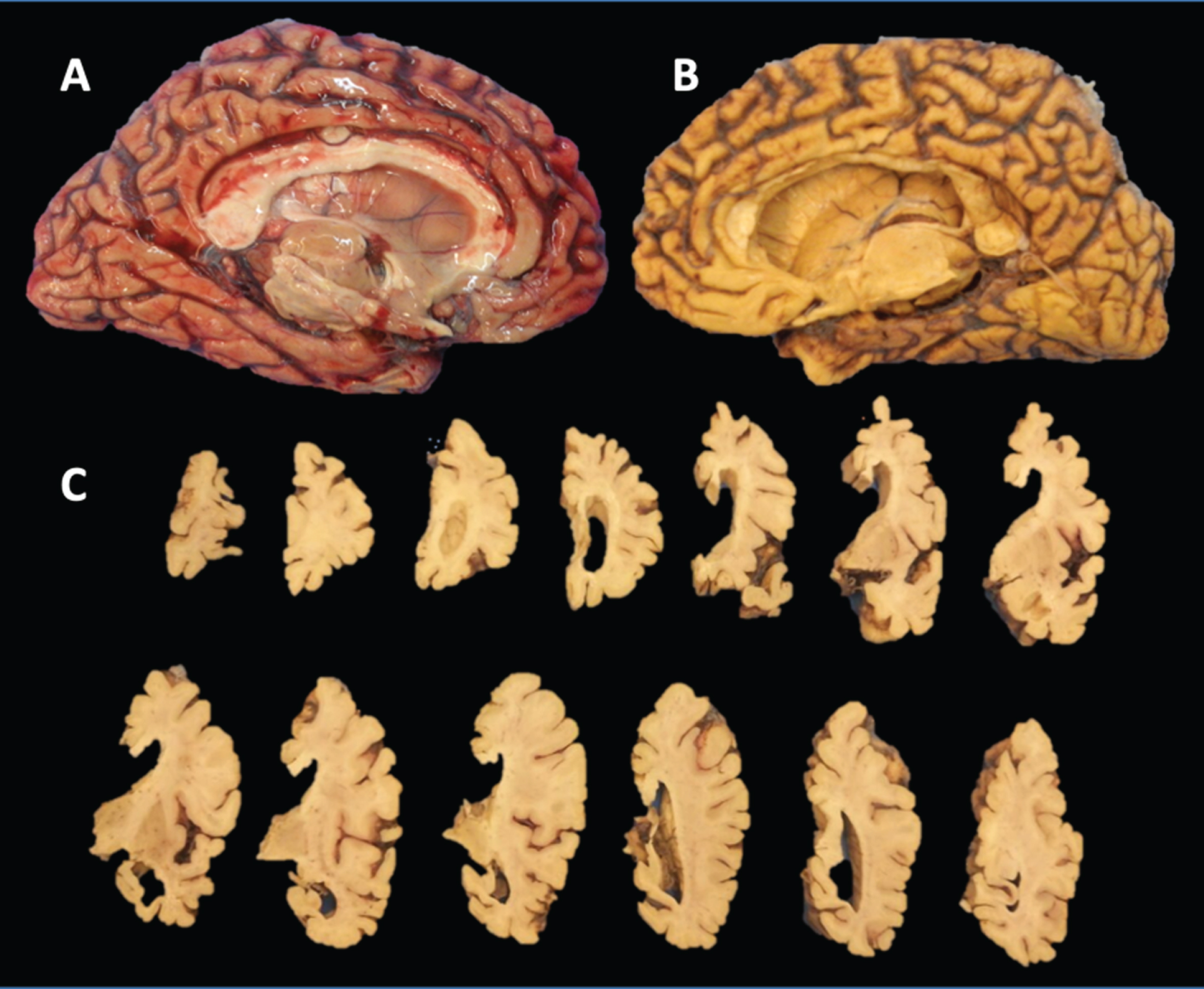
Brain with Alzheimer’s disease. A) Fresh left hemisphere, which is cut into coronal sections to a thickness of approximately 1 centimeter and frozen at –80°C. B) Formalin fixed right hemisphere, which is sectioned as shown in image (C).
The CSF can be carried out from the middle ventricle by puncture from the basal aspect of the brain and inserting a plastic pipette, slightly sailing the chiasm to allow its passage without damaging it. pH measurement of the CSF should be recorded. External macroscopic observation, photographic record, and weight measurement should be recorded. Extraction of sensory organs is only carried out in the case of express authorization.
ORGAN DONOR AWARENESS ACTIVITIES
The director of the BND, as well as ungraduate and postgraduate students (Fig. 5), actively participate in the annual Mexican Alzheimer Congress coordinated by Mexican Alzheimer Federation (FEDMA; Fig. 5A), annual Alzheimer’s Association International Conference (AAIC; Fig. 5B), Association Neuropsiquiatric Argentina Congress, Alzheimer’s IberoAmerica Congress, among others, disclosing the results and advances made using tissues from BND. On the other hand, BND, in collaboration with Alzheimer México I.A.P association, held a massive event in Mexico City, on Alzheimer’s Day (21 September), to give information to the population about the attention and care of patients with dementia (Fig. 5D, E), and to explain the importance of organ donation (Fig. 5C, F–I). In this activity, participants could observe immunohistochemical staining for specific markers in neurodegenerative diseases (Fig. 5H). Formalin-fixed tissues contained in glass containers were shown with the aim of analyzing the macroscopic alterations in different neurodegenerative diseases (Fig. 5F, G, I). In addition, a travelling museum called “Brain and neurodegenerative diseases” was developed. This presents information on AD, Parkinson’s disease, Huntington’s disease, and prions; photomicrographs of histopathological lesions of AD and other dementias; brain tissue preserved by plastination; and monitors showing information from clinical experts and researchers from different institutions in Mexico.

Activities carried out by the National Dementia BioBank to raise awareness of donation of fluids, brain, and other organs. A) Promotional activity of the Alzheimer Association Mexico IAP. In it, a massive event is held for the public with the aim of giving information and carrying out activities. A BND student holds tissue in an acrylic box. B) Activity carried out on World Alzheimer’s Day. Attendees are gifted with commemorative shirts and invited to form a big brain. Image taken with a drone. C–G) BND students give detailed information about organ donation and research activities within BND. E) Someone observing the lesions characteristic of Alzheimer’s disease with a microscope showing a peroxidase stain against the tau protein. H) The results obtained from the BND’s investigations are presented by students at international conferences such as the AAIC. I) The dissemination of research and donation of fluids, brain, and other organs are also carried out in forums of National congresses for people with relatives with Alzheimer’s disease. Events carried out by the Mexican Alzheimer Federation (FEDMA).
PAST, PRESENT, AND FUTURE DIRECTIONS
The first Brain Bank in Mexico and Latin America was founded in 1994 by Dr. José Raúl Mena López, with the aim of promoting research on the pathological processing of proteins involved in AD [43]. Dr. Raúl Mena, surgeon and PhD in cell biology and a native of the state of Yucatan, devoted much of his life to describing the sequence of aggregation of the tau protein and the Aβ peptide in the formation of neurofibrillary tangles and dystrophic neurites, respectively, using thiazine red [23, 44–46]. Mena and Luna-Muñoz (director of the BND) proposed that thiazine red, a red fluorescent dye with an affinity for the beta-folded conformation in aggregated proteins, could serve as a method for the rapid and accurate detection of the pathogenic lesions [45]. This allowed the differentiation of each one of the stages in the processing of tau [47, 48], which starts with the diffuse intracellular granular deposit (pre-neurofibrillary tangle) and ends with the formation of an extracellular tangle [44, 49]. This method is still used to show the characteristic fibrillar aggregates of AD [23]. In an extensive neuropathological study, the combination of different fluorescent markers for this disease allowed Raul to see multicolored structures (corresponding to neuropathological lesions). In a poetic way and for a better understanding, Raul called it the “rainbow of dementia” [43].
Currently, BND continues with the original approach of understanding the pathological molecular mechanisms of tau protein in neurodegenerative diseases [31, 49]. The pathological processing of the tau protein tauopathies is being focused on the search for a specific biomarker for AD [9, 50–53]. The phosphorylation of the tau protein in AD has been suggested as a possible protection mechanism that allows neurons to function longer, in the presence of a 92–95 amino acid fragment that terminates with Glutamic (Glu)-391. This fragment called the paired helical filament “core” [54–56] is highly toxic [49]. We have also carried out a deeper understanding of the role of tau protein in physiological conditions. Physiologically, tau protein has specific phosphorylation sites to carry out its various essential functions. However, it has not been possible to fully understand the new physiological roles of the tau protein [49, 57]. Finally, the presence of tau protein in non-neural tissue was demonstrated over 20 years ago [58]. Therefore, the distribution and function of the tau protein in this type of tissue has been analyzed in heart, liver, intestine, lung, and pancreas. We have found that this molecule is phosphorylated at different sites depending on the tissue being analyzed; and above all, these post-translational events can arise at both pathological and non-pathological sites. Simultaneously, in recent investigations we have identified the two characteristic lesions of AD in other brain structures, such as the cingulum, optic chiasma, and olfactory bulb; which would impact the emotional state, vision, and smell in affected persons.
Awareness of donation of fluids, brain, and other organs has been treated favorably by the Mexican public with the development of the itinerant museum “The brain and neurodegenerative diseases” in collaboration with Lic. Ricardo Cerón Plata, Héctor de la Peña and Efren Díaz Millan of Centro de Investigación y Estudios Avanzados (CINVESTAV). This itinerant museum has been exhibiting for 4 years both in Mexico City and in several states of the Mexican Republic. Socio-academic-scientific activities have brought recognition of the BND both nationally and internationally. Such is the case that Universidad Nacional Pedro Henríquez Ureña (UNPHU), in Santo Domingo, Dominican Republic, have created and developed a National Brain Bank with advice and guidelines from Dr. José Luna Muñoz, under the direction of Dr. Daisy Acosta and Dr. José Guillen Sarita.
FUTURE CHALLENGES
Due to the stigmatization of brain tissue donation for research, obtaining tissue has been difficult in the past. However, this obstacle is gradually changing thanks to the engagement of researchers with society and through multidisciplinary collaboration [59] between groups of clinical specialists and scientists passionate about dementia and other neurodegenerative diseases [60–62]. Thus the BND has been able to collect fluids, brain, and other organs from patients with AD and other dementias, as well as those with no neurological conditions [34, 37].
Collaboration and agreements in progress with FEDMA and different states of the Mexican republic have allowed a greater awareness for the public, especially for the families of patients suffering from the disease. However, there are still many people who have difficulty with the concept of “donation for research”. It is through dissemination of information through various Alzheimer’s associations where we hope that the concept will gradually become accepted. When we talk about these donations, it is necessary to emphasize that not only “sick” brains are required, since, “we can all be donors of brain tissue for research, whether we suffer from any neurological or psychiatric disease, or if we are perfectly healthy donors” [63]. Healthy neural tissue is as important as its counterpart, since it serves as a control to better understand the differences between normal and pathological molecular processing in these tissues [64]. This is the basis of the motto of the BND. We consider that “science offers a new life to the brain: to be a key piece in one of the puzzles of current medicine, the origin and development of dementias and other diseases of the nervous system”. In the BND, the collection, classification, preservation, and distribution of samples for research adheres to the ethical and legal stipulations of Mexico [65].
BND and Latin-American network of neurobanks
The emergent international collaboration among brain banks (México, Dominican Republic, Colombia, Argentina, Brazil) fosters networking interactions among researchers, standardization of criteria and protocols, and access to diverse tissue samples for robust research. This ultimately strengthens the field and fosters the generation and exchange of knowledge as well as developing skills andexpertise.
CONCLUSIONS
BND is a first repository of fluids, brain, and other organs in Mexico. It focuses on the study of the human brain through biochemical and molecular biology techniques to deepen the knowledge about neurodegenerative disorders. From a legal and ethical point of view, BND is a non-profit company, in which human fluids, brain tissue, and other organs are donated voluntarily and are not marketed. Unique potential discoveries and research advances will benefit the Mexican population and the international community.
Footnotes
ACKNOWLEDGMENTS
This work was supported by Fondo Nacional de Ciencia y Tecnologia, FONDOCyT, from the Ministry of Higher Education, Science and Technology, Dominican Republic (2015-3A2-127 to M.P-H) and (2018-2019-2A3-208 to J. L-M and M. P-H).
The authors want to express their gratitude to the following: Dr. P. Davies (Albert Einstein College of Medicine, Bronx, NY, USA) and Lester I. Binder† (North Western, Chicago, IL, USA) for the generous gift of mAbs TG-3 and Alz-50, and Tau-1, Tau-5 and Tau-7, respectively; Tec. Amparo Viramontes Pintos for handling of the brain tissue; Samadhi Moreno-Campuzano for her technical assistance; and the confocal microscopy unit of CIIDIR Durango, Instituto Politécnico Nacional, Union Medica University Clinic, Dominican Republic, for their support and collaboration in the development of this research project. We also want to express our gratitude to the Mexican families who donated the brains of their loved ones affected with Alzheimer’s disease and made our research possible. This work is dedicated to the memory of Professor Dr. José Raúl Mena López†.
