
Abstract
Since electroencephalogram (EEG) signals contain a variety of physiological and pathological information, they are widely used in medical diagnosis, brain machine interface and other fields. The existing EEG apparatus are not perfect due to big size, high power consumption and using cables to transmit data. In this paper, a portable real-time EEG signal acquisition and tele-medicine system is developed in order to improve performance of EEG apparatus. The weak EEG signals are induced to the pre-processing circuits via a noninvasive method with bipolar leads. After multi-level amplifying and filtering, these signals are transmitted to DSP (TMS320C5509) to conduct digital filtering. Then, the EEG signals are displayed on the LCD screen and stored in the SD card so that they can be uploaded to the server through the internet. The server employs SQL Server database to manage patients’ information and to store data in disk. Doctors can download, look up and analyze patients’ EEG data using the doctor client. Experimental results demonstrate that the system can acquire weak EEG signals in real time, display the processed results, save data and carry out tele-medicine. The system can meet the requirement of the EEG signals’ quality, and are easy to use and carry.
Introduction
The EEG (electroencephalogram) signal is a bioelectrical signal that reflects the internal information of the brain. Brain is the most complex and functional super-characteristic organ in human tissues and organs. Therefore, the EEG signals generated by the brain are very complex, and they have the characteristics of weak amplitude, strong randomness, non-stationarity and nonlinearity. EEG signals are the outward reflection of the neurons in human brain. They contain abundant, pathological, physiological and mental information. EEG signals provide important evidences of medical diagnosis, cerebral consciousness and cognitive research and so on [1]. It is the basis of the brain science to extract the effective information in EEG signals effectively.
Nowadays, the EEG acquisition instruments developed by some foreign companies have been accepted and applied in many fields [2, 3]. There are many famous companies overseas, such as the Nebraskan and EGI of American, the Brain Products of German, Nihon Cohen of Japan and other companies. Similarly, Chinese corporations also have representative producers of EEG equipment, such as Beijing Xintuo, Nanjing Vishay, Shenzhen Mindray and so on. These Chinese companies are still in the developing stage due to late start, and they have not conducted in-depth research on epilepsy [4, 5]. In terms of performance indexes, the precision and stability of Chinese equipment are far away from meeting requirements for more accurate and deep research [6, 7]. Due to lack of original and independent research and development, it is necessary to buy the mature technology and core components [8, 9]. Thus, Chinese EEG apparatus have more cost and less application. For the collection of brain electrical signals, comfort is important because it increases wearing time [10, 11, 12, 13, 14, 15, 16]. The main difficulties in dealing electroencephalogram is that the existing EEG apparatus are not perfect due to big size, high power consumption and using the cable to transmit data. In this paper, a portable real-time EEG signal acquisition and tele-medicine system is developed in order to improve performance of EEG apparatus.
The main contribution of current work includes the following: EEG signals are the reflection of electrophysiological activities of cerebral nerve cells in the cerebral cortex [17]. EEG signals can provide diagnosis and treatment for certain brain diseases [18, 19]. Because EEG signals have the characteristics of non-stationary and strong background noise, the analysis and processing of EEG signals is a challenging issue [20]. Classical processing methods of EEG signals include spectrum analysis, power spectrum analysis, correlation analysis, bispectrum analysis [21], etc. These methods are simple and easy to implement. In recent years, various new methods have vigorously promoted the development of EEG signal analysis techniques. The research of EEG signals, for example, the methods of nonlinear dynamics, approximate entropy, complexity, correlation dimension, maximum Lyapunov exponent, etc. At present, with the joint efforts of Chinese and foreign research scholars, the research and application of EEG signals have achieved important results in many fields.
Chinese and overseas EEG acquisition equipment performance parameters comparison table
Chinese and overseas EEG acquisition equipment performance parameters comparison table
As research work on EEG becomes deeper step by step, more and more instruments recording EEG signals emerge [22, 23]. Unfortunately, the traditional EEG acquisition algorithms are bulky [24]. EEG signals used to be sampled and converted into digital signals by collecting circuits. Then, digitized EEG signals are transferred to PC by either wired or wireless channel and conducted digital filtering [25, 26]. This method leads to bad instantaneity and mobility, and tele-medicine service cannot be provided. The geographic scope between doctor and patient is limited and medical expense becomes more expensive [27]. The need to develop accurate EEG signals detection scoring techniques to reduce the burden on users has recently increased. Therefore, many research institutions have been established to make research efforts. Gain amplification and signal extraction are the most important step in any developed EEG signals detection technology. The measurement and analysis methods of time domain and frequency domain are widely used in EEG. However, EEG signals are complex non-stationary signals, and all statistical characteristics of EEG signals are not available only in the time or frequency domain. Thus with the development of modern medical career, EEG equipment, which has such performance as real-time, strong anti-interference ability, easy to carry, long distance transmission, will have a great commercial value. The comparison of Chinese and overseas EEG acquisition equipment performance parameters is as follows. Therefore, a kind of portable low-power-consumption, DSP-based EEG acquisition system with remote medical service was designed in this paper.
EEG signals have such features as follows: The range of amplitude is 5
Comparison of theoretical simulated and actual values of positive
Comparison of theoretical simulated and actual values of positive
The overall structure diagram of system.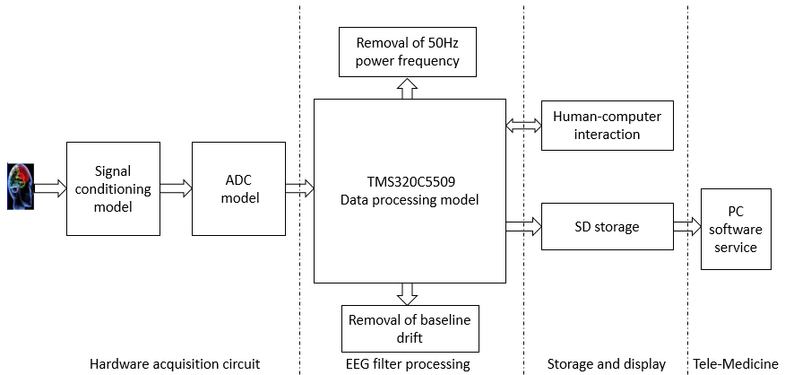
The system is composed of EEG signal acquisition system and tele-medicine system. The EEG signal acquisition system includes hardware acquisition circuits, EEG digital filter processing model, displaying and saving model and other modules. The major mission of hardware acquisition circuit is inducing weak EEG signals to signals conditioning circuits with bipolar leads. Then, these signals will undergo multi-level amplifying and filtering. In order to obtain EEG signals with less noise, EEG signals digital filter module transforms the signals that have finished ADC (analog to digital conversion) to DSP (digital signal processor) firstly. The total input noise can be rewritten as Eq. (1). Then the baseline drift from electrodes and the power interference varied with power grid will be removed. The filtered EEG signals are displayed on LCD and stored in SD card. Finally, users can use the tele-medicine service software based on PC to upload EEG data to the server by network communication. Personal information of patients is managed by SQL Server database on the server, and patients’ EEG data files are saved on the server
The EEG signal acquisition system is composed of hardware model and software model. The hardware circuits can be divided into analog circuits (pre-processing circuit) and digital circuits (DSP and its peripheral circuits). The major function of the software part is to acquire EEG signals and store EEG data on SD. Generally, the gain of amplifier used for EEG ranges from 5000 to 30000. To avoid serious distortion of the single stage amplifier, we use multi-level amplifier and filter in this paper. With shield guard and right leg drive, the CMRR (common mode rejection ratio) and input impedance can be both improved. Then, the analog signals are converted into digital signals by ADS8320. And the digitized signals will be transferred to the DSP to filter the power interference and physiological artifacts (EOG (Electro-Oculogram) and EMG (electromyography) artifacts). Finally, the EEG signals are displayed on LCD and saved in SD card.
Signal acquisition and conditioning circuits
In this system, the medical ECG (electrocardiogram) electrodes are used to obtain EEG signals [17, 18] with bipolar leads. The signal conditioning circuit is the precondition for getting EEG signals effectively, which includes multistage amplifier and filter circuits, common mode interference suppression circuit and other circuits. Figure 2 shows the overall structure of pre-processing circuit.
The overall structure diagram of pre-processing circuits.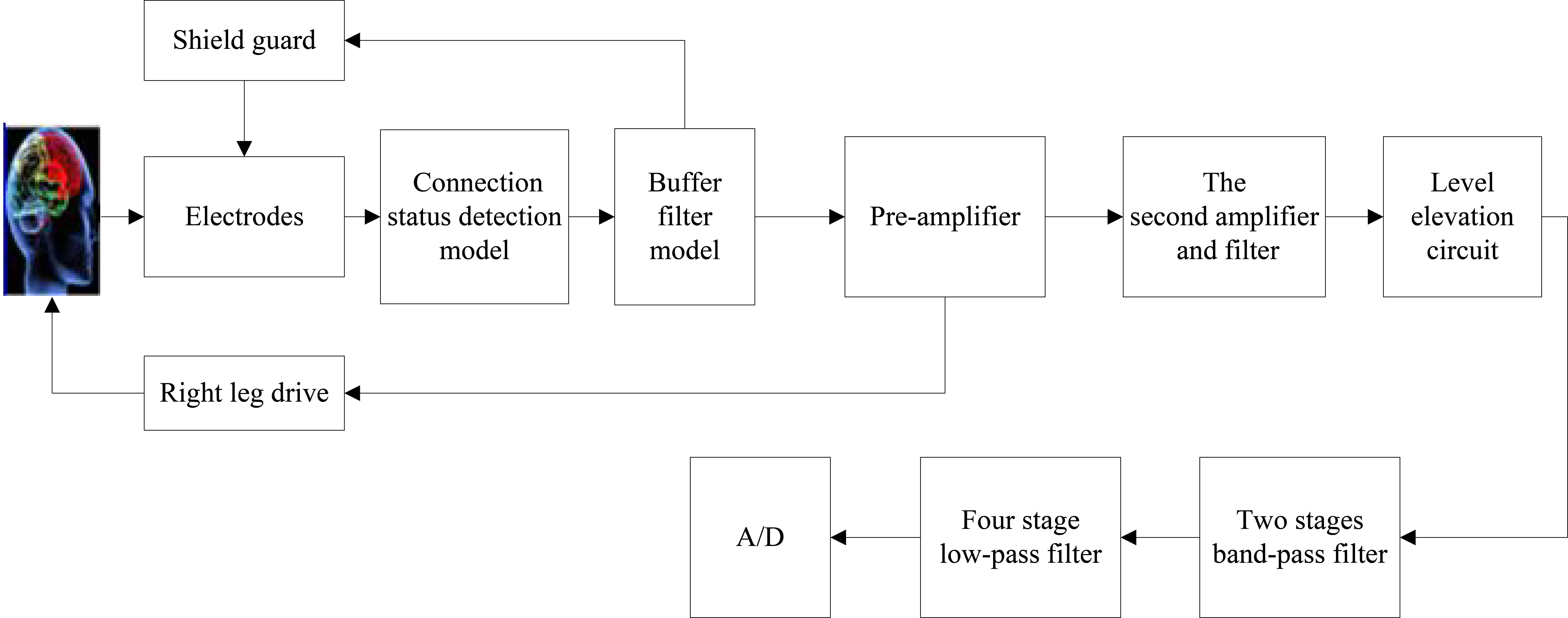
As is shown in Fig. 2, electrode state detection circuit detects the contact status between patient’s skin and electrodes. EEG signals will be induced to pre-processing circuits when electrodes have good contact with skin. These signals will pass through guard circuit, multi-level amplifier and filter circuits, common mode interference suppression circuit and level elevation circuit. Then, effective analog-digital conversion will be conducted.
Microprocessor
In this system, TMS320C5509 is chosen as the microprocessor. Its highest frequency can be up to 200 MHz. The data bus is 16 bit. The core voltage is 1.6 V and the IO port voltage is 3.3 V. CCS (Code Compile Studio) is used to program so as to implement dispatching tasks and processing signals when acquiring signals. It has strong readability and transferability of program. TMS320C5509 can control the entire system. The minimum system of the DSP consists of the power supply circuit, clock and reset circuits. Compared with other systems, this system has such advantages as simple structure, small size, low power consumption and cost.
Circuits design of interfaces between SD card and DSP
The circuit Schematic to connect DSP and SD card is illustrated in Fig. 3. The four general purposes I/O pin of DSP, SPISTEA, SPISIMOA, SPICLOKA, SPISOMIA connect to CS, DI, SCLK, DO of the SD card respectively. When the SPISTEA port outputs low electrical level, the SD card will work effectively. DSP sends data and commands to the SD card through SPISIMOA pin, reads the SD card data through SPISOMIA port and provides clock signal for SD card through SPICLOKA pin.
The interface circuits of SD card and DSP.
Schematic of interface between LCD and DSP.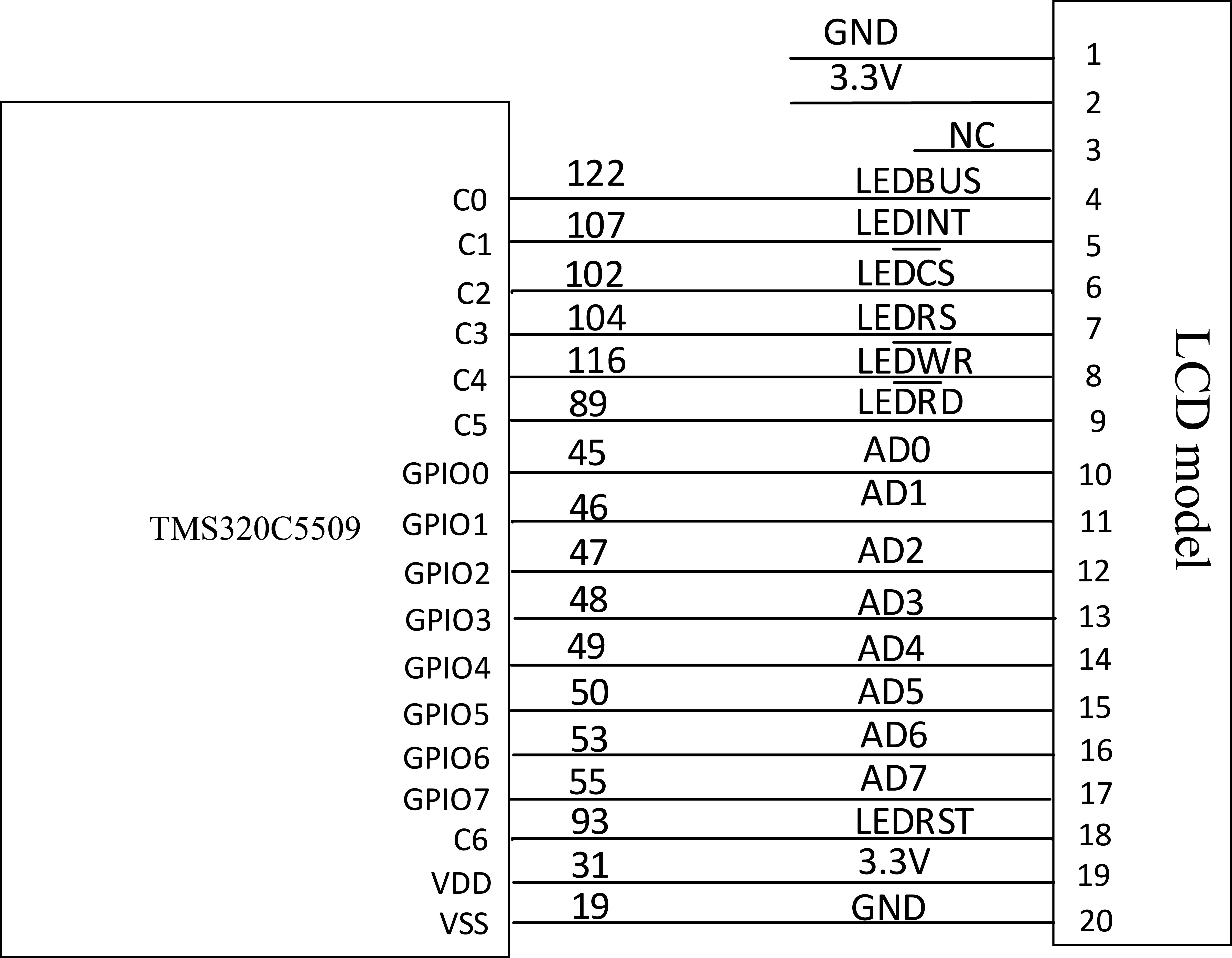
The data display circuits of EEG signal acquisition system are mainly composed of LCD module. The system uses 320
The software design of lower computer
The main function of lower computer software is to collect EEG signals. Firstly, the LCD, A/D, SD card and other modules are initialized [19]. Then the system checks whether the electrodes are in good contact with scalp. When the reminder light is on, it indicates bad contact of electrodes. On the contrary, the system collects signals and then converts them into digital signals. After filtering, data are displayed on LCD and saved on SD. The SD card will delete the first 30 minutes of files and records when the SD card is full.
The overall flow diagram of lower computer software design.
The telemedicine software installed on PC transmits data on the SD card to the server by the patient client. Patients’ information and EEG data can be managed on the server. Doctors query and download patients’ information on the doctor client.
The overall running flow diagram of patient client.
The running flow chart of doctor client.
When the server receives the authentication packet which sent by the patient client, it will check whether there is the user ID in the database table. If the ID exists, the server will reply the patient client with response packet and insert the patient’s information according to user ID as index. At the same time, it is conducted to search whether there is the file folder named after the user ID on the disk of server. If not, a new folder will be built on the disk. After patient client receives a successful response packet from the server, the button to import patients’ data becomes bright from gray and is clickable. The over running flow charts of patient clients are shown in Fig. 6.
All of patients’ information tables have been stored on the server. Each doctor manages a list of his patients’ information. The list of patients’ information on the server will be sent back to the doctor client when the doctor’s ID is verified successfully. Doctors can double click and choose a patient which has not been given diagnosis results. And the patient’s EEG file can be acquired from the server. Doctor client receives the basic information of all files in the folder, then doctors can click to select a file and reply to the server. The server will transfer the file until it receives the response of which file is wanted from the doctor client. The over running flow charts of doctor clients are illustrated in Fig. 7.
EEG displaying on LCD.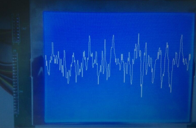
EEG display interface of doctor-patient client.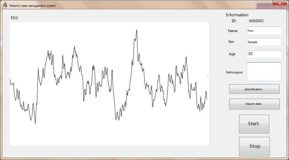
As demonstrated in Fig. 8, the EEG signal acquisition system designed in this paper can collect the brain waves and display it on the LCD screen in real time. The patients’ EEG data are shown on the doctor-patient client on PC as illustrated in Fig. 9. Doctor screen and patient screen are the same, as shown in Fig. 9. Experimental results demonstrate that the system meets the design requirements. When the medical electrode is in good contact with the head, the system uses a bipolar lead to induce a weak EEG signal to the signal conditioning circuit. After multi-level filtering, amplification and suppression of common-mode interference, the signal conditioning and other operations improve the performance of EGG apparatus. This improvement is significant compared to other portable and simple systems.
Conclusions
In this paper, we do some research on the collection technology for EEG signals and have built the EEG signals condition and acquisition circuits based on TMS320C5509. At the same time, digital filtering of EEG signals is realized on DSP. After filtering, data are displayed on LCD and saved on SD. The EEG data and personal information of patients are uploaded to the server and managed by the doctor-patient client in order to implement the tele-medicine service. As far, the function test of the system has been finished. It provides a good technical support for research on brain science, real-time mobile monitoring on EEG and the development of family-based and community-based EEG products. Experimental results show that, in view of the different characteristics of each basic rhythm and characteristic wave in EEG signals. The method used in this measuring system can effectively measure the relative intensity of each basic rhythm of EEG signals, as well as measure and extract various characteristic rhythms in the signal. It is believed that accurate measurement of the system can provide a more complete and accurate diagnosis for the clinical diagnosis.