
Abstract
Adenomyosis is an abnormality in the uterine wall of women that adversely affects their normal life style. If not treated properly, it may lead to severe health issues. The symptoms of adenomyosis are identified from MRI images. It is a gynaecological disease that may lead to infertility. The presence of red dots in the uterus is the major symptom of adenomyosis. The difference in the extent of these red dots extracted from MRI images shows how significant the deviation from normality is. Thus, we proposed an entroxon-based bio-inspired intelligent water drop back-propagation neural network (BIWDNN) model to discover the probability of infertility being caused by adenomyosis and endometriosis. First, vital features from the images are extracted and segmented, and then they are classified using the fuzzy C-means clustering algorithm. The extracted features are then attributed and compared with a normal person’s extracted attributes. The proposed BIWDNN model is evaluated using training and testing datasets and the predictions are estimated using the testing dataset. The proposed model produces an improved diagnostic precision rate on infertility.
Introduction
Adenomyosis is diagnosed by the cracks or breaks found along the inner lining (endometrium) of the muscular wall of the uterus (myometrium). The symptoms of adenomyosis are menstrual cramps, bloating and lower abdominal pressure before menstrual periods that can lead to heavy bleeding during periods. The symptoms can be found in the uterus in a localised spot or throughout the myometrium. Though adenomyosis is considered as the initial stage (not life-threatening), the frequent pain and continuous heavy bleeding associated with it reduces women’s confidence in life. The rate of occurrence of adenomyosis can be between 5% and 70%. This disease can affect 2 out of 10 women specifically aged below 40 years; however, the count increases to 8 out of 10 women aged between 40 and 50 [1].
Adenomyosis is a common condition in middle-aged women, and women who have given birth are often diagnosed with adenomyosis. Some studies reveal that women who have undergone uterine surgery may be at risk of adenomyosis. In view of the fact that the causes of adenomyosis are not known, studies reveal that a range of hormones like prolactin, follicle-, progesterone- and oestrogen-stimulating hormones may be responsible for the condition. Nevertheless, the prevalence of adenomyosis cramp is not easily ascertained because of the requirement for a cohesive description and investigative criteria relying on non-invasive analytic trials [2]. Presently, there are no standards either for dealing with pathognomonic-reliant proven features to identify adenomyosis cramp or any laparoscopic-reliant criterion that might become the basis for diagnosing them [3].
Until now, the only way to identify adenomyosis has been by performing a hysterectomy and examining the tissues under a microscope. However, emergent technologies like imaging technology have helped doctors to diagnose adenomyosis without performing surgery. Doctors use MRI to see the characteristics of the disease in the uterus. If the presence of adenomyosis is detected, the first thing the doctor carries out is a physical examination. By performing a pelvic exam, the enlarged and tender uterus can be identified using ultrasound resonance on its walls and its linings. Ultrasound does not help to diagnose adenomyosis completely, but it can identify a few similar symptoms. Sometimes techniques like sonohysterography help in the detection of symptoms similar to adenomyosis. In sonohysterography, saline solution is injected through a tiny tube into the uterus as an ultrasound is given.
Magnetic resonance imaging can be used to verify the analysis of adenomyosis in women from their typical uterine blood loss. Sometimes symptoms similar to adenomyosis are misdiagnosed as uterine fibroids. However, the two conditions are not similar. While fibroids are considered as the beginning of tumours in the wall of the uterus, adenomyosis is a mass of cells present within the uterine wall. Accurate diagnosis is the key in selecting the proper treatment.
In the past, adenomyosis cramp was specifically identified in premenopausal women using pathological investigation, which can be done after a hysterectomy test [4, 5]. Today, the analysis is done using techniques like magnetic resonance imaging or transvaginal ultrasound scan [6]. In most cases, the adenomyosis cramp is asymptomatic. Frequent medical indications include menorrhagia cramp, metrorrhagia cramp and dysmenorrhea cramp, with additional medical criteria such as an inflated uterus [2, 6]. In an investigation among 945 patients affected by hysterectomy cramp, an important positive relationship was discovered while diagnosing adenomyosis cramp, assessing the evidence of earlier pregnancy and dealing with leiomyoma cramp. However, cramps such as in ovarian endometriosis and endometrial hyperplasia cannot be caused by normal delivery, smoking, etc. [7].
Endometriosis is a painful event, where the tissue that normally lines the interior of the uterus (endometrium) grows outside the uterus. It is a condition that mainly involves the ovaries, fallopian tubes and the tissue lining the pelvis. In rare cases, endometrial tissue spreads away from pelvic organs. With endometriosis, the displaced endometrial tissue continues to act as it generally does in the uterus, thickening, breaking down and bleeding with every menstrual cycle. The tissues which are displaced have no way out and are trapped inside. When the endometriosis pierces the ovaries, cysts called endometrial cysts form. Neighbouring tissues can become irritated, finally creating scratch marks on the tissue and adhesions on abnormal bands of fibrous tissue, which can be the source of pelvic tissues and organs becoming fixed to other parts, causing pain.
Endometriosis is sometimes mistaken for other conditions that can cause pelvic pain, such as pelvic inflammatory disease or ovarian cysts. It may be confused with irritable bowel syndrome, a condition that causes bouts of diarrhoea, constipation and abdominal cramping. These can also accompany endometriosis, which complicates the diagnosis.
Background - Diagnosis of adenomyosis cramp was not easy in the past; it was associated with multiparous women with infertility issues. In fact, women habitually impediment their first pregnancy and the issue of adenomyosis cramp is naturally discovered in those aged above 30. When women are advised to undergo nonsurgical analysis such as MRI, the impact of adenomyosis cramp on infertility becomes predictable. Limitations - Many methods have been used to deal with the impact of adenomyosis and endometriosis in women. Each method has produced its own prediction rate, but the prediction of adenomyosis and endometriosis cramps using neural networks has rarely been discussed [24]. There is actually a strong association between infertility problems and adenomyosis and endometriosis cramps in women.
Figure 1 shows the process flow of the proposed BIWDNN method, which has the target of appraising the health condition of women suffering with either adenomyosis or endometriosis cramp. The BIWDNN workflow begins with finding and collecting the necessary dataset, which is then segmented for better forecasting from the prediction model. Then, the BIWDNN extracts vital features from the segmented blocks. Next, the feature selection process is applied to obtain a feature set that denotes the entire dataset collected at the start. Lastly, training of the proposed neural network model helps to envisage the present health status of a women suffering with either adenomyosis or endometriosis cramp. This is done by determining the probability of having an impact on infertility.
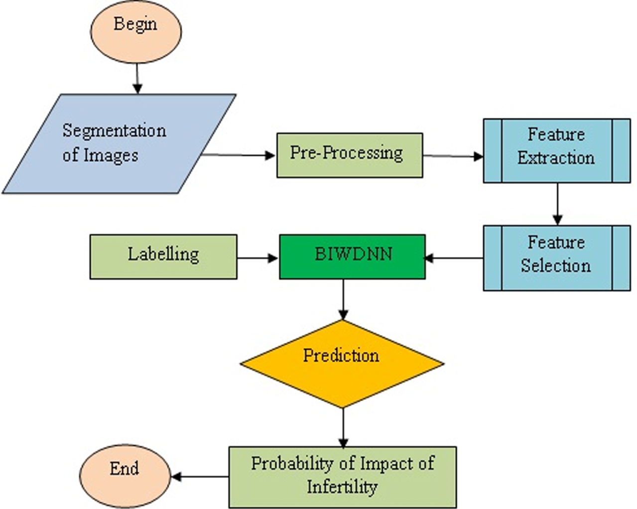
Process flow of the proposed method.
The key contributions of this article are as follows. A new approach named BIWDNN is proposed for early discovery and prediction of infertility issues in women by utilising the water drop-based back-propagation neural networks technique. Initially, vital features from MRI images are identified and then segmented and classified by applying the fuzzy C-means algorithm. The mined features are next matched with the attributes extracted from a normal person. Finally, the proposed model is assessed using different training and testing datasets to determine the final accuracy rate.
Alves et al. 2011 [8] stressed the importance of the analysis of adenomyosis through MRI and also listed various categories of adenomyosis in the human body. On the other hand, another approach reviewed the existence of adenomyosis and its impact in non-human species. A detailed review of the fuzzy based C-mean segmentation method was presented by Balafar et al. 2014 [9] for brain-associated MR images. In this context, the authors also listed various algorithms and their comparative analysis. A study about indicative patients who were analysed with a leiomyoma and adenomyosis through transvaginal ultrasonography and underwent myomectomy amid expurgation of the myometrium is presented in [10].
Nilesh et al. 2017 [11] conducted a study on improving the performance of MR images during segmentation and reducing their complexity. The authors applied the Berkeley wavelet transformation technique for brain tumour-based image segmentation and the support vector machine technique to enhance its quality and accuracy. Tasuku et al. 2016 [12] discussed the impact of diagnosing adenomyosis among infertile patients. The authors presented a detailed study on the epidemiology and hypothesis of adenomyosis, the use of diagnostic techniques for optimisation, the relationship between infertility and adenomyosis, and different treatment strategies and reproductive effects on adenomyosis. Ghazala et al. 2013 [13] presented a study on the correlation between leiomyoma and adenomyosis as the reason for atypical uterine bleeding in women in different age categories in Kuaon state.
Asha et al. 2011 [14] presented a hybrid neural network model for diagnosing diabetes in India. This approach integrates two techniques, namely a back-propagation network for data processing and a genetic algorithm for the purpose of optimising data associated with the back- propagation network. However, the accuracy of their method needs to be improved. A method for classifying glioma-linked MR images by extracting two different vital features, namely wavelet and statistical features, was presented in [15]. After extraction, these features were given as input to the multilayer perceptron module with the aim of classifying glioma-based MR images.
Basem et al. 2013 [16] designed a water drops algorithm for inferring vital features with the inclusion of a rough set. The proposed method was appraised based on various datasets and evaluated against existing methods, and the authors claimed that their method outperformed the other methods. Jayadevappa et al. 2009 [17] proposed a hybrid MRI segmentation method by combining the watershed algorithm and the gradient vector flow technique to reduce the computational intricacy of noise in MR images. The proposed method aims to improve the boundaries of tumour-affected areas in MR and CT images. However, this method failed to handle all classes of images.
As the pre-processing of a patient’s MRI image is one of the vital operations, Rajeswari et al. 2013 [18] presented an article on enhancing the resolution and quality of MR images. To conserve the contour and edge information of MR images, this approach concentrates on enhancing the resolution of the MR images through Wiener filtering, improving the median of such images, and discrete wavelet transform. A similar method, but with infrared-based image segmentation using wavelet transform modulus maxima, was presented by Luo et al. 2015 [19] for superior segmentation outcome.
Chapron et al. 2017 [20] carried out a statistical analysis to find the relationship between endometriosis and adenomyosis phenotypes through magnetic resource imaging. The data analysis was carried out in terms of the mean and standard deviation for categorical and continuous variables. Oliverio et al. 2016 [21] used an artificial intelligence system to find the probable causes of the adenomyosis condition to help clinicians in diagnosing the process. This method uses a supervised learning algorithm with multi-label problem modelling techniques and attributes selection algorithms to achieve the desired goal.
A method presented in [22] named smart home-based cognitive assessment is used to determine the capability of smart homes during the execution of lightweight to multifaceted tasks carried out in day-to-day life based on pre-calculated scores by an expert. Through a supervised classification technique, this method is also able to determine the value of each task carried out by the participants. Szubert et al. [23] presented a detailed review on clarification of the comparative association between infertility and adenomyosis and explains different methods of offering treatments for an affected patient. In addition, this review presents a critical study of the dataset associated with the effects of adenomyosis on the reproductive system.
Proposed methodology
The proposed method comprises of vital processing modules as listed below.
The MRI images are preprocessed with enhanced progressive-based equalization and the noises are removed. The image is segmented using the GSO-based fuzzy C- means algorithm. The segmentation assists in finding the existence of adenomyosis. The features are extracted and are classified by a bio-inspired intelligent water drop-based back- propagation neural network. Once the features are extracted and classification is done, the severity of the adenomyosis is found based on the levels in the classification. MRI scanning is done on women affected by adenomyosis to find the existence of endometriosis. The significance values are calculated to determine the chance of infertility in women.
The various tasks involved in attaining improved prediction on infertility issues is depicted in Fig. 2. Adenomyosis occurs where endometrial cells stay alive and develop into the walls of the uterus. Like endometriosis, the cells continue to behave as they would normally, and are affected each month with a woman’s period. The result of these menstrual cycles can be an enlarged uterus, pelvic pain and heavy bleeding. Figure 3 shows symptoms caused by adenomysis and endometriosis.
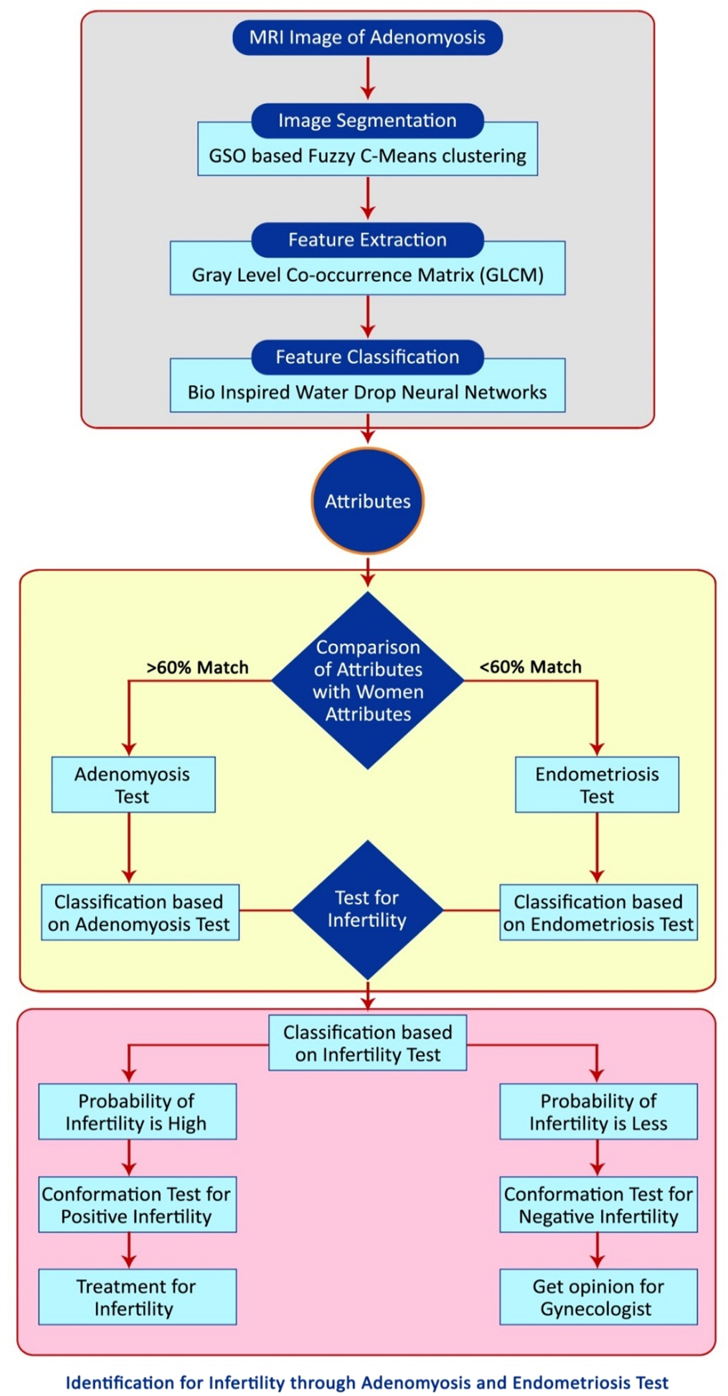
Identification for Infertility through Adenomyosis and Endometriosis Test.
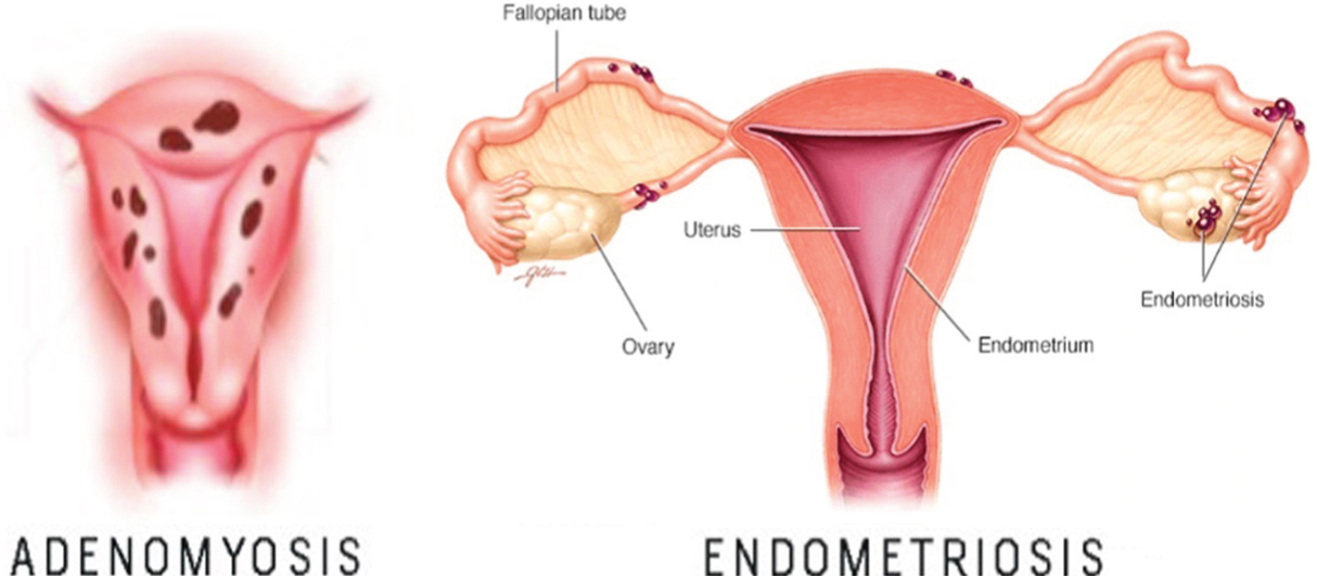
Adenomyosis and Endometriosis Symptoms [25].
As shown in Table 1, endometriosis occurs when cells from the uterine lining are planted in areas outside the uterus. The cells then act in the same way as if they had remained inside the uterus: they thicken, break down and bleed each month. The problem then is that these implants have no way to leave the body. The uterus is designed to funnel the broken down menstrual tissue out of the body with each cycle. However, debris from the endometrial implants remains in the body, potentially causing other problems, including pelvic adhesions or infertility.
Common symptoms of endometriosis and rate of occurrence
Adenomyosis and endometriosis are different and they do not represent the same condition. It is possible for both to occur together; endometriosis being when endometrial cells (the lining of the uterus) are found in a location outside the uterus and adenomyosis when these cells exist or grow into the uterine wall. Although both can cause pain, endometriosis does not often cause heavy bleeding. Women who suffer from gynaecological conditions know the interruptions that arise from pelvic pain and heavy bleeding. While there are multiple conditions that can cause these symptoms, endometriosis and adenomyosis are fairly common, although that is not to say that they are normal.
In essence, infertility is the inability to get pregnant (conceive) after one year of unprotected sex. Women who suffer from irregular menstrual cycles or who are older than 35 years and have not conceived within six months of trying should make an appointment with an endocrinologist - an infertility specialist. Doctors may sometimes detect two or more losses (miscarriages). Pregnancy is the result of a process that has many steps are listed below. Eggs are released from the female ovary (ovulation). Male sperm fuse with the female egg (fertilization). The fertilized egg moves down the fallopian tube (womb). The fertilized egg attaches to the wall of the uterus (implantation).
Infertility may occur if the above steps do not take place properly. Impaired fecundity is a situation connected to infertility and indicates women who have difficulty getting pregnant.
People affected with endometriosis will have great fertility problems. Fortunately, after undergoing treatment such as IVF, women have a chance of getting pregnant. If the patient is affected by endometriosis that is still in the initial stage, she can become pregnant after proper monitoring. Some patients are treated with high-level treatment like IVF OR GFIT. Breastfeeding helps in the reduction of endometriosis. Figure 4 depicts th causes of endometriosis and infertility issues.

Endometriosis and Infertility [25].
There only exists a limited dataset for determining the indicative accuracy of adenomyosis and endometriosis cramps. This might cause redundant adverse side-effects after undergoing complete treatment. The parameters are set based on the literature review done. Figure 5 shows the comparison of the performance accuracy rate of different classification models including BIWDNN.
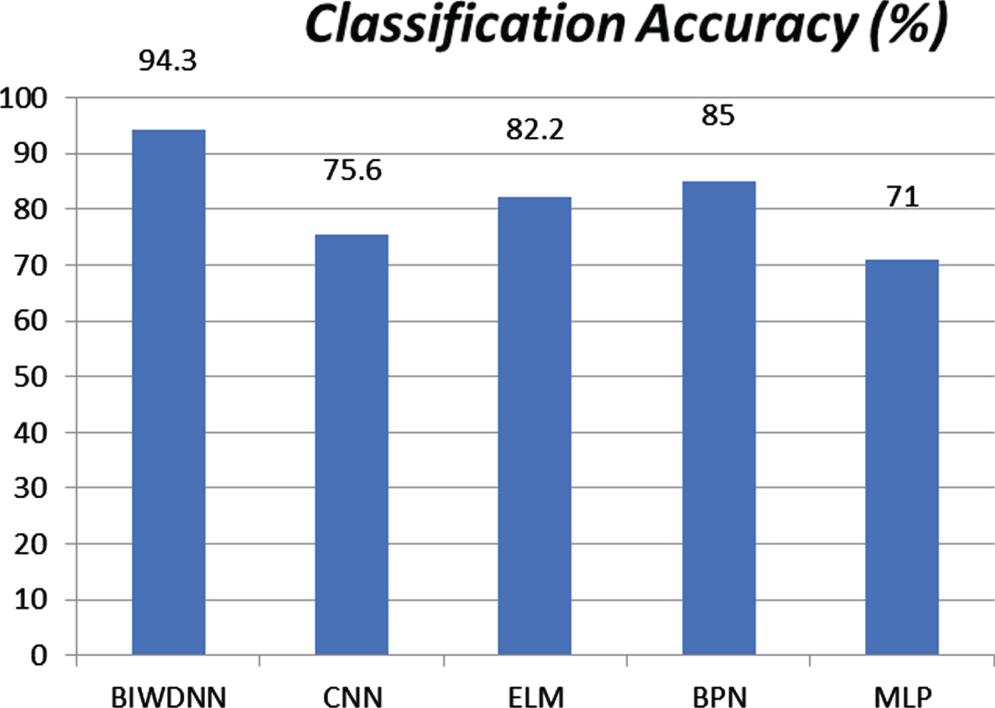
Comparison of Accuracy for various Classification Techniques.
The best prediction model is the BIWDNN. GLCM texture is used to find the connection between two pixels at any given moment, these being called the reference pixel and the neighbouring pixel. GLCM is considered as an estimate of the joint PDF of a grey-level image pair, which is expressed as given in Equation 1.
Varying one value determines the direction and distance of any two pixels. It is usually chosen as 0, 45, 90, or 135 as the four directions. The extraction of the GLCM matrix provides the directional orientation and patterns distance, and textural features from a grey scale image. In the proposed method, contrast, energy, entropy, correlation, and homogeneity are extracted from the images using Equations 2–6, and it is then analyzed using the average value of all four distances and directions., d = 6.
Algorithm 1 is used to extract the features using entropy. Let S1, S2, Sn be the probability distribution of the grey-levels. Two probability distributions are derived: one for discrete values from 1 to m and the other for values from (m + 1) to n. Equations 8 are the two different distributions.
The entropies associated with each distribution are as determined from Equation 9.
Thus BIWDNN classification technique is applied on the segmented images to identify the infertility caused by Adenomyosis and endometriosis.
A computer can discover unique patterns from huge multifaceted datasets without human intervention and apply such patterns to build predictions. However, the intelligent neural network technique aims at designing an automated medical decision system for resolving infertility issues and envisaging pregnancy results, to help physicians make accurate decisions instead of recording the present conditions of such disease.
Infertility in women can change their social value and can degrade their lifestyle. There may be many causes of infertility, of which adenomyosis is just one. Determining the extent to which it contributes to infertility is the purpose of the prediction done using BIWDNN. MRI scan images are taken from a random sample with the constraint that the patient is suffering from adenomyosis. The features are extracted from the MRI images and are classified using the Naive Bayes classifier. A patient with a positive significance is vulnerable to infertility. The features are extracted from the MRI images of 5 samples and the values are tabulated and the significance is calculated. The patients with adenomyosis are tested for infertility and the results show that the patients with a positive significance of adenomyosis have a higher probability of infertility. Figure 6 shows the probability of accuracy of infertility and the significance od adenomysis against different samples.
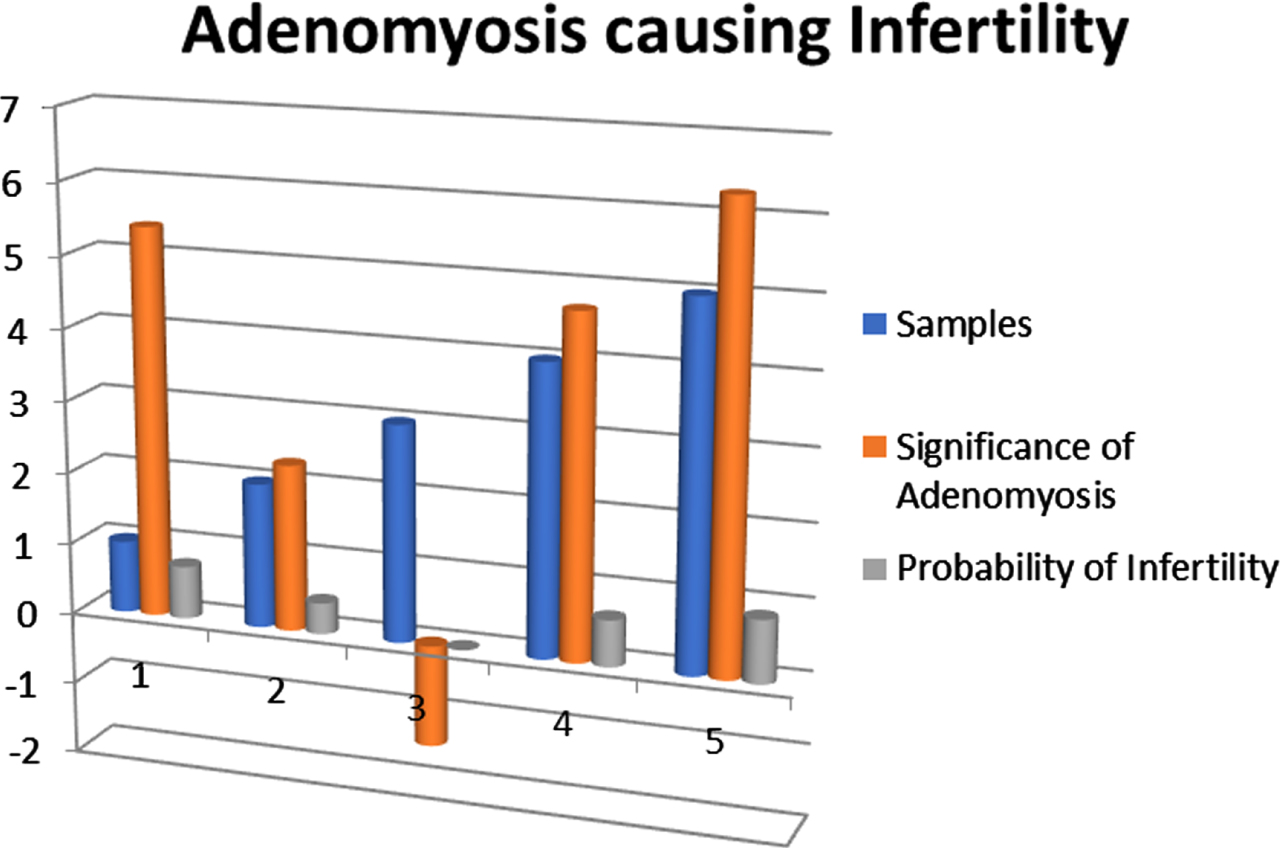
Adenomyosis causing Infertility.
Endometriosis is the condition where the uterine wall becomes thick, resulting in heavy bleeding. This affects the regular functioning of the body. Endometriosis is most common in women between the ages of 32 and 40. The MRI images of women with endometriosis are analysed with BIWDNN and the significance values are tabulated with features extracted from the samples of five women. The values are tabulated as shown in Table 2 and modelded in Fig. 7.
Adenomyosis causing Infertility
Adenomyosis causing Infertility
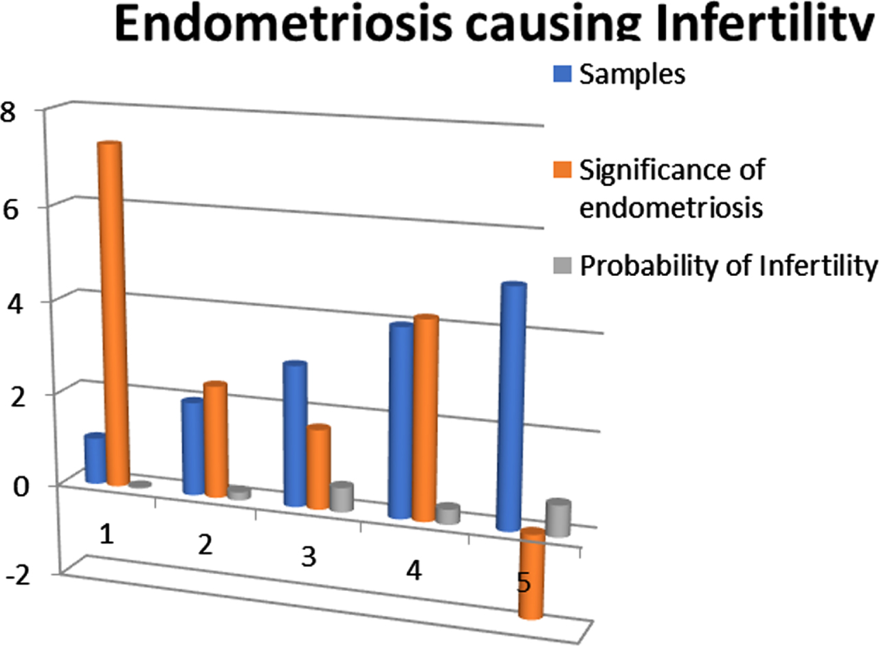
Endometriosis causing Infertility.
The causes of endometriosis are numerous but the significance values tabulated do not show endometriosis to be a cause of infertility. The probability of infertility is independent of the significance of endometriosis. From Fig. 7, it is inferred that women affected with endometriosis are examined for infertility; the results revealed that not all the women affected by endometriosis suffer from infertility, which clearly supports the classified values from the MRI images.
Table 3 list the impact of infertility against different samples. From the image extraction, it is clear that women affected with adenomyosis can suffer from infertility but women with endometriosis are not affected by infertility. Hence, adenomyosis affects fertility in women who may have endometriosis, but endometriosis itself does not affect fertility.
Adenomyosis and Endometriosis causing Infertility
From Fig. 8, it is inferred that there were five patients samples are chosen for analysis. After a significant study is conducted, it indicates that women aged 30 and above are mainly affected, but endometriosis does not affect fertility in women.
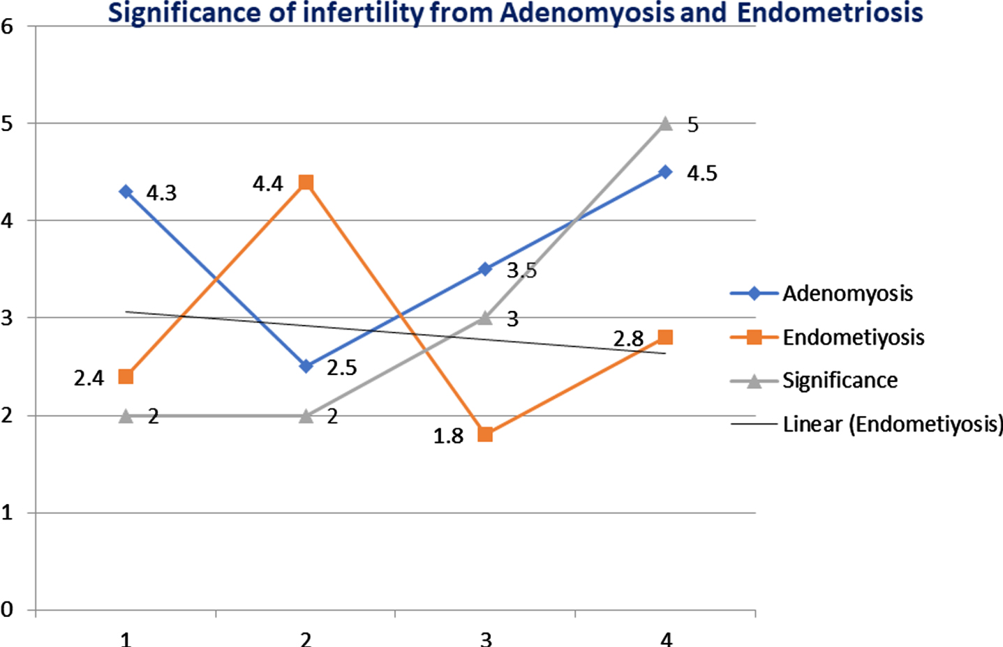
Significance of infertility from Adenomyosis and Endometriosis.
The cause of infertility may not be determined with MRI images of the uterus alone. But the features extracted from the MRI images are classified and may then be used for predicting infertility. Thus, we proposed a bio-inspired intelligent water drop back-propagation neural network-based method to improve the prediction accuracy rate of the impact on the fertility of women suffering from adenomyosis and endometriosis cramps. The proposed neural network-based image analysis results in an improved detection rate compared to other prediction algorithms. A higher significance of adenomyosis will result in infertility and hence it can be treated at the initial stage without many complications.