
Abstract
The present research investigates the size effect of Deferasirox, “4-[3,5-Bis(2-hydroxyphenyl)-1H-1,2,4-triazol-1yl]-benzoicacid”, to reduce lead(II) poisoning in rat bodies. The lead(II) ions as PbCl2 was given orally to the rats in 40 mg/kg dose for 100 days. Deferasirox in nano scale was prepared by sonochemistry method. The effects of initial substance (0.025, 0.05 and 0.1 g) and reaction time (15, 30 and 45 min) on deferasirox size were investigated. Chemotherapy by deferasirox in two scales, nano (16 nm) and bulk (6 μm), was done during 15 days. The lead(II) concentration in various tissues such as heart, liver, spleen and kidneys was determined by graphite furnace atomic absorption spectroscopy (GFAAS). The results show by decreasing in deferasirox particle size; more lead(II) amount were removed from rat’s organs.
Introduction
For centuries, lead(II) has been an environmental pollutant and serious threat to human health [1, 2]. Exposure to lead(II) causes a wide range of adverse physiological effects in humans [3, 4]. The most susceptible are the nervous systems of the fetus, infant and child [5–8]. Increasing soft heavy metals stores such as Cd, Hg and Pb, in the brain has been implicated in neurodegenerative diseases and impaired emotional behaviors [9]. Also, increasing in some metal-ion transporter activities may cause heavy metal accumulation and chelation therapy would be a protective method against these effects of heavy metals [10].
Deferasirox (ICL670 or Exjade) is a poorly-water-soluble chelating agent that is used to remove transfusion overload iron in thalassemia patients or some toxic elements such as lead(II) as chelation therapy [11–17]. Deferasirox or any chelators have multi binding sites, and are flexible enough to surround the metals. Crystallographic structure of some deferasirox complexes show this ligand coordinate to metals with two oxygen atoms of phenolic groups and one nitrogen of central ring. The studies show Defrerasirox has composed a 2:1 complex with Iron(II) and 1:1 complexes with some metals such as vanadium and chromium [18–20]. Chelation therapy is a medical treatment in which a ligating agent (chelators) is added to the blood through a vein or administered orally for removing toxic elements that may be potentially fatal [21–24]. These chelators can mobilize toxic element from different tissue compartment and therefore better overall results would be expected [25]. Furthermore, poorly-water-soluble drugs have limited dissolution rate and decreasing in absorption. So, administered drug must be enhanced to reach the effective blood drug concentration. This dose escalation causes toxicity in the body and the manufacturing cost increases. One of the effective and broadly applicable approaches of poorly-water-soluble drugs is particle size reduction [26]. This approach leads to surface area increasing, dissolution rate enhancement, easier absorption and the reduction of unwanted side effects [27, 28]. Recently, chelation therapies by combining two chelators; deferasirox and deferiprone, for removing lead(II) ions has been reported. The results show two chelators can be more effective for removing lead(II) ions from the biological systems than single therapy [29, 30]
The aim of this research is the investigation of the chelator size effect in decreasing lead(II) concentration from some manually infected rat’s organs; heart, liver, kidneys and spleen. The results show decreasing in particle size in single therapy would be as effective as combining chelator therapies. Deferasirox size was decreased in nanometer by sonochemical method [31–33] and two parameters; initial substance amount and irradiation time, were optimized.
Experimental
Reagents
Dimethyl sulfoxide (DMSO), lead(II) chloride and nitric acid were purchased from Merck Co. (Germany). Deferasirox (m.p: 262°C) [34] was purchased from Novartis Co. (Basel, Switzerland). Double distilled water was used in all experiments.
Methods
The nanoparticle was prepared by using ultrasonic bath (operating frequency 35 kHz, 560W, Sonorex Digitec, Bandelin). Characterization of the particle size was estimated by scanning electron microscopy (SEM) (S4160-Hitachi Japan). FT-IR spectrum was recorded as KBr pellets on a Bruker tensor 27 spectrometer and X-ray diffraction (XRD) technique using Cu radiation at 40 kV and 30 mA and data were collected in the range of 2θ= 10–80° (Advance Bruker D8). Also, atomic absorption spectrometer (GFAAS) Varian model was used for measurement of Pb(II) concentration in organs.
Maintenance of the animals
25 Male Wistar rats were obtained from Kerman Neuroscience Research Center, Kerman, Iran. The rats were maintained under a controlled light:dark (12h:12 h) schedule at 22°C and humidity of 55%. The rats were kept in well-cleaned sterilized cages and were assigned randomly to control and treated groups.
Preperation of deferasirox as bulk and nano-scale
Double distilled water (5 ml) as antisolvent was added drop wise to three various solutions (10 ml, DMSO) of deferasirox (0.1, 0.05 and 0.025 g). These solutions have been poured in a round-bottom flask under ultrasonic irradiation before water addition. Finally, a white precipitate was obtained, filtrated and dried at room temperature. The above procedures were done in three different irradiation times (15, 30 and 45 min). To assess deferasirox size effect on lead(II) release from rat organs, bulk-scale (6 μm) and smaller deferasirox nano-scale (16 nm) were selected. Bulk form in micrometer scale was prepared from initial substance (deferasirox, 0.1 g) solution as the above procedures without any irradiation.
The investigation of lead(II) removal from heart, liver, spleen and kidneys
In this study, 25 male Wistar 6 week old rates were used. Rats were classified as following groups: 1) control 2) before chelation therapy 3) without chelation therapy 4) chelation therapy with bulk deferasirox 5) chelation therapy with deferasirox in nano size (Table 1). Control group had common diet (normal food and double distilled water) but another four groups received 40 mg/kg lead(II) ion during 100 days for manually infection. The lead(II) ions as a solution of PbCl2 (LD50: 1947 mg/kg) were used to contaminate drinking water.
Classification of rats
Classification of rats
Chelation therapy was done by deferasirox (187.5 mg/kg body weight) in nano and bulk scales during 15 days on two final groups: 4) chelation therapy with bulk deferasirox 5) chelation therapy with nano size. Two first groups were anesthetized with ether vapors, immobilized by cervical dislocation and killed after 100 days. Also, the other three groups were sacrificed after 115 days to detect the lead(II) concentration. Then, their heart, liver, spleen and kidneys tissues were collected, weighted and digested by 3 ml HNO3. The residue was diluted with double distilled water to 10 ml volume. The lead(II) concentration was determined by graphite furnace atomic absorption spectroscopy (GF AAS).
FT-IR and XRD analysis
The FT-IR spectra for both (bulk and nanoscale) are similar, and revealed an absorptions bands at 3319 and 3407 cm-1 (OH, stretching), 1680 and 1660 cm-1 (acid, conjugated C = O stretching), 1608 and 1611 cm-1 (C = N stretching), 1450 and 1463 cm-1 (aromatic, C = C stretching), 1279 and 1272 cm-1 (C-O stretching), 1036 and 1030 cm-1 (C-N stretching) [35]. These results indicate that the bulk and nano size of deferasirox have the same structure and ultrasonic irradiation exposure has no structural changes in nano scale (Fig. 1).
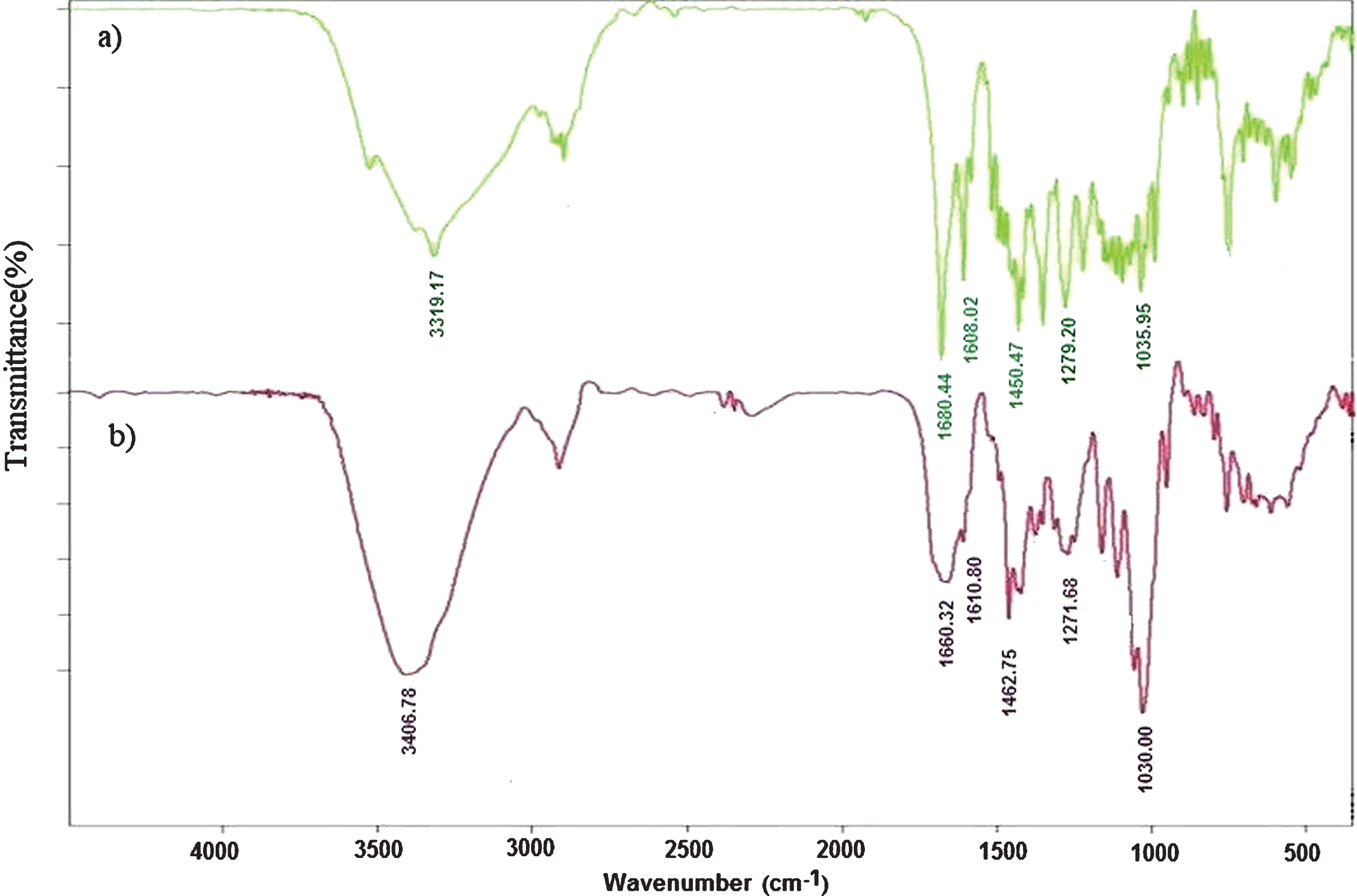
FTIR spectra of deferasirox in a) bulk and b) nano size.
X-ray powder diffraction (XRD) patterns for the bulk and nano forms of deferasirox are shown in Fig. 2. The images show similar patterns and confirm the same crystallographic structure in both sizes and no decomposition during ultrasonic exposure. The crystallite sizes were calculated from FWHM using Scherrer’s formula. The average grain sizes of nano and bulk samples from this equation are 11.8 and 17.68 nm, respectively.
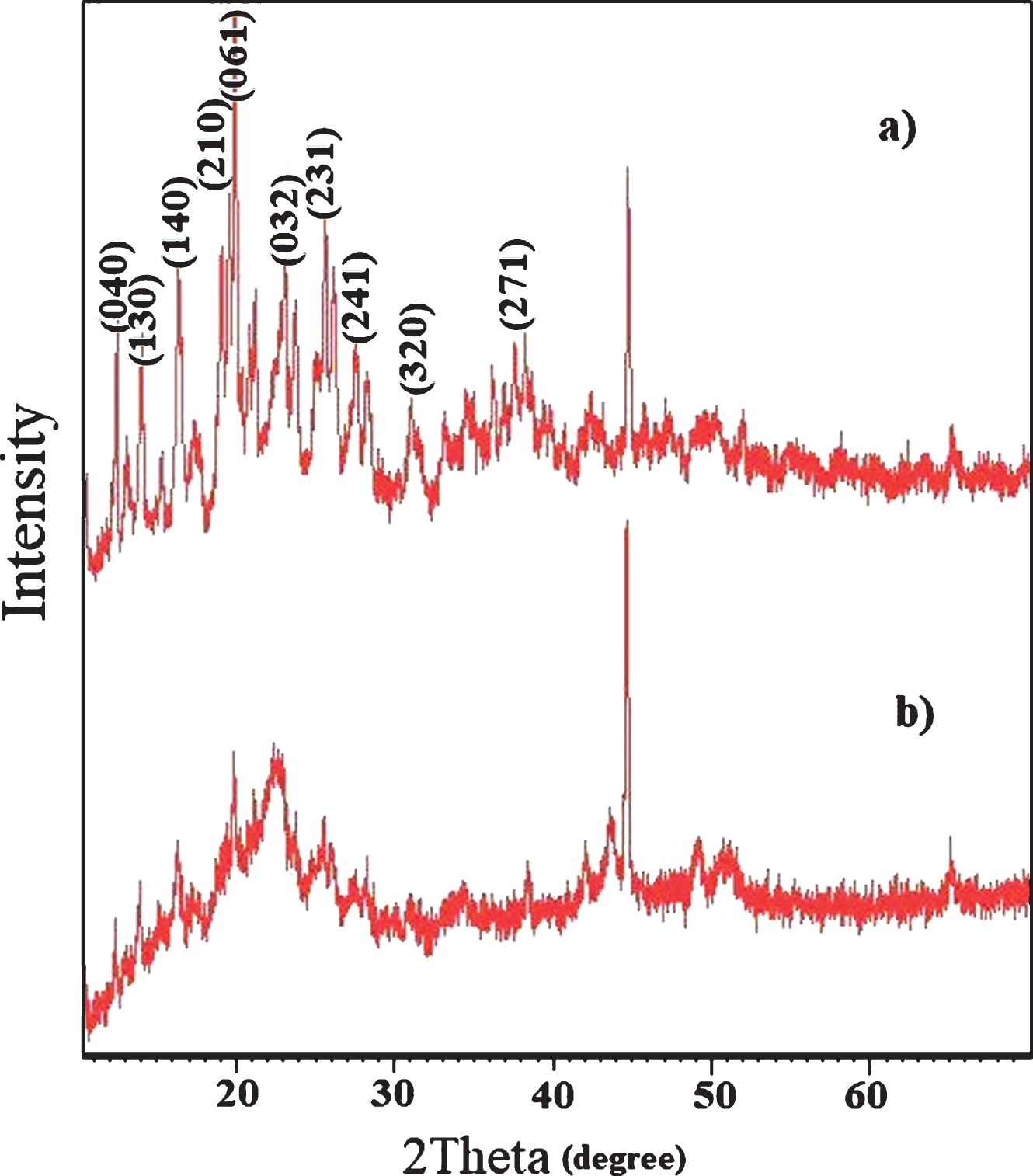
X-ray diffraction patterns of deferasirox in a)bulk and b)nano particle size.
The structure of deferasirox (Fig. 3) was obtained from X-ray crystallographic data (CCDC = 237451) [18]. single-crystal structure of deferasirox shows this structure has crystallized in monoclinic system with these parameters; a = 8.900(2) ringA, b = 26.946(5) ringA, c = 7.558(2) ringA, β= 94.77(3)°, and space group P21/c. Also, the collected data from XRD analysis using CuKα1 radiation have confirmed this crystalline structure, perfectly [34].
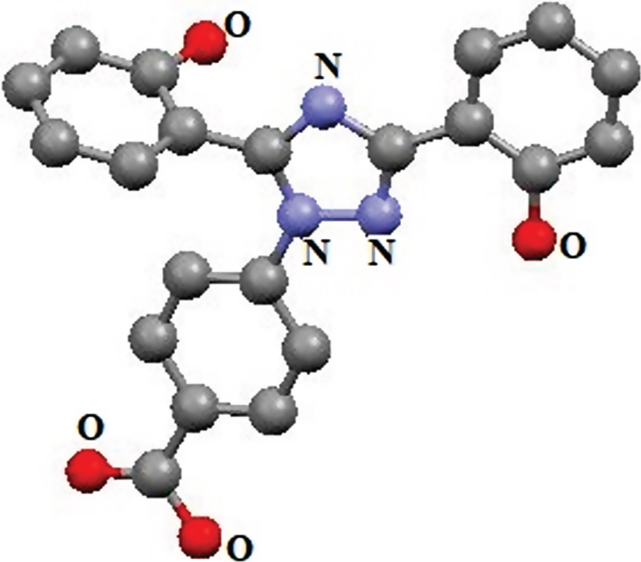
ORTEP diagram of deferasirox molecule, hydrogen atoms were omitted for clarify.
In this study, the effects of initial substance (0.1, 0.05 and 0.025 g) and reaction time (15, 30 and 45 min) were investigated. The SEM images show an increasing in the irradiation time or in the precursor amount, decrease the particle size (Fig. 4). As the images show, these prepared nano particles (16–40 nm) have smaller size compared with the other synthesis method (5–500 μm) [26]. So, all the nano structures were obtained from 0.1 g initial sample and after 45 minutes irradiation. The product was collected as nanoparticles with agglomeration. The Fig. 5 shows deferasirox was obtained as large microstructure after precipitating from solution. A comparison between two Figs. 4 and 5 shows the role of ultrasound waves on decreasing particle size.
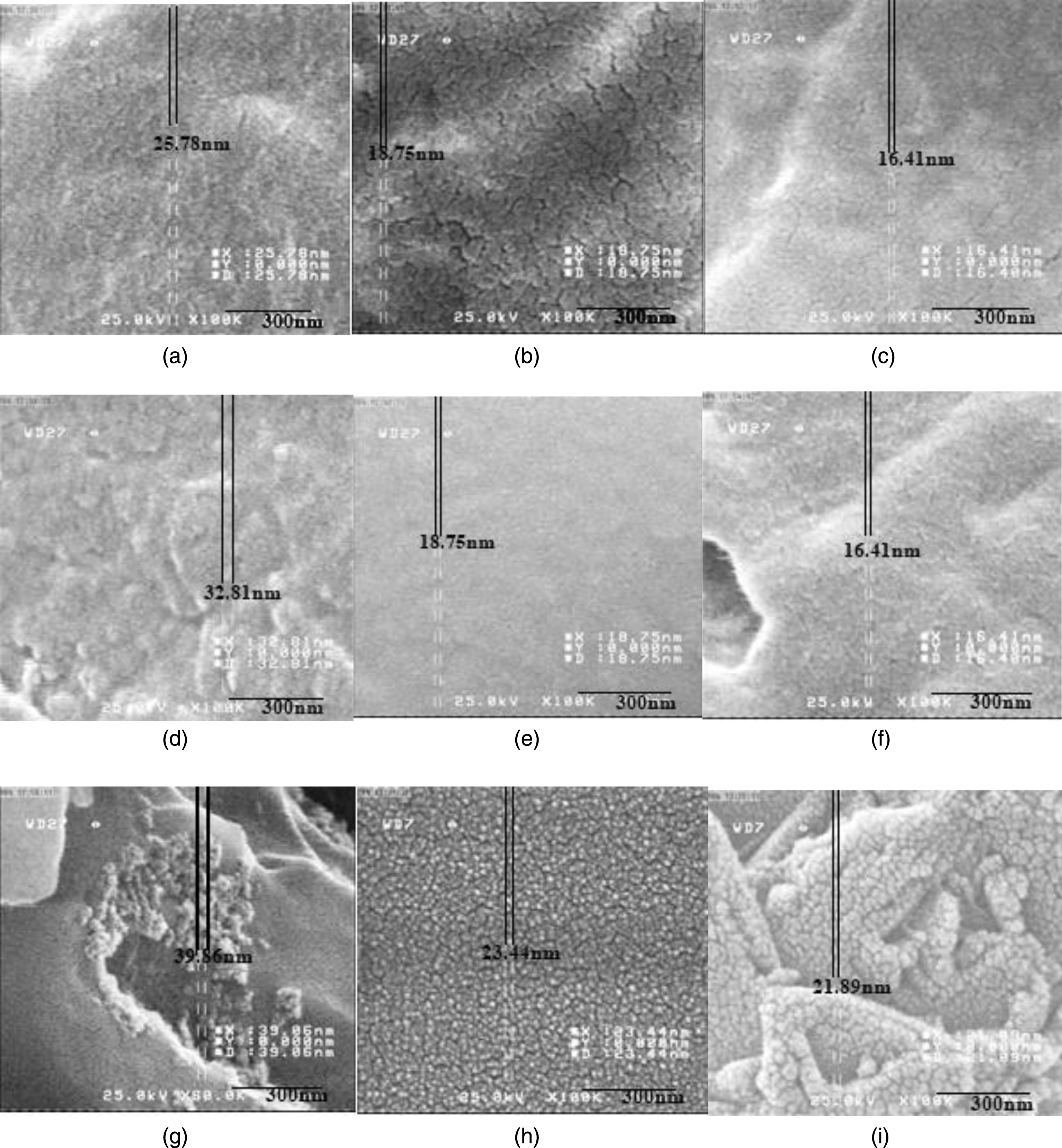
SEM images of prepared nano-particles at various amounts of deferasirox and different reaction times: (a) 0.1 g, 15 min, (b) 0.1 g, 30 min, (c) 0.1 g, 45 min, (d) 0.05 g, 15 min, (e) 0.05 g, 30 min, (f) 0.05 g, 45 min, (g) 0.025 g, 15 min, (h) 0.025 g, 30 min and (i) 0.025 g, 45 min.
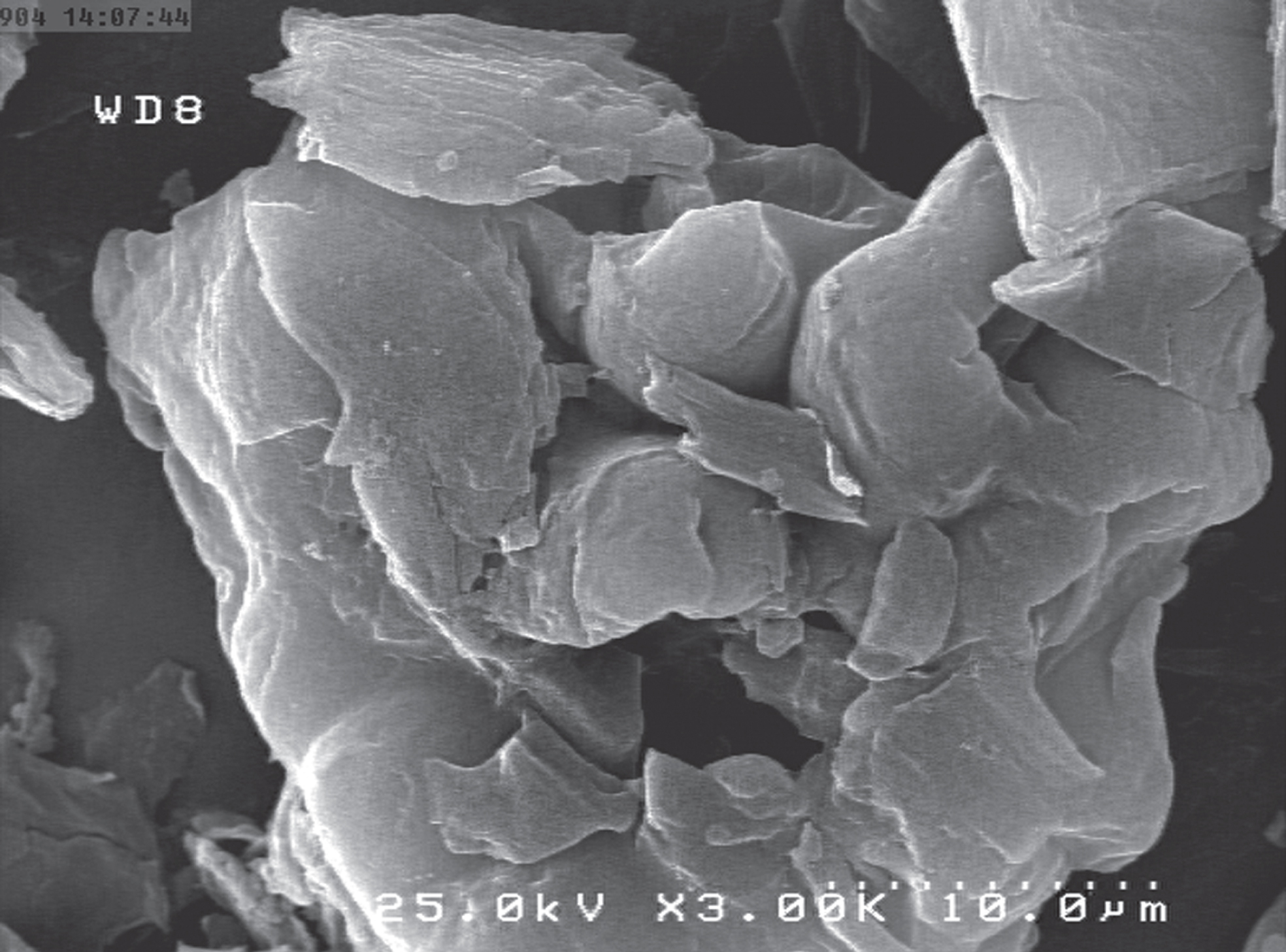
SEM image of deferasirox in bulk form.
After 100 days of lead administration, all rats except control group showed significant weight loss (Table 2). Some other abnormal clinical signs were observed such as irritability, weakness, spots on the liver and loss of hair. During administration, lead(II) concentration increased in heart, liver, spleen and kidneys tissues.
The rats weight changes within 100 days of lead feeding or control diets
The rats weight changes within 100 days of lead feeding or control diets
aMain of five determinations±standard deviation.
After chelation therapy, lead(II) level in tissues and the symptoms of toxicity were significantly reduced. Table 3 shows the concentration of lead(II) before and after chelation therapy in some organs. The lead(II) level in third group, 3) without chelation therapy, indicate lead(II) ions didn’t exit from the body over the time, spontaneously. But, the results show significant decrease at lead(II) level in both final chelation therapy groups: 4) chelation therapy with bulk deferasirox and 5) chelation therapy with nano size. Also, the results show by decreasing in deferasirox particle size, lead(II) level would be decreased in selected organs.
The results of lead(II) level before and after chelation therapy
In the present work, deferasirox in nano scale was successfully prepared in the presence of ultrasounic waves. Different physicochemical parameters such as reaction time and precursor amount brought up a signification changes in the size of nano particles. The results show by increasing in reaction time and precursor amount, the particle size would be decreased. The deferasirox was used as a chelator to remove Pb(II) ions from infected rat bodies. Chelation therapy was done with two different size (6 μm and 16 nm) of deferasirox in bulk and nano scales, respectively. The results show deferasirox was able to remove lead(II) ions from the selected rat’s organs and this chelator in nano scale reduce more ions from these biological systems.
Footnotes
Acknowledgments
The authors are thankful to the head and director of Kerman Neuroscience Research Center for their support of these investigations.