
Abstract
Commiphora gileadensis is commonly used in Saudi Arabia for oral hygiene. A lack of data about its biological activity encouraged us to evaluate the antioxidant and antibacterial activities of its leaf and stem extracts. Ethanol, methanol, acetone and deionized water were tested as extraction solvents. 80% methanol gave the highest extracted concentrations of phenolic and flavonoid substances. The leaf and stem extracts were respectively evaluated for their radical scavenging activity with DPPH (EC50 = 3.39, and 1.06), ABTS (EC50 = 0.690, and 0.55), and peroxide scavenging activity (EC50 = 2.43, and 1.28). GC-MS identified a wide range of compounds that may be responsible for these activities of the results observed. The highest levels of chlorophyll, carotenoids, and lycopene were found in the leaf extract while level of proanthocyanidins was found in the stem peels extract. The peroxidase and catalase activities of stem peel extract were higher than those of the leaf extract. The findings showed that the leaf and stem peel extracts of C. gileadensis exhibited significant antibacterial activity against the test organisms. The minimum inhibitory concentrations for the plant extracts were compared with the standard reference drug Augmentin but the time–kill curves for the C. gileadensis extracts showed that they were less effective than Augmentin. Moreover, the stem peel extract exhibited stronger antibacterial activity than the leaf extract. In conclusion, C. gileadensis can be an important source of natural antioxidants, used as a healthy chewing stick for teeth brushing and oral hygiene purposes.
Abbreviations:
2,2-diphenyl-1-picryl-hydrazyl-hydrate
2,2’-azino-bis(3-ethylbenzothiazoline-6-sulfonic acid)
Effective concentration
Gas chromatography–mass spectrometry
Introduction
Commiphora gileadensis (Burseraceae family, known locally as besham) is an aromatic plant that is widely cultivated in Saudi Arabia and Yemen and is used as a traditional medicine in the Middle East for many ailments, such as pain and inflammation, and has a strong antiproliferative effect on cancer cell lines [1]. Its leaf and flower decoctions are used as an analgesic for cancer and diuresis in traditional Arabic medicine [2]. C. gileadensis is still an important therapeutic plant in Saudi Arabia [3], and is used to treat fever and swelling [4]. In Arab Gulf nations, chewing sticks made of C. gileadensis in addition to Salvadora persica are used for dental purposes, particularly for oral hygiene. Moreover, C. gileadensis is used by Muslims throughout the day for oral care. The World Health Organization recommends the use of chewing sticks as a powerful tool for oral cleanliness in countries where such use is commonplace [5]. C. gileadensis and other related plants are believed to be active against microorganisms that cause tooth decay [6]. An extract of C. gileadensis was found to repress development and actuate apoptosis in two lymphocytic tumor cell lines [7]. An extensive review of the applications, pharmacology, and photochemistry of C. gileadensis was published recently [8].
Antioxidants are substances that either directly or indirectly protect cells against the unfavorable effects of xenobiotics, medications, cancer-causing agents, and harmful radical responses [9]. Many natural products and their mixtures have been shown to have antioxidant activity, including vitamin C, polyphenols, flavonoids, peroxidases and catalases [9–11]. Antioxidants are able to reduce oxidative stress by eliminating free radicals. They are also considered to be beneficial for the cell reinforcement movement of plants. Antioxidants act as hydrogen donors, eliminating free radicals before they can damage cells and other organic components. Accordingly, they are important for health and wellbeing [12]. The high levels of antioxidants in herbs and spices are responsible for their medical advantages [13]. The free radicals created as a result of the various metabolic processes occurring in the body are one of the critical causes of illnesses such as cancer, diabetes, dementia, and myocardial localized necrosis as they interact with cell DNA and cause mutation [14]. The objectives of this research were to assess the total phenolic and antioxidant enzyme content and antimicrobial activity of C. gileadensis stem and leaf extracts. The phenolic compounds in the stem and leaf extracts were Identified by gas chromatography-mass spectrometry (GC-MS) analysis.
Method and materials
Chemicals
Gallic acid, 2,2-diphenyl-1-picryl-hydrazyl-hydrate, Folin–Ciocalteu reagent, vanillin, and 2,2′-azino-bis(3-ethylbenzthiazoline-6-sulfonic acid) were purchased from Sigma–Aldrich. catechin, ferric acid, hydrogen peroxide, catechol, and guaiacol were purchased from Extrasynthese (Lyon, France).
Sample collection
C. gileadensis was collected from Makkah city, Saudi Arabia. The leaves and stem peel were manually collected, cleaned, and dried at room temperature, then stored in a dry place until further analysis.
Preparation of plant extracts
Dried C. gileadensis leaves and stem peel (10 g of each) were extracted by shaking at 120 rpm for 24 h with 30 mL of solvent (distilled water, 80% and 40% methanol, 80% and 40% ethanol, 80% and 40% acetone). The obtained extracts were then filtered and stored in a refrigerator until needed.
Total phenolic content determination
The method described by Velioglu was adapted to evaluate the total phenolic content (TPC) in the C. gileadensis leaves and stem peels using Folin–Ciocalteu reagent (FCR) [15]. At room temperature, 800μL of distilled water and 0.1 mL of obtained extract were mixed with 0.1 mL of FCR for 5 min. The mixture was then supplemented with 0.5 mL of sodium bicarbonate (20%) and then left for 20 min before measuring the absorbance at 750 nm. The results are reported as the mg gallic acid equivalent/g dry matter (mgGAE/g DM).
Total flavonoid content
The total flavonoid content in the plant extracts was determined using the method reported by Zhishen [16], with slight modification. Catechin was used as the standard. 1.25 mL distilled water and 0.250 mL plant extract were mixed with 0.075 mL of 5% NaNO2 solution and allowed to stand for 6 min. The mixture was supplemented with 0.150 mL of AlCl3 solution (10%), 0.275 mL of distilled water and 0.5 mL of 1 M NaOH, and allowed to stand for 5 min. Thereafter, the absorbance was measured at 510 nm. The results are reported as the mg catechin equivalent/g dry matter (mgCE/g DM).
Proanthocyanidin content
The method described by Broadhurst was adapted to quantify the proanthocyanidin content in the C. gileadensis leaves and stem peels [17]. 0.4 mL of plant extract 3 mL of vanillin (4% methanol) were blended with 1.5 mL of hydrochloric acid concentration. After 15 minutes of incubation at ambient temperature, the mixture’s absorbance was recorded at 500 nm. The Proanthocyanidin content was expressed as μg catechin/g DM.
Determination of chlorophyll, lycopene, and carotenoids
The chlorophyll, lycopene, and carotenoid contents were determined according to Wang’s method [18]. One g of every fresh plant portion was ground to 10 mL (60:40) of hexane: acetone. The supernatant was moved into an ice-capped tube. The remaining aqueous layer was re-extracted using 10 mL of the same solvent, and move the organic layer into the same tube. This process was carried out until the aqueous layer become colorless.At 450, 502, 645, and 663 nm, 1 mL of the organic extract was then used for absorbance determination. Carotenoid, lycopene, and chlorophyll concentrations were calculated using the following formulae:
Antioxidant assays
DPPH radical scavenging activity
The method described by Ao was adapted to evaluate the free radical scavenging activity (FRSA) in the C. gileadensis leaves and stem peels using the DPPH assay [19]. 0.1 mL of obtained extract was mixed with 0.9 mL of 0.1 mM DPPH reagent and left for 30 minutes in the dark. The mixture’s absorbance was recorded at 517 nm and the scavenging activity was calculated by the equation:
The results are reported as the percentage scavenging activity relative to the sample concentration. The EC50 value (the concentration needed to produce 50% FRSA) was interpolated from the plots.
ABTS radical cation decolorization assay
The ability of the extracts to scavenge ABTS•+ was determined according to Re method [20]. 1 mL of diluted ABTS•+ in 0.1 M phosphate buffer, pH 7.4 was mixed with a 0.1 mL crude extract. At 1 minute after mixing, the mixture’s absorbance was recorded at 734 nm. Sample scavenging activity was calculated using the equation below:
Hydrogen peroxide-scavenging activity
The method of Runch was used to determine the H2O2 scavenging activity [21]. A solution of 2 mM H2O2 was prepared in 0.5 M sodium phosphate buffer, pH 7.4. 0.5–2.5μg/mL of extracts was added to 0.5 M phosphate buffer, pH 7.4 to give a volume of 0.4 mL, which was made up to 1.0 mL using H2O2. The absorbance was recorded over 10 min at 230 nm and the scavenging activity was calculated using the following equation:
Antioxidant enzymes determinations
Crude enzyme extract
Fresh stem peel and leaves (10 g of each) was ground using a mortar with 0.2 M Tris-HCl buffer (pH 7.2). The homogenates were centrifuged at 13 000 rpm and 4 °C for 12 minutes. The supernatant was used as the crude enzyme extract and was stored at –18 °C until needed.
Peroxidase assay
The peroxidase activity was estimated according to the Mar’ia, method [22]. The crude extract (0.1 mL) was blended with H2O2 (8 mM), guaiacol (40 mM), and 50 mM acetate buffer (pH 5.5), to give a total volume of 1 mL. The mixture’s absorbance was measured every 1 min at 470 nm.
Polyphenol oxidase assay
Catechol was used as the substrate to measure the polyphenol oxidase activity according to the method reported by Siddiq and Cash [23]. 0.9 mL of catechol reagent (20 mM) in sodium phosphate buffer (10 mM, pH 6.8) was mixed with 0.1 mL of the crude extract. The mixture’s absorbance was then recorded for 3 min at 420 nm.
Catalase assay
The catalase activity was measured using the method described by Bergmeyer and Gawehn [24]. 2 mL of 25 mM H2O2 prepared in sodium phosphate buffer (75 mM, pH 7) was mixed with 0.5 mL of crude extract. The mixture’s absorbance was then recorded for 1 min at 240 nm.
Gas chromatography-mass spectrometer (GC-MS) analysis
The methanolic extracts of C. gileadensis were analyzed using a TRACE GC ULTRA system with a setting described by previous work [25]. GC-MS analysis of C. gileadensis methanolic extracts was described after comparing mass spectra with databases and enumerating compounds along with their retention time, molecular formula, and peak area. The peaks were identified using library searches with the NIST database and Wiley Registry of Mass Spectral Data Version 8.
Antibacterial activity
Pathogenic bacteria strains
E. coli, S. sonnei, K. pneumoniae, P. aeruginosa, S. aureus, and S. epidermises were obtained from the Microbiology Laboratory, Department of Biology, King Abdulaziz University, KSA.
Agar well diffusion assay
Bacterial isolates were set to match the 0.5 McFarland standard. An organism volume of 0.1 mL was distributed over the surface of an agar plate. In each of the cultivation plates, three holes were punched. Next, 10μL of Augmentin was added in the center hole as a positive control; 0.15 mg of the plant extract was put in each of the remaining two holes. The cultivation plates were incubated for 24 h at 37 °C. The inhibition zone was measured in mm. The tests were carried out in triplicate [26].
Micro broth dilution assay
The minimum inhibitory concentrations (MIC) of each of the C. gileadensis samples against the bacteria described above were determined by micro-diluted broth method as described by NCCLS [27]. Augmentin was used as the control. To each well was added 0.1 mL of Muller Hinton broth (MHB), 0.005 mL of inoculum and the 0.5 McFarland suspensions (1×10⁸ CFU/mL). Additional bacterial suspension dilutions were done in MH broth to achieve a final inoculum dilution of 5×10⁵ CFU/mL. The tests were carried out on the same day using three similar wells in triplicate.
Susceptibility testing (Time-kill kinetics)
For each bacterial strain, the highest MICs for C. gileadensis and Augmentin were measured in Muller Hinton broth to determine the time–kill kinetics. Cultures were aerobically incubated at 37 °C and the C. gileadensis extracts and/or asolution of Augmentin were added to each well to determine antibiotic concentrations. At 0, 2, 4, 6, 8, 10, 12, and 24 h following the addition of the test solutions to the wells, 100μL of the culture was withdrawn aseptically using a sterilized loop and then streaked evenly on a blood agar plate and incubated at 37±1 °C for 24 h. Bacterial colonies were counted using a plate counter [26].
Statistical analysis
Data were analyzed by the Student’s t-test and one-way ANOVA using OriginPro 2018 The results are expressed as mean±standard deviation (SD). The results were significant when P < 0.05.
Result and discussion
In view of the beneficial impacts of phenolic compounds on human health, including anti-inflammatory, anti-cancer, and anti-atherosclerosis activities [28], the present study aimed to evaluate the antioxidant and antibacterial activities of extract of C. gileadensis, which is an important plant in the Middle East, where it is widely used for oral hygiene. The TPC of the crude extracts of C. gileadensis leaves and stem peel were evaluated using the Folin–Ciocalteu assay. For this assay, phenolic groups are deprotonated under alkaline conditions and form phenolate ions in the presence of Folin–Ciocalteu reagent owing to the reduction of the phosphotungstic–phosphomolybdic complex, generating a blue color.
The optimum solvent must be selected for the extraction of phenolic and flavonoid compounds. Ethanol (40, 80%), methanol (40, 80%), acetone (40, 80%), and water were investigated as solvents for the extraction process. Table 1 demonstrates the ability of the various solvents to extract phenolic and flavonoid compounds from the different parts of C. gileadensis. Among these solvents, 80% methanol provided the best extraction of phenolic compounds (34.98 mg GAE/g DM for stem peel and 20.97 mg GAE/g DM for leaves). On the other hand, the total flavonoid contents in the stem peel and leaves of C. gileadensis were 10.49 and 6.90 mg CE/g, respectively. Methanol extraction was previously used to evaluate antioxidants and flavonoid compounds in Portulacaceae, Phoenix dactylifera, and Coleus forskohlii [25, 29–30]. Most natural antioxidants are multi-functional. Consequently, a robust antioxidant assessment protocol needs specific antioxidant activity assessments to account for specific antioxidant action mechanisms [31]. Consequently, several methods were used to evaluate the antioxidant capacity of the C. gileadensis methanol extracts. DPPH free radical scavenging is commonly used to calculate antioxidant activity. The degree of coloration reveals the scavenging ability of the antioxidant extract owing to hydrogen donation [32]. The C. gileadensis stem peel and leaf extracts exhibited concentration-based DPPH radical scavenging, which is attributed to DPPH’sability to accept hydrogen. The results showed that the C. gileadensis extracts significantly (p < 0.05) inhibited the generation of free radicals based on the concentration and assay used (Table 2). The DPPH radical scavenging (%) increased in the leaves and stem peels of C. gileadensis extracts as the extract concentration increased from 2 to 10μg/mL. The highest DPPH radical inhibition value was observed in the stem peel extract at a low concentration of 2μg/mL (46%), whereas the highest inhibition value (32%) was reported at the initial concentration (2μg/mL) for the leaf extract. In addition, the maximum inhibition (97%) was observed in the stem peel extract at 10μg/mL, followed by the leaf extract (86%) at a final concentration of 10μg/mL. The DPPH radical scavenging results show that the C. gileadensis stem peel extract is the most effective DPPH radical scavenger.
The phenolic and flavonoid contents of the leaf and stem peel extracts of Commiphora gileadensis obtained using various solvents
The phenolic and flavonoid contents of the leaf and stem peel extracts of Commiphora gileadensis obtained using various solvents
Parameters are presented as means±SD (n = 4). GAE, gallic acid equivalent, CE, catechin equivalent.
Antioxidant activities of Commiphora gileadensis leaf and stem peel extracts as measured by DPPH and ABTS assa
The values are presented as mean±SD (n = 4). The values followed by different superscript letters (a–e) within the same column are significantly different (p < 0.05).
The inhibition percentages in both DPPH and ABTS assays were determined using the effective concentration (EC50), which is defined as the amount of antioxidants needed to reduce the initial scavenging radical’s concentration by 50%. The EC50 values for the DPPH assay were found to be 1.06 and 3.39μg/mL for the crude methanol stem peel and leaf extracts, respectively. The correlation coefficients (R2) for the leaf and stem peel extracts and the DPPH radical scavenging activity were found to be 0.981 and 0.903, respectively, indicating high correlation. High EC50 values were also reported for the medicinal plant S. persica (4.8μg/mL) [33], and Portulacaceae stem peels and leaves (20.56 and 25.26μg/mL crude extract, respectively) [25].
On the other hand, the extracts from C. gileadensis leaves and stem peels also showed a significant (p < 0.05) increase in radical scavenging activity with different leaf and stem peels concentration (0.24–1.20μg/mL) using the ABTS method (Table 2). At an initial concentration of 0.24μg/mL, the stem peel extract exhibited higher inhibition (21%) than the leaf extract (12%). Our results showed that the stem peel extract had the highest scavenging activity (98%) at the maximum concentration (1.20μg/mL), followed by the leaf extract (96%), with a statistically insignificant difference at p < 0.05. The ABTS assay EC50 values were found to be 0.690 and 0.550μg for the leaf and stem peel extracts, respectively. The correlation coefficients (R2) for the leaf and stem peel extracts and the ABTS scavenging activity were found to be 0.967 and 0.965, respectively. The EC50 values of 2.86 and 3.70μg/mL were reported the leaf and stem peel strip crude extracts, respectively, of Portulacaceae, 1.16μg/mL for C. forskohlii leaf extract [29], 1.6μg/mL for S. persica [33], 2.23% and 12.02% for the skin and flesh extracts of Kyoho grapes, respectively [34], 2.6μg/mL for safawi (Phoenix dactylifera) [30], and 1.7μg/mL for Raziqi Yemeni grapes [35].
Scavenging of hydrogen peroxide by plant extracts can be due to their phenolic compounds, which contribute electrons to H2O2, reducing it to water. Table 3 demonstrates the scavenging activities of the methanol extracts from C. gileadensis on H2O2. These are compared to gallic acid. The EC50 values required to eliminate 50% of hydrogen peroxide were found to be 2.43 and 1.72μg gallic acid equivalents for the leaf and stem peel extracts, respectively. In comparison, with 1.28μg of gallic acid the removal of H2O2 was 50%. The highest H2O2 hydrolysis scavenging activity was observed for gallic acid (standard compound) (33.4%), whereas the lowest hydrolysis scavenging activity was recorded for the C. gileadensis leaf extract (15%) at the initial concentration (0.5μg/mL). In addition, at a final concentration of 2.5 mg/mL, the standard solution (gallic acid) exhibited the highest hydrolysis scavenging activity (79%), followed by the stem peel (68.9%) and leaf (51.6%) extracts. Since H2O2 does not have unpaired electrons, it is not a radical. However, it appears to be lethal to cells because it can administer hydroxyl radicals to cells [9].
Antioxidant activities of Commiphora gileadensis leaves, stem peels extracts and gallic acid as measured by the H2O2%
The values are presented as mean±SD (n = 4). The values followed by different superscript letters (a–e) within the same column are significantly different (p < 0.05).
Chlorophyll is an essential component of the thylakoid membrane pigment–protein complexes, which represents the photosynthetic potential of plants. This component is essential for capturing and transmitting light energy in photosynthesis [36]. In the current study, chlorophyll a, chlorophyll b, and total chlorophyll (chl) were evaluated in the fresh leaf and stem peel extracts of C. gileadensis, as shown in Table 4. The chl a, b and chl (a + b) concentrations in fresh leaves were 741.6, 240.71, and 982.31μg/g, respectively, which were higher than those found in the stem peel (160.08, 87.36, and 247.44μg/g, respectively). These results are comparable with those reported for other plants, such as Portulacaceae.
The total concentrations of some antioxidant components in Commiphora gileadensis plant parts
The values are presented as mean±SD (n = 4).
Carotenoid was found in the fresh leaf and stem peel extractsof C. gileadensis at 363.12 and 112.96μg/g, respectively (Table 4). Mendelová et al. demonstrated that the carotenoid content in tomato fruits varied from 44.1 to 785μg/g [37]. The highest lycopene content was observed in the leaves (99.03μg/g) compared to 37.07μg/g in the stem peels (Table 4). Some studies have shown that the total carotenoid and lycopene contents are significantly influenced by the degree of maturity, variety, and growing position [38–40].
Proanthocyanidins, also referred to as condensed tannins, are oligomeric and polymeric end-products of the biosynthetic flavonoid pathway [41]. They play a protective role against microorganisms and fungi in plants [42, 43]. They are found in the stems, leaves, fruits, and seeds of many plants [25, 44]. The proanthocyanidin content in the leaf and stem peel extracts of C. gileadensis is shown in Table 4. The highest proanthocyanidin content was observed in the stem peel (13.99 mg CE/g), whereas the lowest value was recorded for the leaf extract (6.81 mg CE/g).
In general, the antioxidant ability of a plant tissue is closely linked to the quality of its antioxidant substances, primarily phenolic compounds, carotenoids, ascorbic acid, and tocopherol, and the activity of free radical enzymes [45]. In addition to phenolic compounds, the antioxidant enzyme activities were measured in the crude leaf and stem extracts from C. gileadensis (Table 5). As expected, the level of peroxidase in the stem peel extract was about 1.9 times higher than that in the leaf extract. Polyphenol oxidase was only found in the crude leaf extract (506.7 units g–1 min–1). The catalase activity in the crude leaf and stem extracts from C. gileadensis was also investigated and was found to be about 5 times higher in the extract from the leaves than from the stem peel. The purification and biochemical characteristics of peroxidase from C. gileadensis have been reported previously [46]. Peroxidase, polyphenol oxidase, and catalase were screened in other medicinal plants, including Portulacaceae, P. dactylifera [30], C. forskohlii [29], and S. persica [33].
Antioxidant enzyme activities ofCommiphora gileadensis
The values are presented as mean±SD (n = 4).
Plants are a great source of natural bioactive ingredients, many of which are used to promote health and fight diseases, and so many plants are sold as functional foods or herbal medicinal ingredients [47]. The use of herbal medicine for numerous illnesses has increased significantly in recent years [48]. The GC-MS analysis of C. gileadensis stem peels and leaves identified a total of 20 components in the stem peel extract in 80% methanol and 19 components in the leaf extract in the same solvent, as shown in Tables 6 and 7. The GC-MS chromatograms for the methanolic extracts of C. gileadensis stem peels and leaves are shown in Fig. 1.
GC-MS analysis of phytochemical constituents of Commiphoragileadensis stem peel extracted in 80% methanol
GC-MS analysis of phytochemical constituents of Commiphoragileadensis leaves extracted in 80% methanol
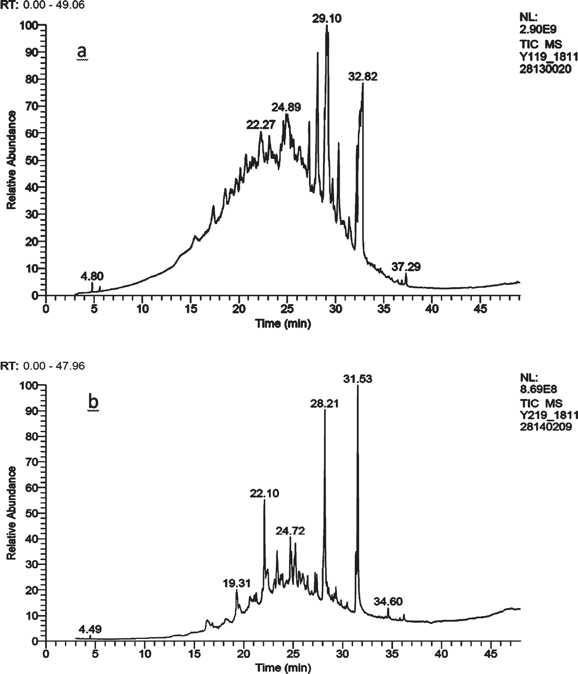
GC-MS chromatograms of C. gileadensis stem peel (a) and leaves (b)extracted in 80% methanol.
The antibacterial activity was investigated by measuring the efficacy of the extracts against various microorganisms. The methanolic extracts of C. gileadensis were tested against six pathogenic bacteria by measuring the diameters of the inhibition zones, as shown in Table 8. The stem peel extract showed better antibacterial activity than the leaf extract. Among these bacterial strains, the stem peel extract showed the largest inhibition zone against E. coli (18±1.5 mm) while the smallest inhibition zone was observed for the stem peel extract against S. epidermises (13±0.5 mm). The largest inhibition zone for the leaf extract was against E. coli (16±0.5 mm) while the smallest inhibition zones were against S. epidermises and K. pneumoniae at 12±1.3 and 12±0.5 mm, respectively. Similar to our results, the methanol extracts of some medicinal plants such as Caesalpinia pulcherrima, Casuarina equisetifolia, and Nepata cataria showed significant inhibition of Gram-negative and Gram-positive bacteria [49, 50]. According to Aligiannis et al. [51], the extracts can be classified based on the MIC results as strong inhibitors: MIC less than 100.0μg/mL: moderate inhibitors; MIC between 00.0 and 500.0μg/mL or weak inhibitors: MIC above 500.0μg/mL. The methanol extract of C. gileadensis stem peel was found to be the most active against bacteria. The wells containing extract concentrations of 128–256μg/mL inhibited the visible growth of all the bacterial species (Table 9). The lowest concentration that inhibited any visual growth was considered to be the MIC[52]. For the C. gileadensis leaf extract, concentrations of 128–512μg/mL inhibited the visible growth of all the bacterial species (Table 10). The time–kill curves for the C. gileadensis extracts showed that they were less effective than Augmentin (Fig. 2). Moreover, the time–kill curve values for the stem peel extract show that it was more effective than the leaf extract (Fig. 2). After incubation for 6–12 h, all the bacteria were completely dead. In the present study, in the absence of an antibacterial agent, the bacterial density for all strains increased rapidly to a plateau of 5×10⁵ bacteria/mL. At the MIC, the C. gileadensis leaf and stem peel extracts initially reduced the bacterial numbers for all of the pathogenic bacteria mentioned above. All of the bacteria were killed after 6 h of incubation, whereas bacterial outgrowth was zero between 12 and 24 h of incubation.
Antibacterial activity of Commiphoragileadensis extracts on the growth of human pathogenic bacteria Escherichiacoli ATCC 25922, Klebsiella pneumoniaeATCC 13883, Shigella sonnei ATCC 25931,Pseudomonas aeruginosa ATCC 27853 Staphylococcus aurous ATCC 25923, and Staphylococcus epidermises 12228 under aerobic conditions
Minimum inhibitory concentration (MIC) of Commiphoragileadensis stem peel extract against Gram-negative and Gram-positive microbial species
Minimum inhibitory concentrations (MIC) of Commiphoragileadensis leaf extract against Gram-negative and Gram-positive microbial species

Time–kill curves for Escherichia coli ATCC 25922 (a), Klebsiella pneumoniae ATCC 13883 (b), Pseudomonas aeruginosa ATCC 27853 (c), Shigella sonnei ATCC 25931 (d), Staphylococcus aurous (SAU) ATCC 25923 (e), and Staphylococcus epidermises 12228 (f) for Commiphoragileadensisleaf and stem extracts (80% methanol) and the MIC of Augmentin as control.
In conclusion, C. gileadensis has been shown to be a good source of natural antioxidants that act through various mechanisms, including the elimination of free radicals, H2O2, DPPH, and ABTS scavengers. The results obtained from this study indicate that the C. gileadensis is an important source of natural antioxidants that can play a very significant role in oxidative stress reduction and the prevention of dangerous diseases, such as cardiovascular diseases, liver, and cancer. The antioxidant potential of C. gileadensis was attributed to the involvement of the antioxidant enzymes peroxidase, catalase, and polyphenol oxidase. The combinedactivities of these antioxidant compounds and antioxidant enzymes contribute to the success of C. gileadensis as a healthy chewing stick for the purposes of teeth brushing, oral hygiene, and functional food. The methanol extracts of the leaves and stem peel have been shown to be good sources of antimicrobial compounds. Thus, it can be concluded that C. gileadensis contains various bioactive compounds and further studies are needed to ascertain their biological and pharmacological activities.
Declaration of competing interest
The authors declare that they have no known competing financial interests or personal relationships that could have appeared to influence the work reported in this paper.
Footnotes
Acknowledgments
The work was funded by the University of Jeddah, Saudi Arabia, under grant No. (UJ-02-028-DR). The authors, therefore, acknowledge with thanks the university technical and support.