
Abstract
The two synthesis of Schiff base SB (Indole-3-carboxalidene-1-phenylsemicarbazide) and organophosphorus Schiff base OPSB (Indole-3-carboxalidene diphenylphosphate-1-phenylsemicarbazide) have been prepared and characterized by elemental analyses, IR, 1H-NMR, 13C-NMR, UV–Vis and XRD. A series of complexes of the type [M(SB)2Cl2].2H2O and [M(OPSB)Cl.(H2O)2].Cl, where M = Cu(II), Ni(II) and Co(II) have been synthesized and the chemical structures of them were established by magnetic susceptibility, conductance measurements, elemental analyses, IR, UV–Vis. These results suggest that the metal complexes have octahedral geometry. X-ray powder diffraction analysis of ligands and SB complexes indicate that they are crystalline in nature and within nano range. The molecular docking of [Co(OPSB)Cl·(H2O)2]·Cl is discussed using MOE software to understand the binding pattern of the investigated compound towards target proteins Bacillus subtilis (PDB ID: 2RHL), Staphylococcus aureus (PDB ID: 4URM), Escherichia coli (PDB ID: 4PRV), Pseudomonas aeruginosa (PDB ID: 4JVI). All compounds have been evaluated for their antimicrobial. The ligands and OPSB complexes showed high antioxidant activity.
Introduction
Schiff bases and their complexes are the buildup results of aliphatic or aromatic amine with carbonyl group to form azomethine (–C = N–) as functional group [1]. They were first revealed and named by Hugo Schiff [2]. Schiff bases assumed a significant part as ligands even a century after their discovery in coordination science, through preparation stable structure with various transition metals that are yet applicable to be of extraordinary interest in inorganic science [3]. Various Schiff bases containing the imino group have been displayed to have a wide scope of biological activities [1], including antibacterial, antifungal [4, 5], anti-inflammatory [6], analgesic [7] anticonvulsant [8], antitubercular [9], anticancer [10], antioxidant [2], anthelmintic [11] and so forth. It is accepted that these activates are due the hydrogen bonding through the imino group of Schiff bases with the dynamic places of the cell constituents and meddles in ordinary cell processes [3]. Aside from biological activities. Schiff bases are likewise utilized as catalysts, intermediates in organic synthesis, dyes, pigments, polymer stabilizers [12] and erosion inhibitors [13]. Also, the metal-imine complexes have been widely used as catalytic and herbicidal [3].
The indole nucleus is a typical foundation of numerous naturally active mixtures and possesses a significant situation in restoratively important heterocyclic system [14]. Schiff bases got through the buildup of amines with indole-3-carboxaldehyde have gotten extensive consideration since the disclosure of their cytotoxic movement against disease cell and bacteriostatic impacts [15].
Organophosphorus compounds are synthetic compounds containing carbon-phosphorus bonds that have far reaching use all through the world, for the most part in agriculture as insecticides, herbicides, and plant development controllers [16]. In scholastic examination, organophosphorus intensifies discover significant application in organic synthesis (Wittig, Mitsunobu, Staudinger, organocatalysis etc.) [17]. The utilization of organophosphorus compounds as achiral or chiral ligands for metal-catalyzed changes is useful addition in both lab and industrial production [18]. These Few papers depict the synthesis and characterization of Schiff base of indol, its phosphorus compound and their complexes.
Experimental
Materials
The chemicals, 1-phenylsemicarbazide (Riedel-dehaen, 99%), Indole-3-carboxaldehyde (Merck, 98%), Diphenyl Chlorophosphate (Riedel-dehaen, 98%), Triethyl amine (Riedel-dehaen, 99%), Bipyridine (Aldrich, ≥99%), Ferric chloride (Aldrich, ≥99.9%), Ascorbic acid (BDH, 99%), Sodium acetate trihydrate (BDH, 99 –101 %), Glacial acetic acid (BDH, 99.7–100.5%) and metals chloride hydrate, CuCl2.2H2O (Aldrich, ≥99%), NiCl2.6H2O (Aldrich, ≥99.9%) and CoCl2.6H2O(Aldrich, 98%). The solvents used were of spectroscopic grade.
Synthesis of the compounds
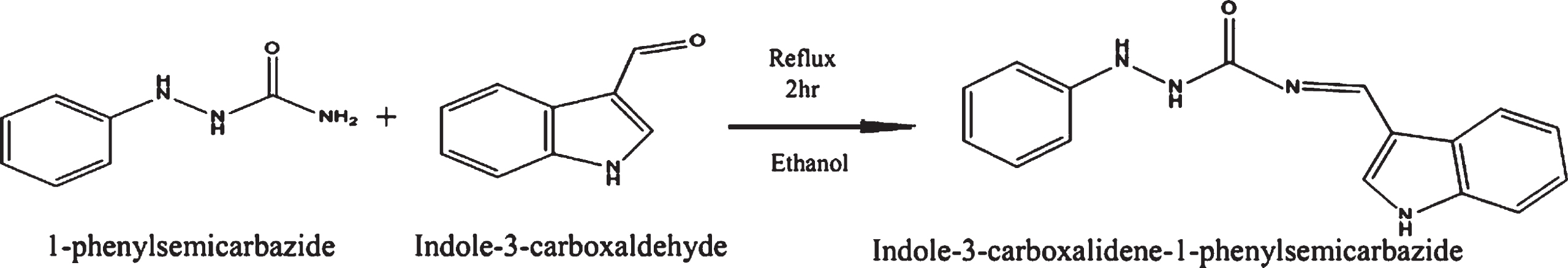
Synthesis of Indole-3-carboxalidene-1-phenylsemicarbazide (SB)
A solution of 1-phenylsemicarbazide (0.05 mole) in 50 ml warm absolute ethanol was added dropwise to a solution of Indole-3-carboxalhyde (0.05 mole) in 50 ml absolute ethanol (Scheme 1). This mixture was allowed to hot reflux under constant stirring for 2-3 h. A solid mass separated out on cooling, which was kept in a refrigerator for better crystallization. It was filtered, washed several times with ethanol, ether, then recrystallized by ethanol and subsequently dried over anhydrous CaCl2 in a desiccator.
Preparation of ligand (SB).
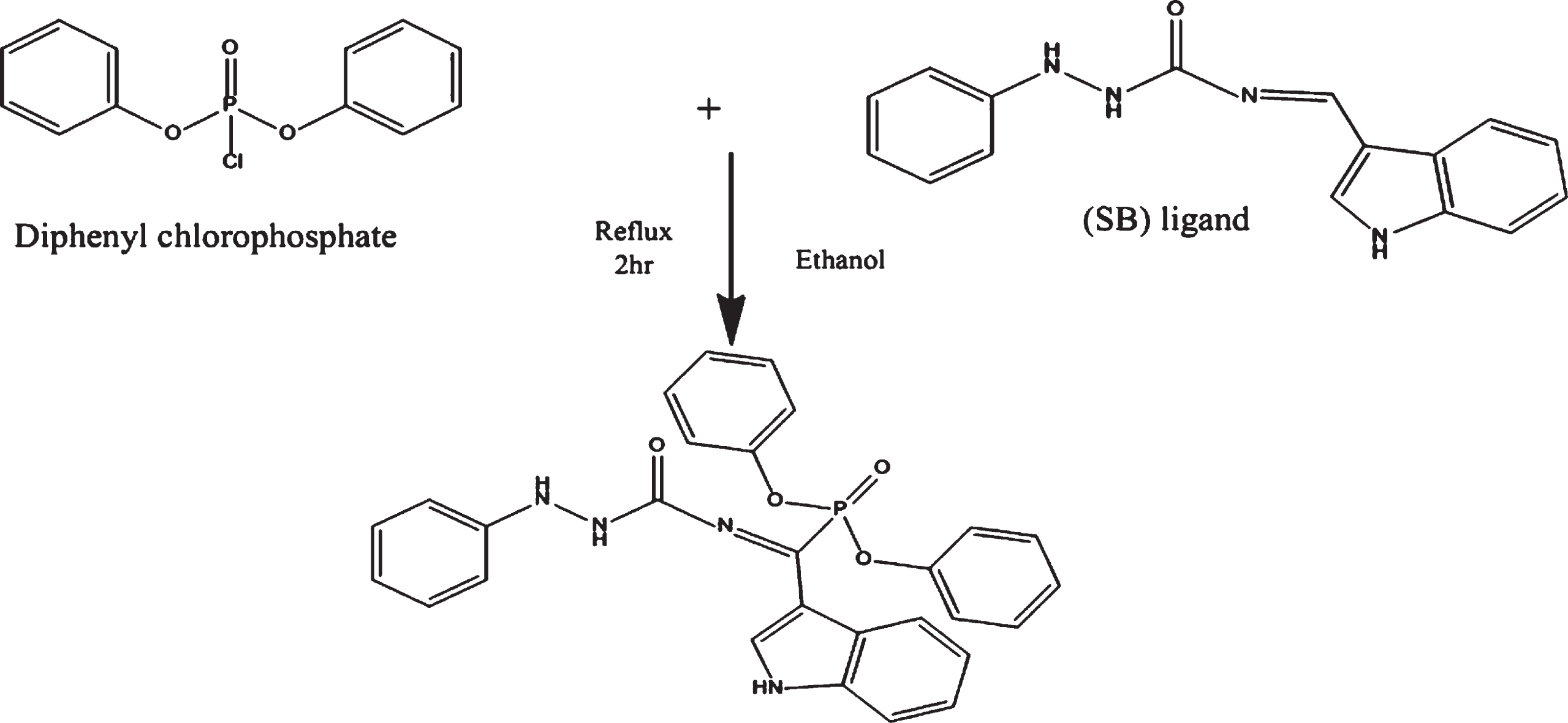
A solution of Diphenyl Chlorophosphate (0.015 mole) in dry benzene (50 ml) was added dropwise to a well stirred solution of Schiff-base (SB) (0.015 mole) and triethyl amine (0.015 mole) in (50 ml) of a dry benzene (Scheme 2). After complete addition, the reaction mixture was heated under reflux for 2 h. The formed solid (triethyl amine hydrochloride) was filtered off and the product was obtained after evaporation on a water bath.
Preparation of ligand (OPSB).
These complexes were prepared by mixing stoichiometric ratios (2L:1M) of a well stirred solution of the ligand in a warm absolute ethanol and metal chloride hydrate in 20 ml absolute ethanol. The mixture was refluxed on a hot plate with stirring for 2–3 h and then left it to cool in room temperature. Fine precipitate which separated were filtered, washed several times with ethanol and dried over anhydrous CaCl2.
Synthesis of Indole-3-carboxalidene diphenylphosphate-1-phenylsemicarbazide complexes
A solution of metal chloride hydrate in 20 ml absolute ethanol was added dropwise to the ligand solution of ligand in a warm absolute ethanol (1L:1M). After the addition, the mixture was heated under hot reflux for three hours. The solution was evaporated, and the solid complexes were collected by using ether.
Instrumentation and measurements
Elemental analyses (C, H and N) were carried out using a Vario EL Fab. CHN Nr, at Central Laboratory, Faculty of Science, Cairo University, Giza, Egypt. Chloride was determined gravimetrically by silver nitrate [19]. The amount of coordinated and uncoordinated water molecules was determined gravimetrically using weight loss method [19]. All melting points reported for the compounds are measured in glass capillary tubes in degrees Celsius. The molar conductance of the metal complexes in DMSO as a solvent were measured on (Jenway conductivity meter model 4510), at Sana’a University. The metals content was measured using Perkin-Elmer 2380 flame atomic absorption spectrophotometer. The infrared spectra of the ligands and their complexes were performed on (FT/IR-140, Jasco, Japan) spectrophotometer with KBr disc (4000–400 cm 1), at Sana’a University. The NMR spectra of ligands were recorded on (varian FT-300 MHz spectrometer) in d6-DMSO solvent using TMS as internal standard, at Cairo University, Giza, Egypt. The UV–Visible spectral studies were recorded on (specord200, Analytilk Jena, Germany) in the rang 200–900 nm, at Sana’a University. The mass susceptibility (Xg) of the solid complexes was measured at room temperature using Gouy’s method by a magnetic susceptibility balance from Johnson Metthey and Sherwood model. XRD patterns were obtained using XD-2 (Shimadzu ED-720) powder X-ray diffractometer at a voltage of 35 kV and a current of mA using CuK(α) radiation in the range of 5° < 2θ < 70° at 1° min–1 scanning rate and a wavelength 1.54056 A °, at Yemen Geological Survey and Mineral Resources Board.
Determination of the stoichiometry of the formed complexes of molecular structure (Molar ratio method)
The molar ratio method was described by Molar ratio method [20]. The concentrations of metal ions (Cu2 +, Ni2 + and Co2 +) were kept constant at (1×10⁻³ M) in methanol and the concentrations of ligands were regularly varied in methanol. The absorbance of the prepared solutions was measured at constant wavelength (λmax). The absorbance values were plotted versus the molar ratio [ligand] / [metal ion]. The intersections of the obtained straight lines indicate the molar ratio of the stable complexes.
Crystallinity and particle size from XRD
The percentage of crystallinity, XC (%) was calculated based on the integrated peak areas of the principal peaks [21]. The crystallinity of the complexes is calculated relative to the crystallinity of the ligands as a ratio:
X-ray diffraction was also used to determine the average particle size (
Antioxidant activity
Free radical liquidation activity of the compounds has been studied using Ferric-bipyridine reducing capacity of total antioxidants (FBRC) [24]. The fixed reaction time and fixed state measurement method were used to find out antioxidant activity of these compounds in methanol as solvent. All spectrophotometric measurements were measured using UV-Vis Spectrophotometer (Specord 200, Analytikjena, Germany) at Sana’a University.
Molecular docking studies
Compound preparation
Among the new synthetic complexes, found that [Co(OPSB)Cl(H2O)2]·Cl complex showed potent cytotoxic activity against microbial strains. First, the 3D structures of both complexes were built and minimized using the MMFF94x force-field with reaction-field electrostatics (Din = 1, Dout = 80). All atoms were tethered with a flat bottom tether (10.0 kcal/ mol, 0.25 A°). MOE software was used for all refinements [25]. Second, in order to utilize as the input for MOE-docking simulation, the database was produced by translating the structures of both complexes into format*.mdb*.
Proteins preparation
As the antibacterial targets, four different X-ray crystal structures were downloaded from the Protein Data Bank (http://www.rcsb.org/pdb/). The protein (PDB ID: 2RHL) Bacillus subtilis [26], Staphylococcus aureus (PDB ID: 4URM) [27], Escherichia coli (PDB ID: 4PRV) [28], Pseudomonas aeruginosa (PDB ID: 4JVI) [29]. To make the simulation easier, deleted the water molecules, ions, and cofactors from the structures, but kept the A chain of each target. The hydrogen atoms were then added once the targeted structures were adjusted for missing atoms [30, 31].
Molecular docking protocol
The interaction of drugs with the active site residues of the target was determined using computational molecular docking. The Molecular Operating Environment (MOE) software [25] was utilized to conduct the docking experiments in our research. Knowing that we used the identical simulation procedures as in earlier studies [32–34]. The following parameters were used for docking: The placement method was Triangle Matcher, and the Rescoring 1 function was London dG.
Antibacterial and antifungal screening
Evaluation of the antibacterial and antifungal activates of the prepared ligands and their metal complexes were carried out in the Lab. of microbiology at Sana’a University. They were Germinated four types of Bacteria (Staphylococcus aureus, Bacillus subtilis, Escherichia. Coli and Pseudomonas aeruginosa) and one fungus (Candida albicans) on Nutrient agar and sabouraud agar solid media respectively. Using filter paper disc method [35], it was Prepared one concentration from each compound (1000μg/ml) in DMSO. Then added (100μl) from each Prepared solution on filter paper disk (Whatman No.l filter paper, 5 mm diameter) which contained the bacteria with agar solid media, all the Petri-dishes were putted in an incubation period at 37 °C for 24 hour. Gentamicin 120μg/ml was used as a reference substance for bacteria and Mycostatin 30μg/ml for fungi. The results was registered by calculated the diameter of the inhibition zone (mm).
Results and discussion
The Schiff base ligand was prepared by the condensation of 1-phenylsemicarbazide with Indole-3-carboxaldehyde (SB) in the molar ratio 1:1. The Organophosphorus Schiff base ligand was prepared by the condensation of diphenyl chlorophosphate with the prepared Schiff base (SB) in the molar ratio 1:1 in the presence of trimethylamine and dry benzene to form (OPSB). The ligand SB was reacted with Cu(II), Ni(II) and Co(II) in the molar ratio 2:1 to form its complexes, the ligand OPSB was reacted with Cu(II), Ni(II) and Co(II) in the molar ratio 1:1 to form its complexes. The molar conductance of SB complexes (1×10⁻³ M, DMF) are in the range 6.77–8.35 Ω–1 cm2 mol–1 indicating that, these complexes are non-electrolytic [36, 37]. These confirmed that, the anions of these complexes were chelated to metal ion, so no precipitation was first observed upon addition of silver nitrate to the solution of the complexes but after digestion with nitric acid, the silver nitrate test gave a positive result. The molar conductivity of OPSB complexes are in the range 78.99–91.53 Ω–1 cm2 mol–1 referring the electrolytic behavior of these complexes 1:1 [36, 38], so the precipitation was observed upon addition of silver nitrate to the solution of the complexes [19]. The ligands aren’t soluble in water but soluble in common organic solvents. All the synthesized complexes are highly colored. The complexes of SB insoluble in water and common organic solvents but soluble in polar solvents such as DMF and DMSO. The complexes of OPSB slightly soluble in water and soluble in common organic solvents.
Physical, elemental, spectral and XRD data were listed in Tables 1–6 which agree with proposed structures (Fig. 1 and 2).
Some physical properties and Elemental Analysis
Some physical properties and Elemental Analysis
The main IR absorption bands of the ligands and their complexes
Electronic Spectral and Magnetic moment data of the ligands and their complexes
Molar ratios for determination of stoichiometry of the SB complexes in methanol
Molar ratios for determination of stoichiometry of the OPSB complexes in methanol
XRD spectra data of the principal values of intensity of the ligands and SB complexes

Proposed structure of SB complexes.
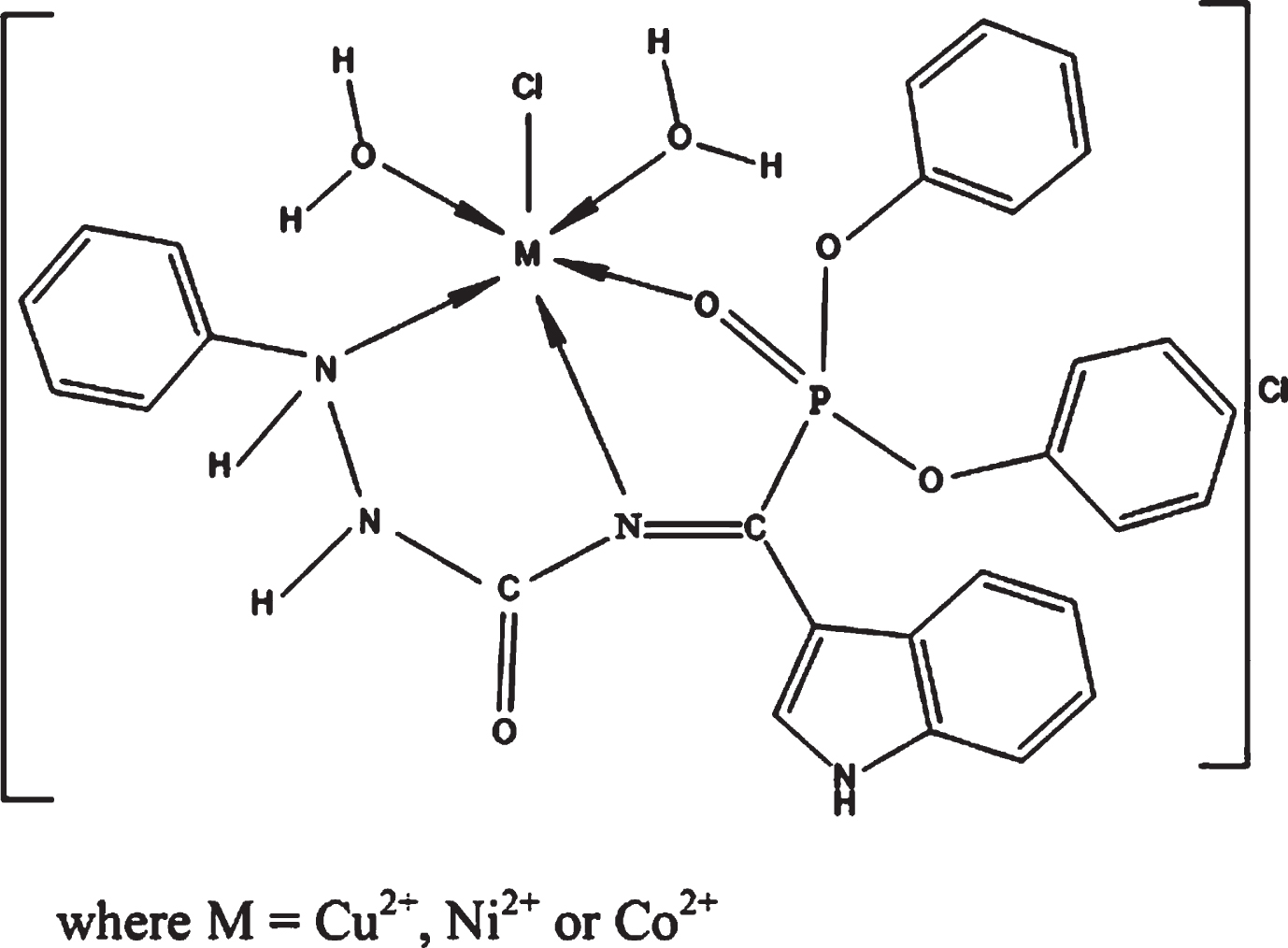
Proposed structures of OPSB complexes.
1HNMR spectra of the ligands
In the 1HNMR spectra of the ligands SB (Fig. 3) and OPSB (Fig. 4), the signals of the NH (indol ring) appeared at δ ∼ 12.144 and 12.400 ppm, while the signal of the HC = N proton appeared at 9.967 ppm in SB [39] and this signal disappeared in OPSB spectrum due to replacement reaction on this group with eliminated HCl and forming P-C bond. The peaks at 6.020, 6.700 and 6.677, 6.706 ppm are assignable to the protons of the NH groups in the ligands SL and PSL, respectively [40]. The peaks appearing in the range 6.724–8.268 and 6.735–8.276 ppm may correspond to protons of the aromatic and indol ring of SB and OPSB respectively [38, 39].

1HNMR spectrum of SB.
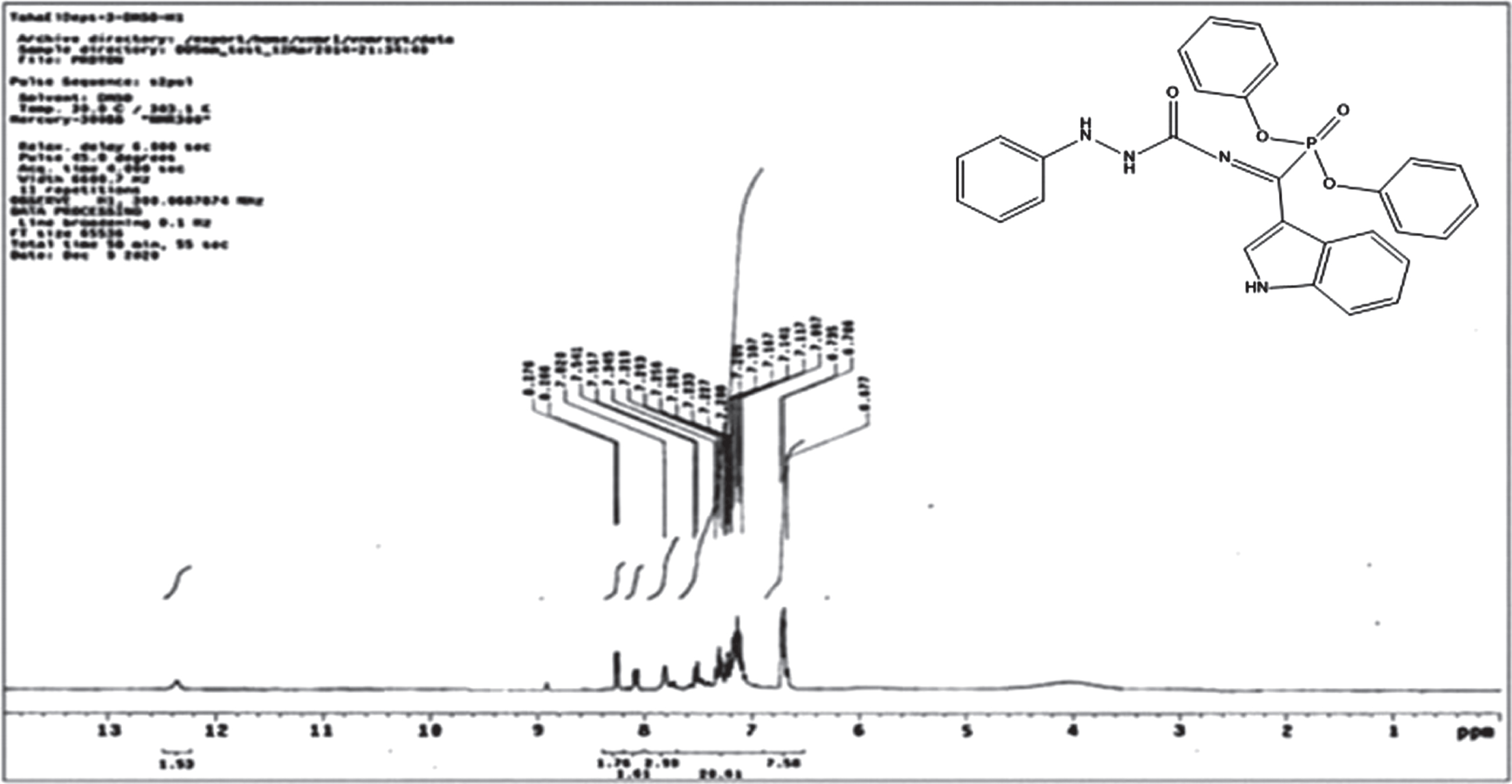
1HNMR spectrum of OPSB.
In the 13 C NMR spectra of the ligands SB (Fig. 5) and OPSB (Fig. 6), the carbon resonance signals of the C = N group appeared at δ= 185.023 and 185.045 ppm, respectively [39, 41]. The signals observed at δ= 160.474 and 160.353 ppm are characteristic for the CO carbonyl group present in the ligands SB and OPSB, respectively [42]. The carbons of the indole and aromatic rings were observed at δ= 112.250–138.437 and 112.182–138.506 ppm in SB and OPSB respectively [43].
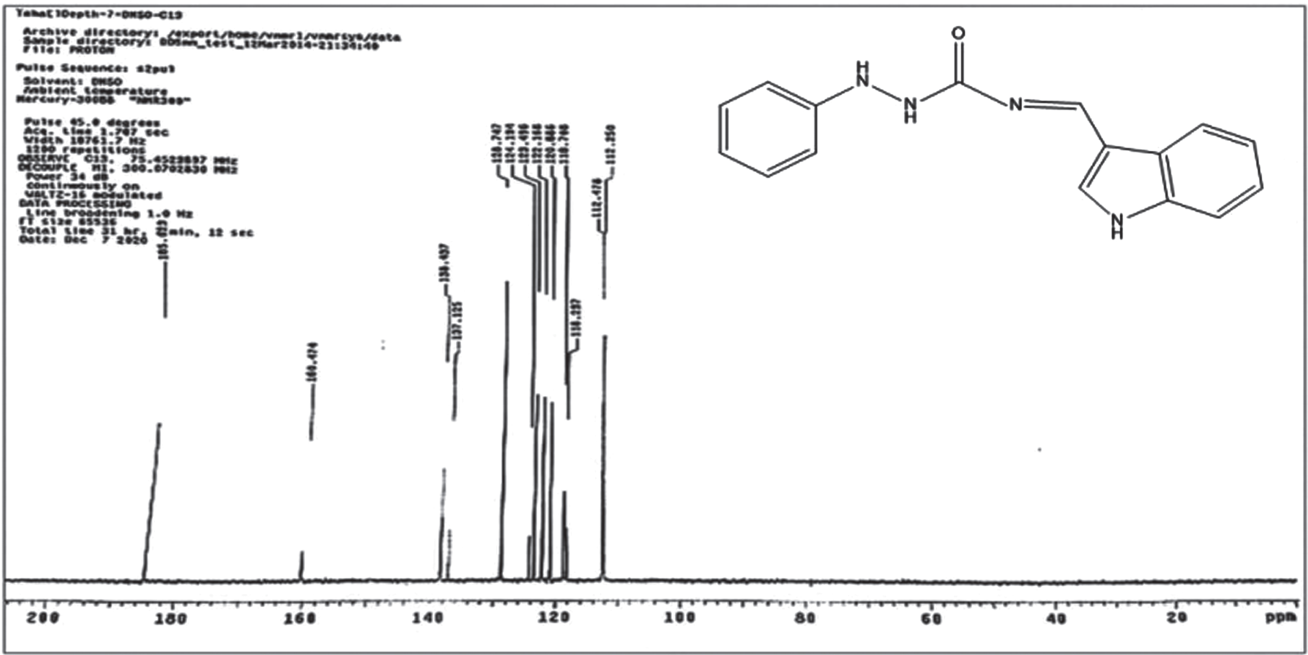
13CNMR spectrum of SB.
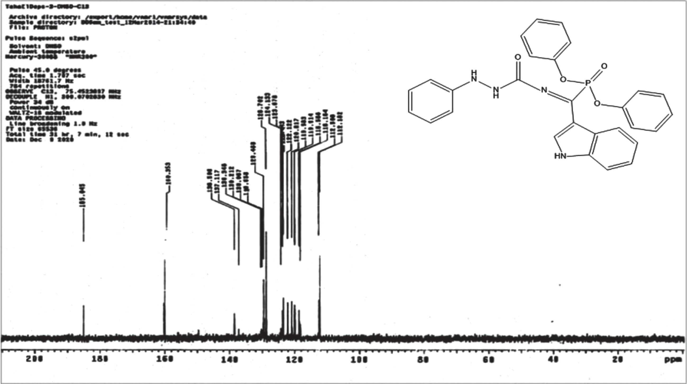
13CNMR spectrum of OPSB.
The comparison of IR spectra of ligands and their metal complexes have been studied in Table 2. In the ligand SB, the bands in IR spectrum at 1685cm–1 and 1605cm–1 correspond to formation of ν(C = O) and ν(C = N), respectively [44]. The strong bands at 3335 and 3399 cm–1 attributed to ν(NH,NH) of this ligand [45]. The bands were showed at 3043, 2978 and 1250 cm–1 are assigned to the aromatic ν(C-H), aliphatic ν(C-H) [46] and ν(-C-N) stretching mode of vibrations, respectively [47]. The band at 3201 cm–1 in its spectrum able to be appointed to ν(NH) of indole ring [48, 49].
The FT-IR spectra of complexes (Fig. 7) described the important characteristics of absorption bands, the broad bands at 3421, 3385 and 3398 cm–1 due to the water molecules in Cu, Ni and Co complexes, respectively [50]. The shift of azomethine vibrations to the lower frequency indicating the coordination of nitrogen with metal ions.The bands of ν(NH,NH) disappeared in complexes due the broad bands of water. New vibrations at 400–600 cm–1 which are not present in the free are attributed to the existence of ν(M-N) and ν(M-NH) [41]. These data suggested that the SB ligand coordinated with metal ions through two sites and acted as bidentate ligand.
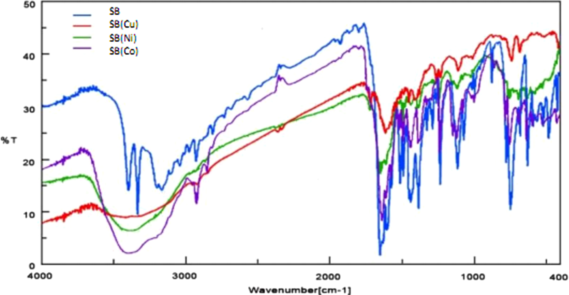
Infrared Spectra of SB ligand and its complexes.
Although presence evidence of ν(M-Cl) could not be brought in the IR data due to instrumental limitation, the insolubility of SB complexes in water and their non-electrolytic nature evinced that 2Cl– coordinated with metal in SB complexes.
In the FT-IR spectrum (Fig. 8) of the prepared ligand OPSB, it can be seen a significant bands at 1653 cm–1and 1627 cm–1 attributed to ν(C = O) and ν(C = N), respectively [14], another two bands at 1171 cm–1 and 1083 cm–1 attributed to ν(P = O) and ν(P-O-C), respectively [47].

Infrared Spectra of OPSB ligand and its complexes.
The bands in the range (3300–3432) cm–1 confirmed the coordination of H2O molecules in the complexes [37]. The IR spectra of the OPSB metal complexes proposed that OPSB ligand is a tridentate with the phosphoryl-oxygen, amine - nitrogen, and azomethine-nitrogen due the shift in the position of ν(P = O), ν(-NH) and ν(C = N) (Table 2).
The UV-Visible spectra of the ligands and their metal complexes were measured in DMSO in the range 200 –900 nm shown in Figs. 9 and 10 and reported in Table 3.

UV-Vis spectra of SB ligand and its complexes.
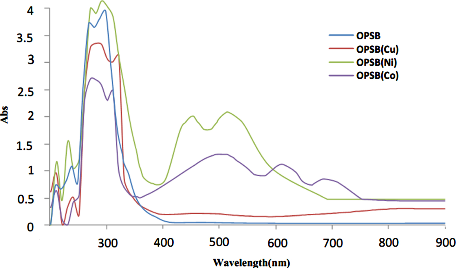
UV-Vis spectra of OPSB ligand and its complexes.
The spectra of the free ligands exhibited two absorption bands in the region 37037.03 –32258.06 cm–1. These bands are attributable to π-π* and n-π* transitions, respectively [51]. The n-π* transition is shifted to a longer wavelength as consequence of coordination when binding with the metal to formation complexes.
New broad bands at 11494.25 and 12195.12 cm–1 in the SB(Cu) and OPSB(Cu), respectively, attributable to the 2Eg⟶2T2 g transition. The broadness of the band indicating distorted octahedral complexes [52]. The magnetic moments were 1.71 and 1.75 B.M., respectively, corresponding to one unpaired electron in an octahedral geometry [53].
The appearance of two bands in the spectra of the Ni (II) d8 complex with SB and OPSB at (19607.84, 16393.44) and (22222.22, 19230.76) cm–1, respectively, were assigned to the 3A2 g ⟶ 3T1 g(P) and 3A2 g ⟶ 3T1 g(F) transitions, respectively [52], these complexes showed (μeff) values 2.88 and 2.65 which are compatible with two unpaired electrons system of octahedral geometry [54].
The Co2 + complexes of SB and OPSB showed bands at (21276.59, 18518.51, 15151.51) and (19230.76, 16393.44, 14705.88), respectively, which imputed to 4T1g⟶ 4T1 g(P), 4T1g⟶ 4A2 g and 4T1g⟶4T2 g(F) transitions [53]. The complexes show the values 4.16 and 3.98 B.M., that is corresponding to octahedral structure [55].
Investigation of the molecular structure of the complexes formed between the metal ions of (Cu2 +, Ni2 + and Co2 +) with ligands SB and OPSB in methanol using molar ratio revealed the formation of (1:2) (M:L) complexes for SB and (1:1) (M:L) complexes for OPSB under investigation. The results are depicted in Tables 4 and 5. The Molar ratio plot for the complexes between metal ions of Cu, Ni and Co with SB and with OPSB in methanol are shown in Fig. 11.

Molar ratio plot for the complexes between metal ions of (a)Cu, (b)Ni and (c)Co with SB and of (d)Cu, (e)Ni and (f)Co with OPSB in methanol.
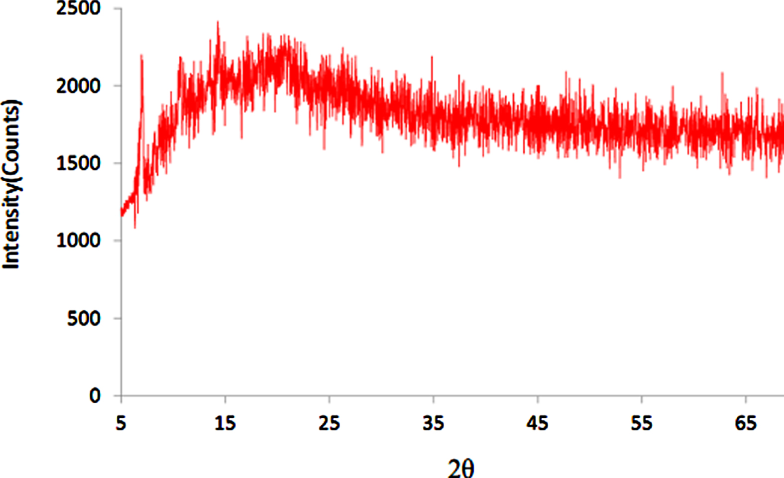
We can study crystalline solid by using the technique of X-ray powder diffraction that exhibit strong peaks, whereas those amorphous ones show diffuse and halo diffraction patterns [21]. The diffraction patterns of SB, OPSB and SB complexes exhibited multiple peaks Figs. (12–16). The XRD patterns for ligands and SB complexes are shown in Table 6. From these results, the noticeable changes in crystallinity, increasing of the XRD principal peaks intensity of SB(Cu) complex compared to SB ligand that can be attributed to the increase in crystallinity. In addition, the high value of crystallinity XC (%) was observed when the crystallinity of SB(Cu) complex (216.762 %) is compared to SB ligand. However, low values in crystallinities are observed in case SB(Ni) and SB(Co) complexes (13.121%and 9.395%), respectively, can be attributed to the variation in the crystal structure of the ligand. The crystallinity calculations are based on the ratio of the principal peaks area of the complex sample to that of the ligand sample obtaining a relative crystallinity [21]. In literature the range between 1–100 nm reported to nanoparticle size [56], so the particle size of the complexes obtained from XRD shows no effects on their nanoparticle size (4.581 –5.344 nm) due to complexation.
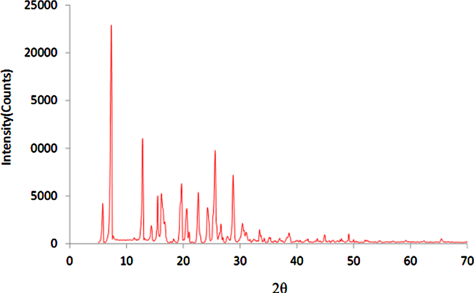
XRD pattern of SB.
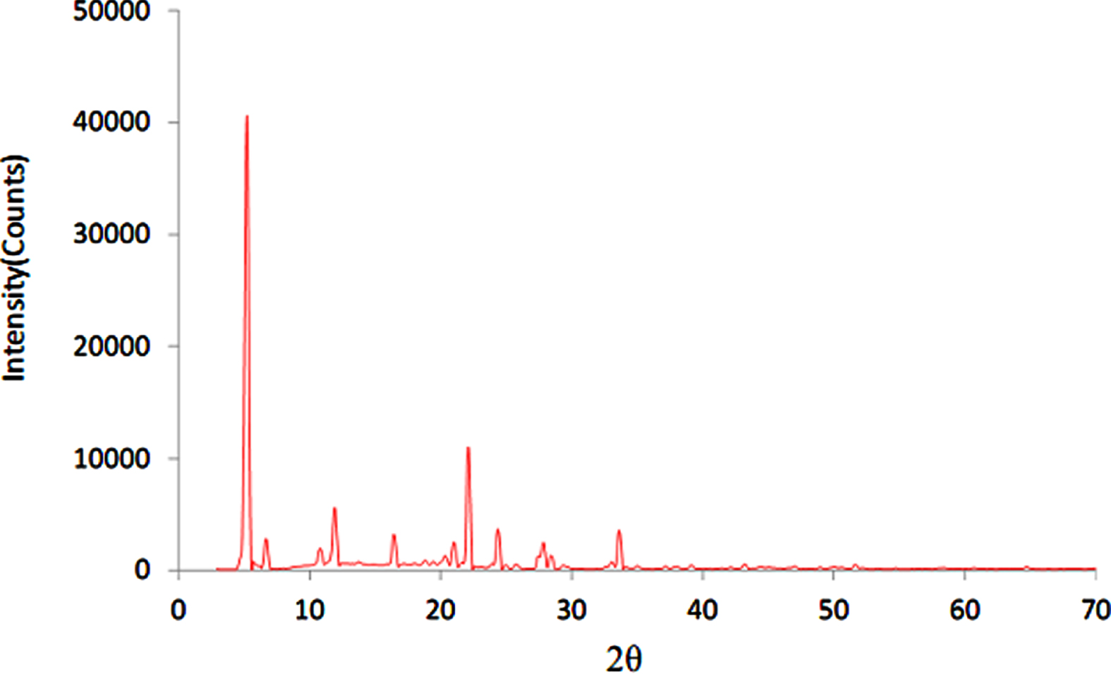
XRD pattern of SB(Cu).

XRD pattern of SB(Ni).
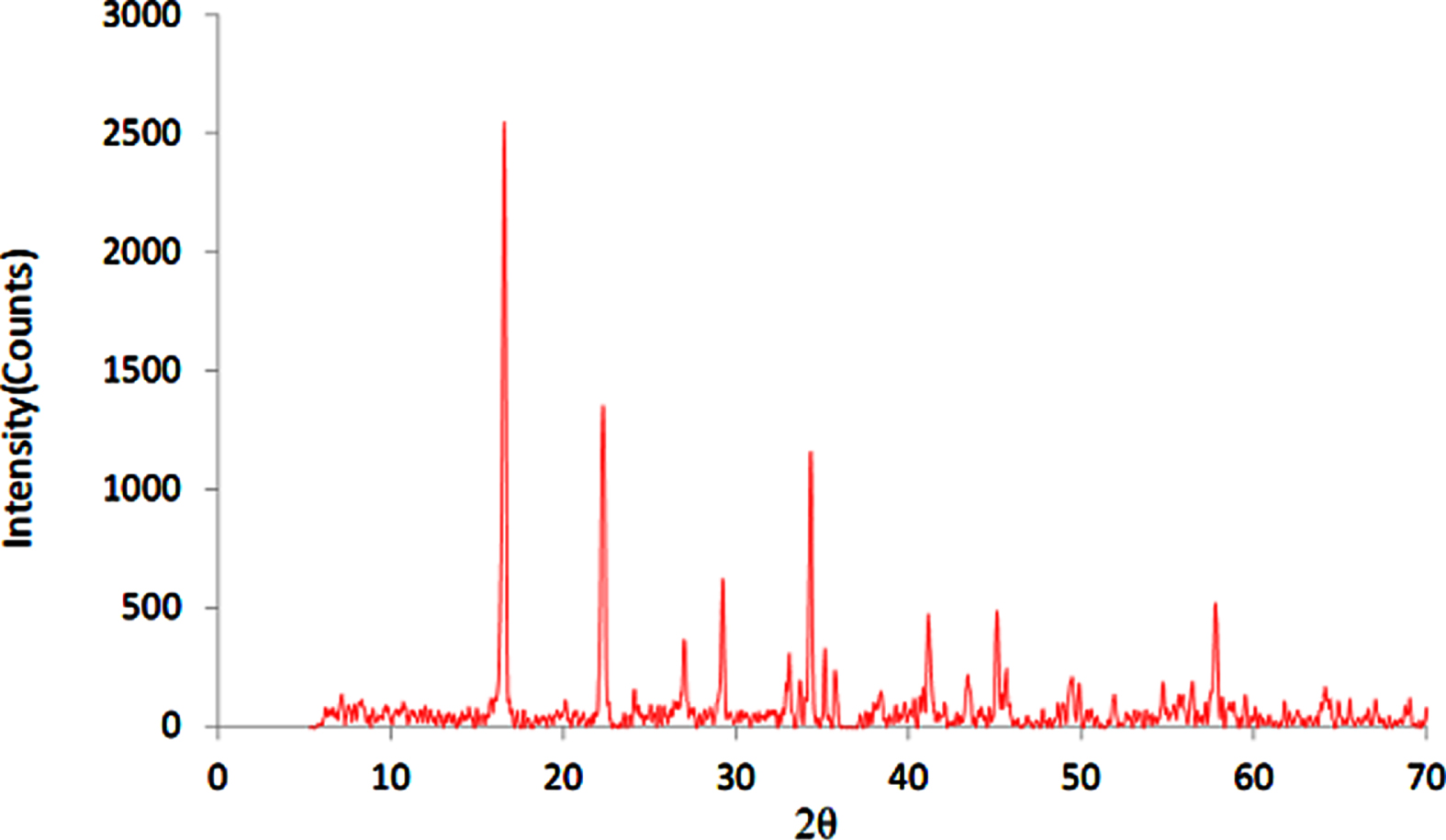
XRD pattern of SB(Co).

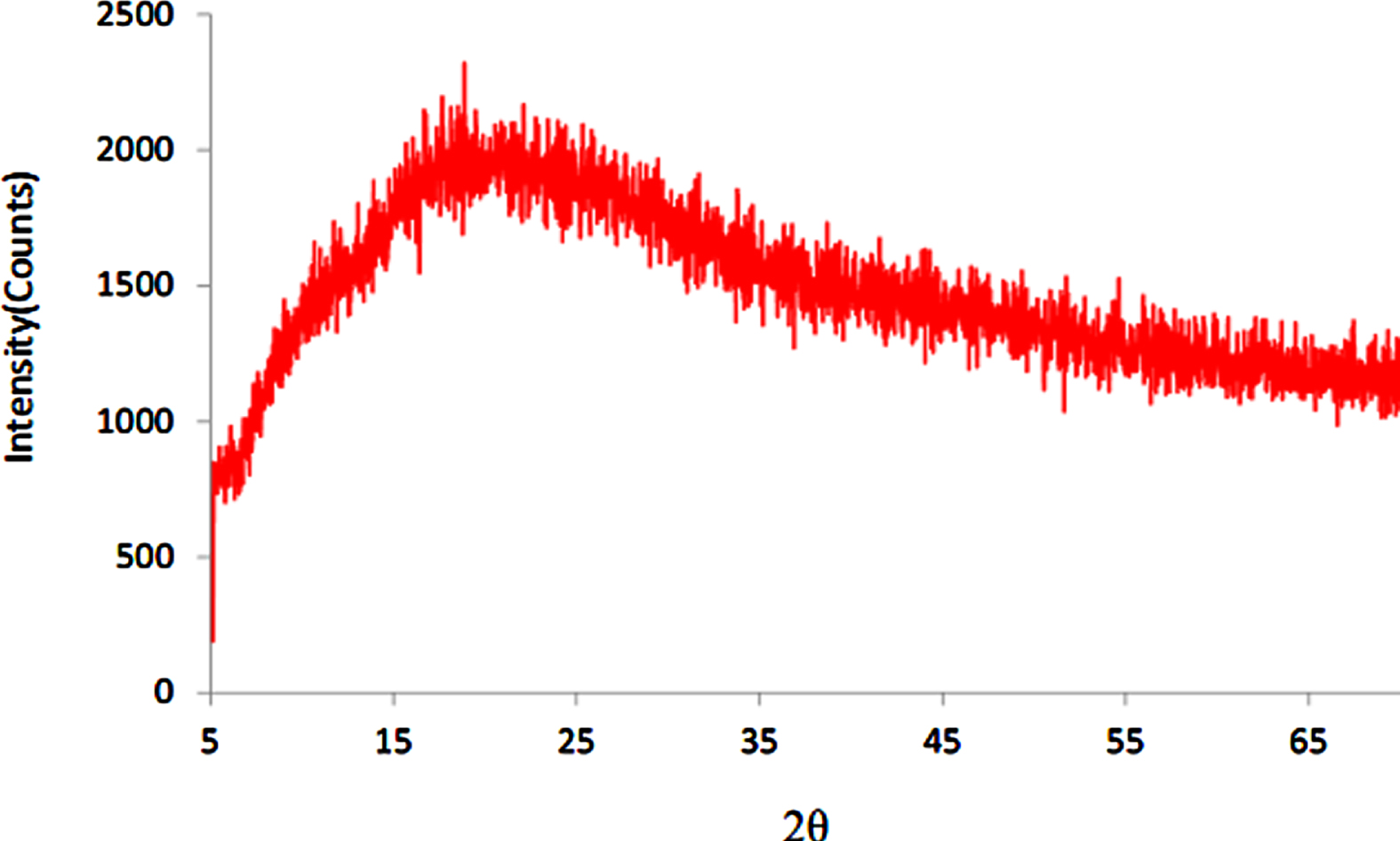
XRD pattern of OPSB.
On the other hand, the particle size of OPSB ligand located within nano range (4.581 nm) as summarized in Table 6, whereas its complexes appeared as amorphous character (Figs. 17–19). From this change, it is expected to improved properties of OPSB complexes as compared with OPSB ligand.
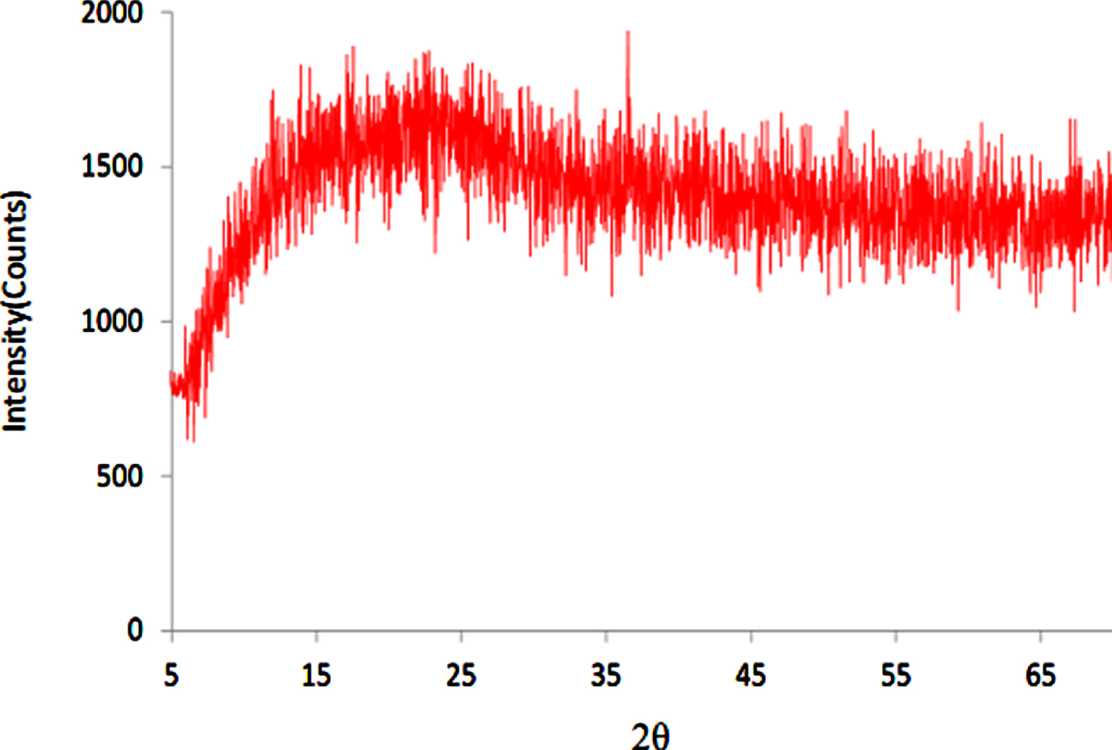
XRD pattern of OPSB(Cu).
XRD pattern of OPSB(Ni).
XRD pattern of OPSB(Co).
The antioxidant activity of all compounds was investigated using (FBRC) technique as published in scheme 3 [24]. 1 ml of 10–2 M FeCl3.6H2O was taken to 10 ml volumetric flasks with different volumes (0.01 ml, 0.02 ml, 0.04 ml, 0.06 ml, 0.08 ml, 0.1 ml, 0.2 ml) of the standard antioxidant ascorbic acid (0.1 g/L). Then 2.0 ml of 0.3M acetate buffer (pH 4) and 1.0 ml of bipyridine (6.4×10⁻³ M) were added. The volume was completed to 10 ml with deionized water. After 10 min of incubation at room temperature the absorbance was recorded against a blank solution at λ= 535 nm. The absorbance values were plotted against the concentrations of the various ascorbic acid solutions (Fig. 20).
Using the same additions,instead of adding ascorbic acid we used 0.02 ml of each tested compound (0.1 mg /ml methanol).

Calibration curve of ascorbic acid with Fe-Bp complex.
Fe(II)-bipyridine complex produced by the reaction of Fe(III) with antioxidant followed by bipyridine exhibited maximum absorption at 535 nm (Scheme 3). Total antioxidant activity is based on the redox reaction between compounds and Fe(III) at room temperature. The initial antioxidant concentration is indicated by the concentration of the oxidizing Fe(III).
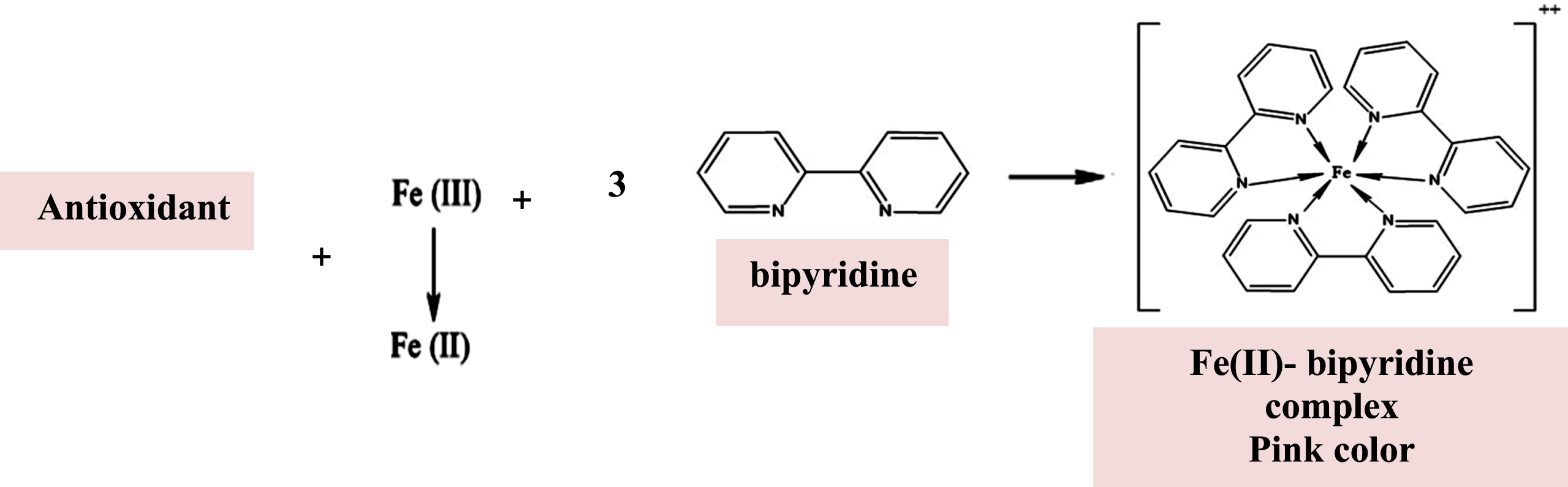
Stoichiometry outline of FBRC method.
These compounds were found to possess potent antioxidant activity better than standard antioxidant Ascorbic acid (vitamin C) due presence a combination of donor sites such amide oxygen and an imine nitrogen [57].
To begin validating the docking method, we re-docked each native ligand into its target and calculated the root-mean-square deviation (RMSD) and binding free energy (S score) for each complex (Table 8). This allows us to compare the affinity of compound and targets, as well as determine the strength of their binds.
FBRC values express as (mg/L) for prepared compounds
FBRC values express as (mg/L) for prepared compounds
Properties of targets, energy score and RMSD values
The [Co(OPSB)Cl(H2O)2]·Cl complex was then docked into the active site residues for the microbial strains’ targets. Finally, for each complex, eight top conformations were obtained, and the optimal pose was chosen based on its energy score. Tables 9 describe the docking findings computation, including energy scores, kind of interactions, and distances between complex and four targets: Bacillus subtilis (PDB ID: 2RHL), Staphylococcus aureus (PDB ID: 4URM), Escherichia coli (PDB ID: 4PRV), Pseudomonas aeruginosa (PDB ID: 4JVI).
Docking score and interactions between [Co(OPSB)Cl(H2O)2]·Cl complex and the active site residues of enzymes target of microbial strains
The interaction created between the complex and the binding site of target proteins was also shown using the Discovery Studio Visualizer v17.2.2.0 software (DSV).
According to the literature [58–60] hydrogen bond distances between 2.5 and 3.1 Å are considered strong interactions, whereas those between 3.1 to 3.55Å are considered weak interactions. The complex demonstrated good inhibition of the target proteins, with the development of hydrogen bonds and electrostatic interactions according to molecular docking computational results (see Figs. 21–24).

2D plot of interaction of [Co(OPSB)Cl(H2O)2]·Cl complex and the active site residues of (2RHL) enzyme of Bacillus subtilis.
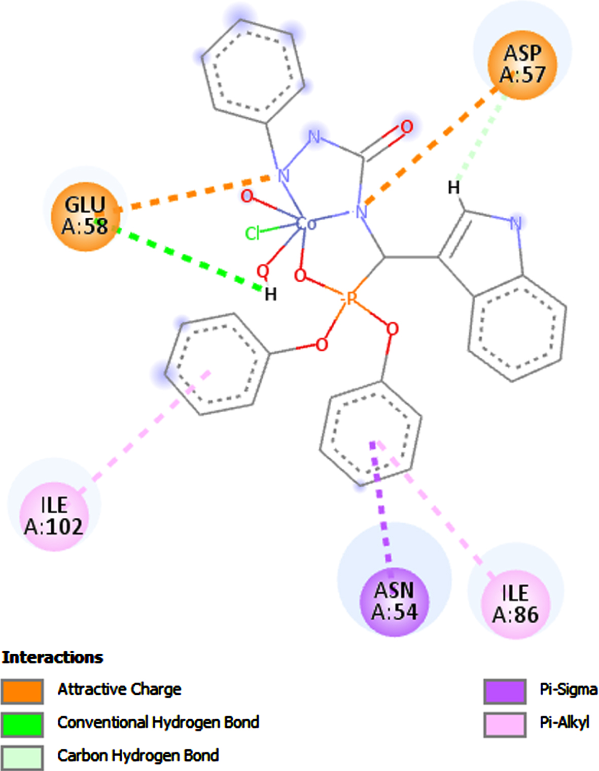
2D plot of interaction of [Co(OPSB)Cl(H2O)2]·Cl complex and the active site residues of (4URM) enzyme of Staphylococcus aureus.
2D plot of interaction of [Co(OPSB)Cl(H2O)2]·Cl complex and the active site residues of (4PRV) enzyme of Escherichia coli.
2D plot of interaction of [Co(OPSB)Cl(H2O)2]·Cl complex and the active site residues of (4JVI) enzyme of Pseudomonas aeruginosa.
It is apparent from the Table 9 that the [Co(OPSB)Cl(H2O)2]·Cl complex has the high affinity with targets of four microbial strains: Bacillus subtilis (PDB ID: 2RHL), Staphylococcus aureus (PDB ID: 4URM), Escherichia coli (PDB ID: 4PRV), Pseudomonas aeruginosa (PDB ID: 4JVI), this is through the formation of stable complexes with energy score: –5.0887, –5.4189, –1.4734 and –6.2250 Kcal/mol, respectively.
We note that in the case of Bacillus subtilis target (PDB: 2RHL), complex was deeply buried in the target binding site and formed six electrostatic interactions (Table 9 + Fig. 21).
Also, [Co(OPSB)Cl(H2O)2]·Cl complex was involved in the formation of two strong hydrogen bonds with the Staphylococcus aureus target pocket (PDB: 4URM): H/GLU58-OE1/bond distance = 2.32 Å and H/ASP57-OD2/bond distance = 2.62 Å (Table 9 + Fig. 22).
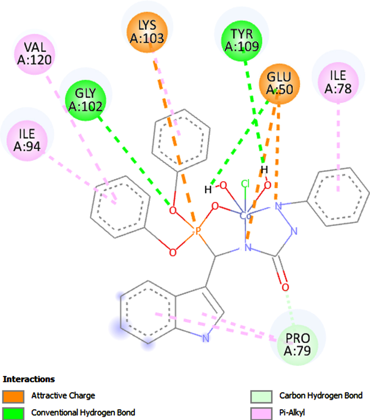
Additionally, this complex and the major residue of the Escherichia coli target (PDB: 4PRV) formed five powerful hydrogen bonds: O/GLY102-H/bond distance = 2.32 Å, H/TYR109-OH/bond distance = 2.44 Å, H/GLU50-OE1/bond distance = 2.72 Å, O/PRO79-HD2/bond distance = 2.62 Å and O/PRO79-HD3/bond distance = 2.45 Å (Table 9 + Fig. 23).
Cl complex form four strong hydrogen bonds with active site residue of Pseudomonas aeruginosa target (PDB: 4JVI): H/ ARG209-O/bond distance = 2.02 Å, H/TYR258-OH/bond distance = 1.87 Å, O/LEU207-HA/bond distance = 2.57 Å and H/LEU207-O/ bond distance = 2.90 Å(Table 9 + Fig. 24).
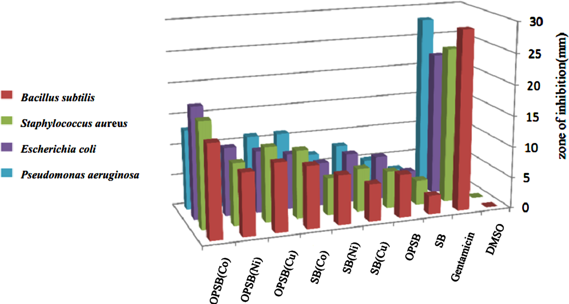
Evaluation of the antibacterial and antifungal activates of the ligands and their metal complexes were screened against four types of Bacteria (Staphylococcous aureus, Bacillus subtilis, Escherichia.Coli and Pseudomonas aeruginosa) and one fungus (Candida albicans) using well diffusion method [25]. The results obtained were presented in Table 10 and Fig. 25. In general, the complexes showed good antibacterial compared to the free ligands. Based on the chelation theory [61], the increased inhibition activity of the metal complexes may be explained on the basis that their structures mainly possess C = N bonds. Besides, the coordination decreases the polarity of the metal ion due to the partial sharing of its positive charge with donor groups and possible π- electron delocalization inside the chelate ring shaped during coordination that makes the complexes more lipophillic. This increased lipophillicity enhances the penetration of the metal complexes into lipid membranes, blocks the metal binding sites in the enzymes and limits further development of the organisms. However, the activity for compounds were found to be lower than the control drug, Gentamicin 120μg/ml. The results showed that No inhibition zone was observed for ligands and their metal complexes against the fungus (Candida albicans).
Biological activities of the ligands and their metal complexes against bacteria and fungus (zone of inhibition in mm)
Biological activities of the ligands and their metal complexes against bacteria and fungus (zone of inhibition in mm)
Inhibition zone dimeter of ligands and their complexes toward of some kind of bacteria.
The order of antibacterial activity for the compounds with Bacillus subtilis was Co(OPSB) > Cu(OPSB) > Co(SB) = Ni(OPSB) > Ni(SB) > OPSB > Cu(SB) > SB, with Staphylococcous aureus was Co(OPSB) > Cu(OPSB) > Co(SB) > Ni(OPSB) > Cu(SB) > OPSB = Ni(SB) > SB, with Escherichia.Coli was Co(OPSB) > Ni(OPSB) > Cu(OPSB) > Co(SB) > Cu(SB) > OPSB = Ni(SB)> SB and with Pseudomonas aeruginosa was Co(OPSB) > Co(SB) > Cu(OPSB) > Ni(OPSB) > Cu(SB)> Ni(SB) > OPSB > SB.
In this work, the new ligands (Indole-3-carboxalidene-1-phenylsemicarbazide