
Abstract
Thymus species are well known as medicinal plants because of their biological and pharmacological properties. Thymus migricus Klokov & Desj. -Shos belongs to Lamiaceae. Young branches of this plant produce an essential oil that is abundant in potent and volatile compounds that have a variety of therapeutic uses. In this study, the natural composition of thymol in this plant’s essential oil was first detected with HPLC and Mass, and then the essential oil was extracted. The increase in the lipophilicity of the synthesized silylated derivatives was then examined by HPLC after numerous silylated derivatives of this phenolic compound were synthesized using various silylation reagents. These derivatives were then analyzed by FT-IR and 1HNMR analysis. The antibacterial activity of thymol with its silylated derivatives against gram-negative and gram-positive bacteria was next tested by diffusion method, after which the antioxidant activity of thymol with its silylated derivatives was assessed by two DPPH and FRAP methods.
Introduction
Essential oils (EO) are secondary metabolites produced by aromatic plants and are volatile, natural, complex molecules with a strong odor. They are used in embalming, food preservation, and as antibacterial, analgesic, sedative, anti-inflammatory, spasmolytic, and locally anesthetic creams. They are well known for their antiseptic, i.e., bactericidal, virucidal, and fungicidal, and therapeutic characteristics as well as their smell. These traits haven’t altered much up to this point, with the exception that we now know more about some of their modes of action, especially at the antimicrobial level [1–5].
Numerous epidemiological and experimental investigations have revealed the therapeutic potential of medicinal plants and their active ingredients for a number of human diseases [6–8]. Many countries and societies’ old cultures and traditions have cited the therapeutic value of plants, and they are thought to be both safe and affordable. Plants and plant products have been utilized in traditional medicine for numerous disorders since ancient times, either as a treatment or as a culinary ingredient [9–11]. Numerous foods have been used extensively for culinary purposes in diets and are thought to be beneficial against a number of diseases, including olives, dates, oregano, thyme, turmeric, and oregano, to name a few. Thyme was a spice that was used by the Greeks, Romans, and Egyptians as a food flavoring, preservative, and odorant. It is a small subshrub that is widely used in western Mediterranean traditional medicine, and its leaves are frequently used as herbal medicines and food additives. Thyme possesses potent antibacterial, antifungal, sedative, antiseptic, antioxidative, expectorant, antispasmodic, antifungal, antivirotic, antihelminthic, carminative and diaphoretic effects [12–15]. Thyme contains abundant amount of terpenoids, flavonoids, glycosides and phenolic acids [16–18]. The EO of thyme is composed of thymol as dominant components, followed by γ-terpinene, pcymene, linalool, terpinen-4-ol and sabinene hydrate [19–21]. Studies on thyme plants in Greece showed that Thymus vulgaris, contained a high amount of EO. The content of EO as high as 8% with thymol as dominant component (95%) was reported for this subspecies [13, 23].
Even though thymol has been the subject of intensive research in recent years, little is known about how it works to kill germs. Thymol’s hydrophobicity, the existence of a free hydroxyl group, and a delocalized electron system have all been linked to its antibacterial properties. Thymol has a significant impact on the cytoplasmic membrane’s structural and functional characteristics, causing the membrane to lose its integrity and become more permeable to protons and ions. It has also been demonstrated that thymol causes bacteria to produce Hsp 60 and inhibit ATPase [13, 24–26]. Natural antioxidants are thought to be effective tools for disease prevention [27–29]. Numerous studies have demonstrated that the ability of phenolic compounds in plant essential oils to scavenge free radicals results in their antioxidant activity [30–32]. Their chemical makeup contained hydroxyl groups, which had an antioxidant effect. Additionally, numerous studies on the antioxidant properties of EOs from different aromatic plants found that the thyme essential oil, which is high in thymol, significantly slows down the process of lardoxidation [33–35].
After 2001, the use of silicon in biotechnology has drawn a lot of interest, and numerous studies on the creation of biocatalytic products from silicon polymers, enzyme release systems, powerful biosynthetic systems, and enzymes have been conducted [36, 37]. Functional groups containing Si covalently linked to an alkoxy group are known as silyl ethers. Silyl ethers work as protective groups in organic synthesis since they are inert to most chemicals that react with alcohols. In chemical synthesis, silyl ethers are typically utilised as protective groups for alcohols. This class of chemical compounds offers a broad spectrum of selectivity for protecting group chemistry because R1R2R3 in R1R2R3-Si-R4 can be combinations of various groups that can be altered to generate a number of silyl ethers. Silyl ethers are the most prevalent protective group for alcohols. Due to the strength of the Si-F bonds, they can be readily formed by treating alcohols with R3SiCl in the presence of a base and then removed with ease using a source of fluoride ion (F-) [38–43]. Silyl ethers, having the general structure OSiR3, have become particularly significant for the protection of alcohols. Variations in the R group lead to major changes in the stability of the protective group. In general, silyl ethers can be cleaved with aqueous base or acid, however the rate of hydrolysis for a secondary silyl ether is substantially slower than that of a primary silyl ether [36]. Alkyl alcohols are also more reactive than phenolic derivatives, and additional silane-protected functional groups are reactive in the following order: COOSiR3 > NHSiR3 > CONHSiR3 > SSiR3 [36, 37]. The relative stability of the OSiR3 group in cleavage reactions is significantly influenced by steric factors. The trimethylsilyl ether [O-Si(Me)3, OTMS] is the model for this class of protective groups [38]. Typically, the group is joined to the alcohol through a reaction with chlorotrimethylsilane (Me3SiCl) and an amine base (such as triethylamine or pyridine). The TMS group is occasionally cleaved during an aqueous workup procedure because this group is sensitive to a variety of aqueous conditions. [39, 40]. The group can be employed with some oxidants but is not very stable to nucleophiles, hydrogenation, hydrides, or organometallics (such as Grignard reagents). The aqueous or acid workup methods necessary for a particular reaction frequently cause the OTMS’ sensitivity to these chemicals. The TMS group can be employed for momentary protection under precisely anhydrous circumstances. Tetrabutylammonium fluoride, for example, rapidly attacks silicon in aprotic circumstances and cleaves the O-Si bond [36].
Silyl derivatization techniques have been widely used mainly in the identification of low- volatility polar molecules such as phenolic analytes. Those chemicals display low sensitivity and tailing in gas chromatographic analysis. Thermally stable and extremely volatile derivatives can be easily synthesized by the silylation procedure. Introduction of a silyl group to highly polar analytes enhances numerous gas chromatographic parameters such as ac- curacy, reproducibility, sensitivity, and resolution by suppressing tailing and boosting thermal stability [41, 42].
In this study, thymol from the essential oil of Thymus migricus Klokov & Desj. -Shos was extracted, isolated and then identified. Then, for this natural composition, Silylated derivatives were synthesized and their antibacterial and antioxidant activity was evaluated.
Materials and methods
Characterization
Fourier transform infrared spectroscopy (FT-IR) analysis were recorded by a Bruker Vector-22 FT-IR spectrometer. Nuclear magnetic resonance (1 H NMR) spectra of the compound was obtained in CDCl3 using a FT-NMR Bruker (400 MHz) spectrometer. Mass spectroscopy (Agilent Technologies 5973 Network Mass Selective Detector, TOF LC/MS, UK), ion source, Electron Impact (EI) 70 eV and at the temperature of 230°C and Quadrupole analyzer at the temperature of 230°C was used to determine the molecular weight of the compounds. HPLC: A Jasco HPLC system, consisted of a PU-1580 isocratic pump, a Rheodyne 7725i injector with a 10–μL loop (Rheodyne, Cotati, CA, USA) and a UV-1575 spectrophotometric detector was used in the experiment. UV-Vis: Double-beam spectrophotometer (UV-Vis T80) company made in England PG1100-190 wavelength range with deuterium lamp background correction equipped with a tungsten lamp as a radiation source. Commercial TLC aluminum sheets were used (0.25-mm silica gel with fluorescent indicator, Alugram SIL G/UV254, Macherey- Nagel).
Materials
Chlorotri-ethyl silane, tert-butyl dimethyl silyl chloride, chloro vinyl dimethyl silane, sodium hydroxide, hydrochloric acid and tri-ethyl amine (Et3 N) were purchased from Merck Co. DPPH (2, 2-diphenyl-1 picrylhydrazyl, free radicals), ferric iron, TPTZ (2, 3, 5-triphenyl-1,3,4- triaza-2-azoniacyclopenta-1,4-diene chloride) and methylen-blue agar (EMB) were purchased from Sigma- Aldrich Co. Acetonitrile, pentane, n-hexane, ethyl acetate, tetrahydrofuran (THF), methanol and all other solvents were obtained from Merck Co.
Collection and preparation of thyme plant
In August 2021, samples of Thymus migricus Klokov & Desj. -Shos were gathered in Arasbaran, Azarbaijan, Iran during the blossoming season. Thyme was allowed to air dry for 10 days at room temperature in a shaded area. The mechanical mill was then used to completely grind the plant sample. After the plant material had dried, it was used for the extraction of the essential oil.
EO extraction
Powdered aerial organs from the previous stage of the plant sample, including leaves and flowers, were obtained by hydrodistillation with the clevenger apparatus. For this purpose, 100 g of herbal sample powder and 400 ml of distilled water were placed in a round balloon, and the clevenger was installed on the balloon. After about 3–4 hours of distillation (until the amount of essential oil stayed constant), the essential oil was separated. The essential oils were derived before analysis by water-free sodium sulfate. After being transferred to the microtube, it was kept in the refrigerator.
EO high-performance liquid chromatography (HPLC) analysis
A volumetric flask containing 10 mg of essential oil was filled with the mixture of acetonitrile: water (80 : 20) (100 g/ml). Three injections were given to each of the six prepared samples. The experiment was conducted using a Jasco HPLC system, which included a PU-1580 isocratic pump, a Rheodyne 7725i injector with a 10-L loop (Rheodyne, Cotati, CA, USA), and a UV-1575 spectrophotometric detector. Using the LC-Net II/ADC interface, Jasco’s HSS-2000 was used to control the chromatographic system. The BORWIN programme was used to process the data (version 1.50). A Perfect Sil Target ODS-3 analytical column (MZ-Analyse ntechnik, Germany) with an ODS-3 pre column (104.0 mm I.D., 5-m), both of which were kept at room temperature, was used for separation. Acetonitrile and water, in a 50:50 v/v ratio, were the components of the isocratic mobile phase, which moved through the column at a constant flow rate of 1 ml/ min. UV detection at a wavelength of 274 nm was used to keep an eye on the effluent. A membrane-type GV filter with a 0.22 m pore size was used to filter the mobile phase (Millipore). The mobile phase was degassed using a 40 kHz, 138 W, temperature-controlled ultrasonic water bath (sonic bath type LBS2 - FALC instruments S.r.l. TREVIGLIO, Italy). A standard acetonitrile-water solution with a thymol content of 500 mg/l was made and kept at 4 °C.
Thymol isolation
1 g of the EO was dissolved in 5 ml of pentane to obtain a fraction of the thymol phenolic component, and the mixture was then extracted with a 20% sodium hydroxide in water solution. The pentane layer’s phenolic components were removed in this way after being treated to this procedure. The hydrochloric acid solution that 10% diluted in water was employed to neutralize the aqueous phase, which contained dissolved phenolic compounds sodium salts. In the end, the thymol was separated out using an extraction procedure that used pentane (5 ml). The efficiency of this separation procedure was verified using thin-layer chromatography (TLC) on silica gel plates (mobile phase: n-hexane: ethyl acetate 85:15 v/v), and the validation of the phenolic compound fraction’s high level of purity was obtained as a consequence.
Synthesis of silyl derivatives of thymol
Triethyl silyl thymol (S2), tert-butyl dimethyl silyl thymol (S3), and vinyl dimethyl silyl thymol (S4) were made in separate experiments. A solution of 2 ml (13 mmol) thymol (S1) was stirred for 24 hours at room temperature separately with 1.84 ml triethylchlorosilane, 1.96 ml tert-butyldimethylchlorosilane, and 1.79 ml vinyldimethylchlorosilane in 1.81 ml Triethylamine base and 30 ml dry THF solvent under Argon (Ar) atmosphere. The progress of the reaction was monitored by TLC. Thin layer chromatography with n-hexane and ethyl acetate solvent systems separated the products after 24 hours. The pure yellow liquid product was obtained by drying the product under vacuum.
Pharmacological properties of thymol and silyl derivatives
Antioxidant activity
Many and common methods for testing the antioxidant activity of samples have been presented, each of which provides information for the antioxidant activity and power of the samples based on their desired parameters, and in this study, the antioxidant activity of thymol and its silylated derivatives was investigated using the DPPH and FRAP methods.
2.8.1.1. 2, 2’-Diphenyl-1-picrylhydrazyl (DPPH) assay. The bleaching of a purple DPPH solution in methanol served as a gauge for the hydrogen atoms or electron donating capacities of the related samples. This test was conducted using the same methodology that has been described elsewhere (19, 20). To a methanol solution of DPPH (2, 2-diphenyl-1 picrylhydrazyl, free radicals), various volumes of samples dissolved in methanol were added until the final volume was 1 ml. After an incubation time of 30 minutes at room temperature, the absorbance was measured at 517 nm in comparison to a blank. The absorbance of the control reaction, which contained all reagents except the test compound, and the absorbance of the test compound are used to calculate the inhibition free radical DPPH, expressed as a percentage (I%): I% = (A control - A sample/A control) 100. The samples were prepared in various concentrations and subjected to a DPPH assay. These samples included thymol (S1), triethyl silyl thymol (S2), tert-butyl dimethyl silyl thymol (S3), and vinyl dimethyl silyl thymol (S4). (Table 1). Microsoft Excel was utilized for the computation of these values.
Different concentrations of samples (v/v) stored at 4–6 °C
Different concentrations of samples (v/v) stored at 4–6 °C
2.8.1.2. Ferric Reducing/Antioxidant Power (FRAP) assay. By using this technique, antioxidants’ capacity to scavenge ferric iron is measured. Its foundation is the reduction of the 2, 3, 5-triphenyl-1,3, 4-triaza-2-azoniacyclopenta-1,4-diene chloride (TPTZ) and ferric iron complex to the ferrous form at low pH. Using a diode-array spectrophotometer, the change in absorption at 593 nm is measured to track this reduction. The FRAP values were computed using the ferrous sulphate standard curve (Fig. 1). The antioxidant activity of thymol along with three synthesized silylated derivatives was investigated using FRAP method.
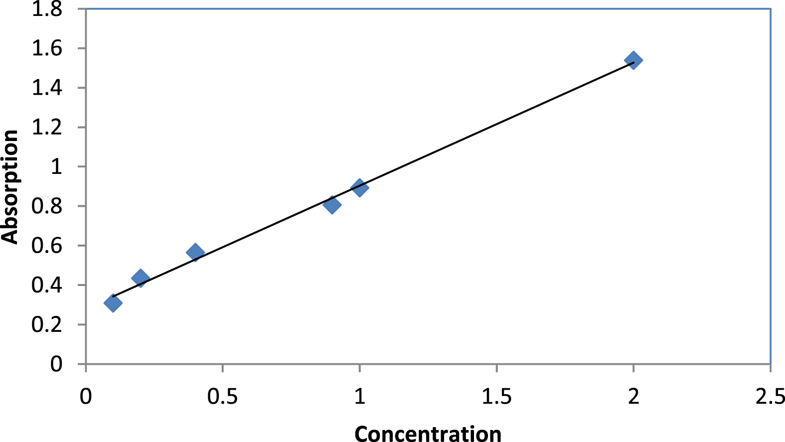
Ferrous sulphate standard curve.
Thymol is a pure natural compound extracted from the Thymus vulgaris, as well as tert-Butyl dimethyl silyl thymol and tri-ethyl silyl thymol, which were both synthesised in the lab. Numerous gram-positive and gram-negative bacteria, including Proteus, Klebsiella, Escherichia coli, and Staphylococcus aureus, were cultured separately in Eosin Methylen-Blue Agar medium. Assays for antibacterial activity were performed using the disc diffusion method. The middle of the inoculated plates was placed with four discs (6 mm in diameter), each of which had been individually impregnated with S1, S2, S3 and S4 (Fig. 2). Inhibition zones were measured in diameter (mm) all the way around the discs after the bacterial cultures were incubated at 37°C for 48 hours.

Bacterial culture and loaded disks.
Isolation procedure
The extraction of essential oil from the thyme plant was done several times with a clevenger, and after collecting the essential oil for dehydrating and obtaining pure essential oil, sodium sulfate was used for dehydrating it in such a way that the addition of sodium sulfate to the essential oil was done slowly until the addition of sodium Sulfate should be made into a powder form of essential oil and should not be lumped. The pure essential oil obtained was stored at 4°C.
Then the natural composition of thymol was extracted from the essential oil, the extracted thymol was identified by HPLC, and its silyl derivatives were synthesized. Silyl ethers are a group of chemical compounds which contain a silicon atom covalently bonded to an alkoxy group. The general structure is R1R2R3Si–O–R4 where R4 is an alkyl group or an aryl group. Silyl ethers are usually used as protecting groups for alcohols in organic synthesis. One of the main features of the silicon-containing material is a dramatic increase in their lipophilic properties. Thymol is a crucial phytochemical, as was already discussed in the previous section, but it also happens to be one of the compounds with hydrophilic properties. Some qualities are anticipated to be improved by the compounds’ increased lipophilicity, which is provided by the addition of silicon-containing groups. The qualities and effects of the medicine are multiplied by increasing its lipophilicity since it passes through the lipid barrier more quickly. Chemical hydrolysis affects the cleavage of the silicon-drug bond and is less prone to interpatient variability than the metabolic mechanism needed to activate conventional prodrugs [43–45]. Pyridyl-substituted 5-Si-soxazolines have been studied for their neurotropic and antitumor properties in other works, and it has been found that these compounds are more effective than their analogues at protecting against hypoxia [46]. Additionally, silatranes are tris(2-hydroxy-alkyl) amine esters of intracomplex organosilicon that have demonstrated a wide range of biological activity [47, 48]. In addition to these compounds other organosilicion compounds were synthesised and their biological activities were studied [49, 50]. Because of this, we synthesise some silyl ethers of thymol (scheme 1).
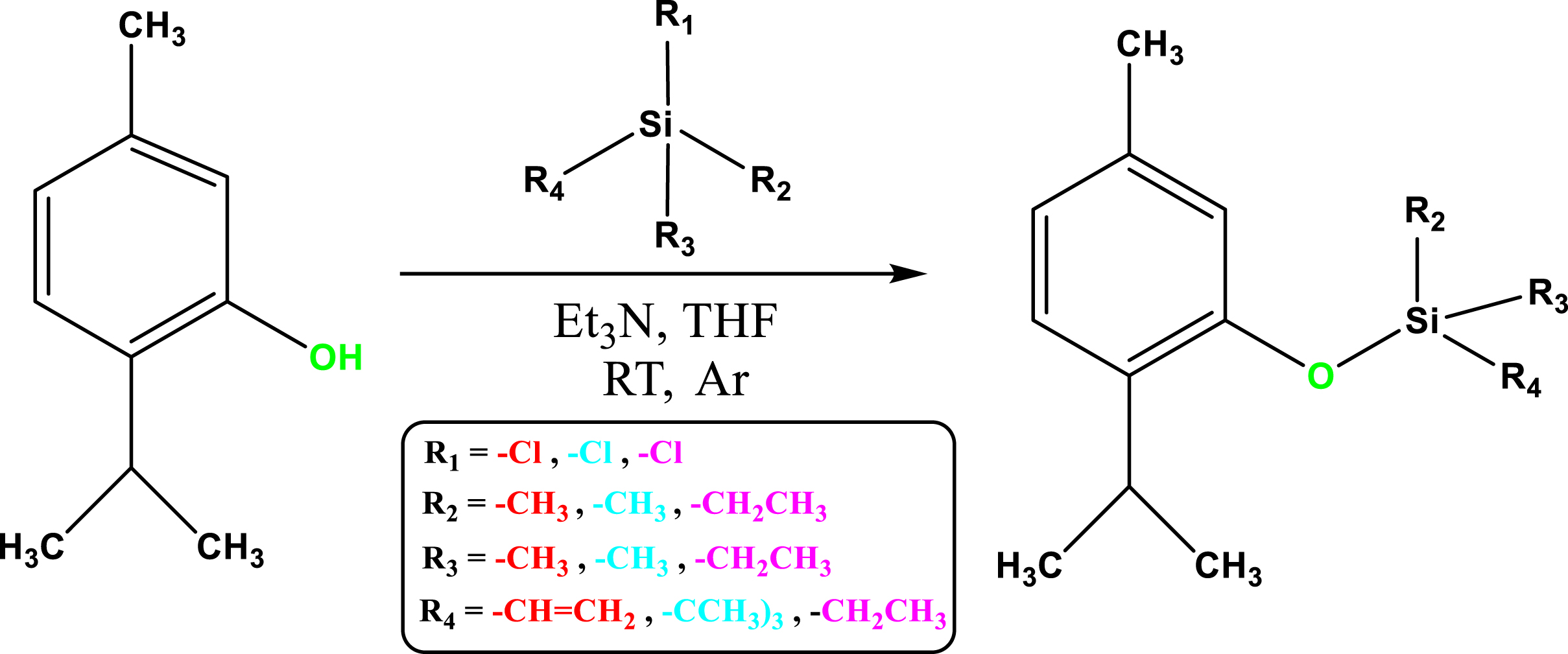
Synthesis reaction of silylated thymol derivatives.
The thymol, natural component in the EO of Thymus vulgaris was validated by HPLC analysis. In this analysis, at a wavelength of 277 nm the absorption peak intensity 22860 inhibitions time at 20.47 reveals that exactly fits the peak of the standard material (Fig. 3a). HPLC analysis was also performed on silylated thymol derivatives. In the results of this analysis, it can be seen that the inhibition time of silylated derivatives is significantly increased compared to thymol, considering that the HPLC column is of the C18 type and non-polar, so substances with a non-polar nature interact more effectively with the column substrate and have a high inhibition time. Based on the results of this analysis, the inhibition time of silylated derivatives is longer than that of thymol, which is due to the increase in lipophilicity of thymol after its silylation. This increase in non-polarity has increased the inhibition time, and this is a confirmation of the correct synthesis of silylated compounds and increase in their property is lipophilic (Fig. 3b).
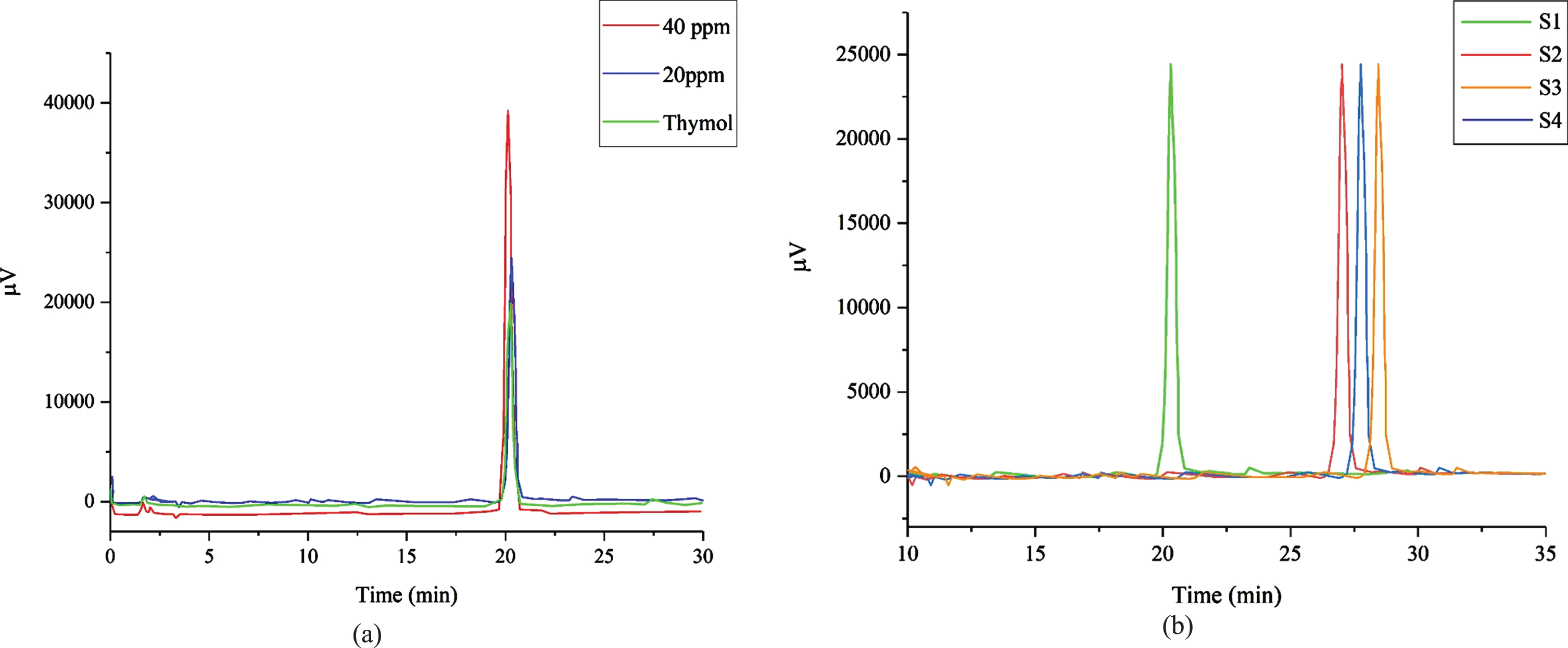
HPLC analysis of (a) extracted thymol and thymol standards and (b) thymol and silyl derivatives of thymol.
In order to determine the mass-to-charge ratio (m/z) of one or more molecules found in a sample, mass spectrometry is a useful analytical tool. The precise molecular weight of the sample’s constituent parts can frequently be determined using these measures as well. Thymol’s natural compound (A) has a 150.28 molecular mass. The fragment of the molecule [A-CH3] that was observed at m/z = 135 and the fragment of the molecule [A-isopropyl group] that was observed at m/z = 107 are connected. The fragment molecule Phenyl is related to the fragments of the molecule’s benzyl carbocation (m/z = 91), phenyl cation (m/z = 77) and cyclopentadienylium (m/z = 65). (Fig. 4).
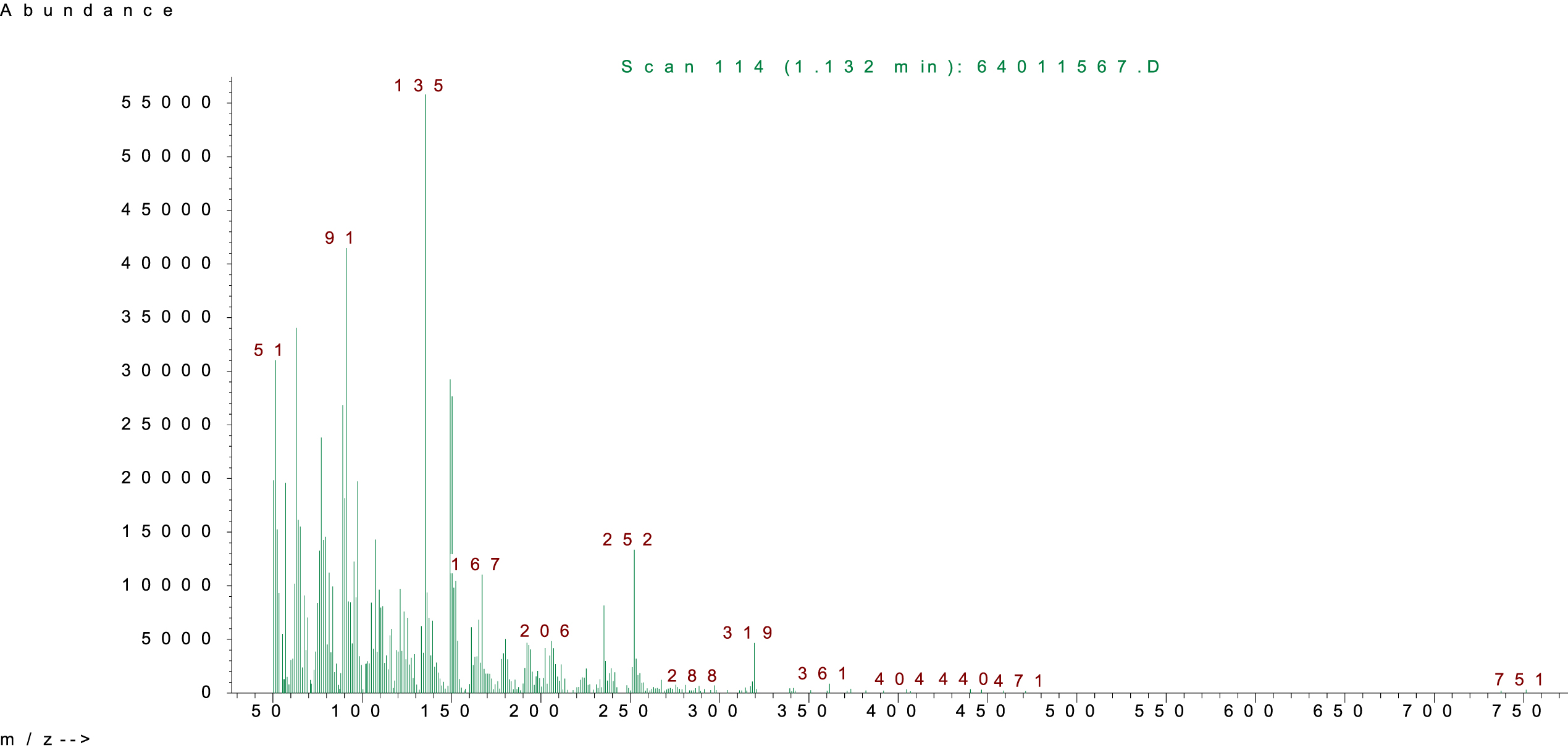
Thymol mass spectrometry analysis.
Thymol’s broad peak in the 3392 cm- 1 region of the FT-IR is connected to the stretching vibrations of O-H phenol. The aromatic C = C stretching vibrations are related to the aromatic C-H stretching vibration peak at 3020 cm- 1, the asymmetric stretching vibrations of aliphatic C-H at 2927 cm- 1, the symmetric stretching vibrations of aliphatic C-H at 2869 cm- 1, and the as harp peak at 1620 cm- 1 (Fig. 5a). The peak around 3040 cm- 1 in the FT-IR spectrum of thymol silyl derivatives corresponds to aromatic C-H stretching vibrations; the peak at 2932 cm- 1 corresponds to aliphatic C-H asymmetric stretching vibrations; and the peak at 2885 cm- 1 corresponds to aliphatic C-H symmetric stretching vibrations. The peak related to Si-C stretching vibrations is at 1270 cm- 1; the peak related to Si-C bending vibrations is observed at 819 cm- 1; the sharp peak at 1612 cm- 1 is related to aromatic C = C stretching vibrations; and the peak at 1056 cm- 1 is related to Si-O stretching vibrations. Finally, the absence of the peak related to thymol O-H in the region of 3392 cm- 1 indicates the synthesis of the desired compounds. Due to the very close similarity of the FT-IR spectra of the derivatives, only one spectrum is given (Fig. 5b).
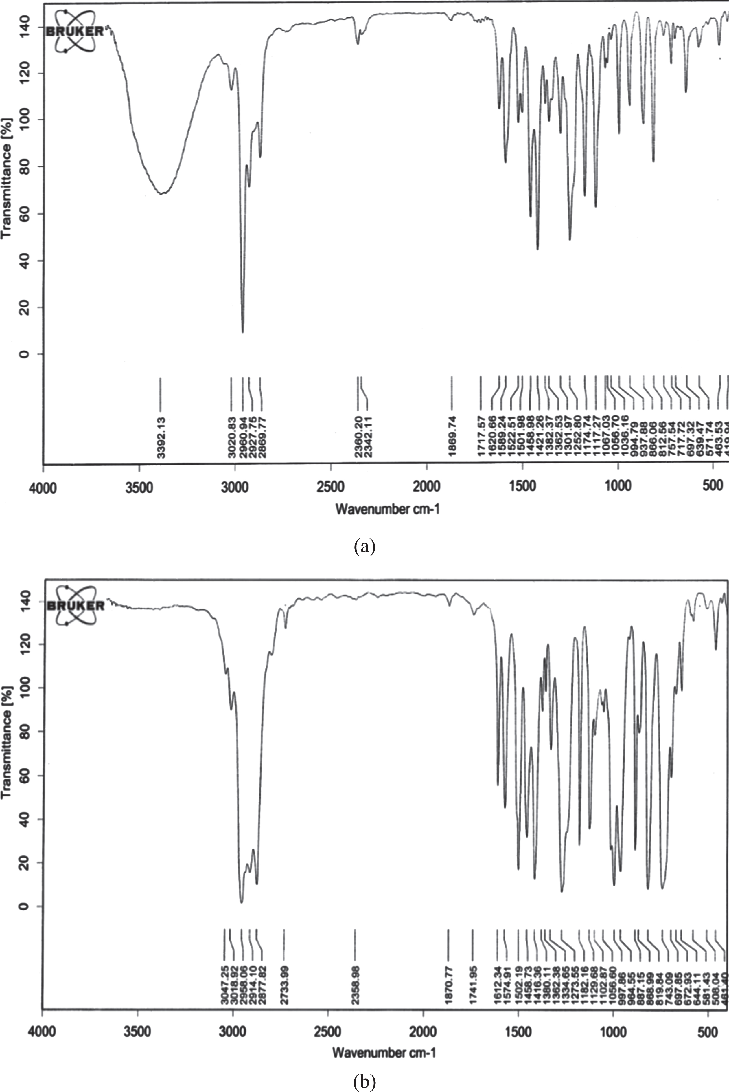
FT-IR of a) thymol, b) thymol silyl derivatives.
Binary peak in the 1.3 ppm range of the 1HNMR spectrum of thymol is connected to the 6 protons of the methyl group with isopropyl bound to the aromatic ring. 3 protons from the methyl group linked to the aromatic ring were responsible for a single branch peak in the 2.3 ppm range. Peak at 2.9 ppm is associated with the isopropyl group’s single proton, which is divided into 7 branches by the methyl group’s 6 protons. A single proton of the hydroxyl group attached to the aromatic ring causes a broad peak with single branches at 5.8 ppm, which is related. Single branches peak at 6.7 ppm in relation to the aromatic ring’s attached proton. One proton attached to the aromatic ring that undergoes a binary split with an adjacent proton causes the peak in the 6.8 ppm spectrum. Peak at 7.1 ppm is caused by a binary split of an aromatic ring proton by an adjacent proton (Fig. 6a).
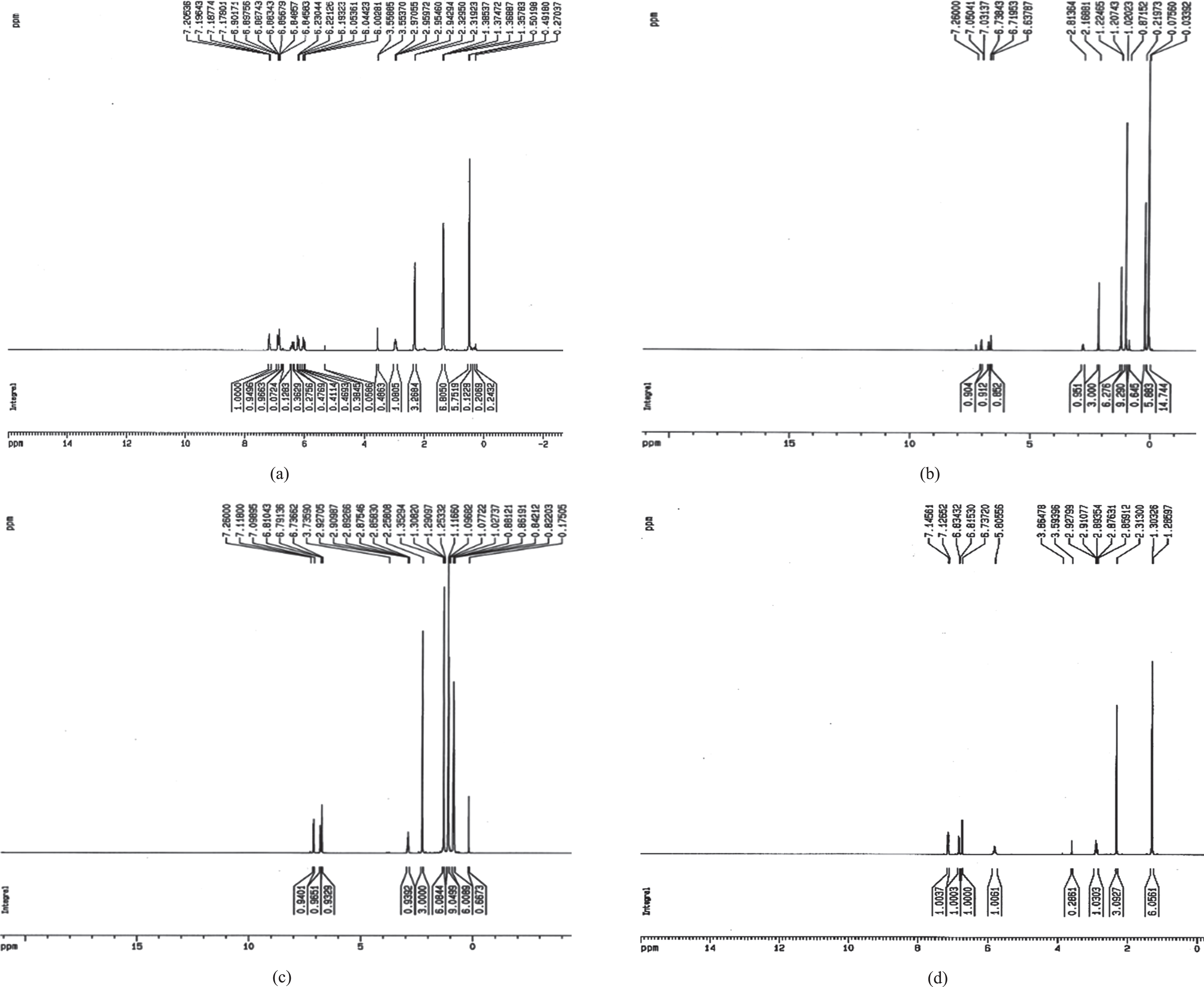
1HNMR of a) thymol, b) triethylsilyl thymol, c) tertiobutyl dimethylsilyl thymol, d) vinyl dimethylsilyl thymol.
In the 1HNMR spectrum of triethylsilyl thymol, the peak at 0.8 ppm corresponds to 6 methylene protons attached to silicon, which are split into 4 by three adjacent methyl protons, and the peak at 1.1 ppm corresponds to 9 methyl protons, which are the side methylene groups are split into 3. The peak in the region of 1.3 ppm corresponds to 6 protons of methyl isopropyl groups attached to the aromatic ring, which is split into 2 by a single proton of isopropyl. The peak of the single branch at 2.25 ppm corresponds to the 3 protons of the methyl group, which is located on the aromatic ring. The 2.9 ppm peak corresponds to an isopropyl group proton, which is split into 7 by 6 methyl protons of the isopropyl group. The single peak in the region of 6.73 ppm, 6.8 ppm, and 7.1 ppm corresponds to the protons of the aromatic ring (Fig. 6b).
In the 1HNMR spectrum of tert-butyldimethylsilyl thymol, a single peak in the 0.22 ppm region corresponds to 6 protons of two methyl groups attached to silicon. The single branch peak in the 1 ppm area corresponds to 9 protons of the tert-butyl group attached to silicon. The double peak in the 1.25 ppm area corresponds to 6 protons of the methyl isopropyl group attached to the aromatic ring. The peak of the single branch in the region of 2.2 ppm corresponds to 3 protons of the methyl group attached to the aromatic ring. The multiple peaks in the region of 2.8 ppm corresponds to single proton of the isopropyl group. The single peak in the area of 6.62 ppm, 6.72 ppm and 7.03 ppm correspond to the aromatic ring protons (Fig. 6c).
In the 1HNMR spectrum of vinyl dimethylsilyl thymol, the peak in the region of 0.5 ppm corresponds to 6 protons of two methyl groups attached to silicon. The double peak in the region of 1.35 ppm corresponds to the 6 protons of the methyl isopropyl group attached to the aromatic ring. The peak in the region of 2.3 ppm corresponds to 3 protons of the methyl group attached to the aromatic ring. The peak in the region of 2.9 ppm corresponds to a proton of the isopropyl group, which is split into a heptad by 6 methyl protons of the same group. The peak in the area of 6 ppm, 6.2 ppm, and 6.4 ppm corresponds to protons of the vinyl group. The peaks at 6.85 ppm, 6.9 ppm, and 7.15 ppm are related to aromatic ring protons (Fig. 6d).
One of the most potent plant essential oil constituents with antimicrobial effects is thymol, which belongs to the monoterpene phenols class. These positional isomers of isopropyl cresol, which have a broad range of bioactive properties, reduce ergosterol content, impair membrane permeability, block efflux pumps, and restore fluconazole susceptibility to antifungal treatment in resistant Candida strains. Exposure to these natural compounds induces a cascade of stress responses, which are important to comprehend their microbicidal mechanisms. The antioxidant properties of thymol have been well documented in various preclinical studies including cell lines and animal models. At high-rate constants, it effectively scavenged the hydroxyl free radicals thereby producing major transient species named phenoxyl radicals. The generated adducts from the phenoxyl radicals undergo dehydration which can be accelerated by an alkaline medium. The addition of hydroxyl radicals at the ortho position (C6 atom) of the phenolic group yields the phenoxyl radical after dehydration. The attack at the ortho position is more favourable energetically while the attack at the para position is also expected to occur. Furthermore, additions at the ortho positions occur without any precomplex formation. The non-toxicity and redox potential of the thymol /thymol couple makes it a promising antioxidant (Venu et al., 2013).
In this study, the DPPH method and the FRAP method were used to assess the antioxidant activity of thymol and its derivatives.
DPPH assay
Each sample was generated at various concentrations (Table 1), and the antioxidant activity of the samples was assessed using the DPPH technique. Results show that thymol has greater free radical scavenging capacity than silyl derivatives (Fig. 7). The hydroxyl group of thymol has been substituted with silyl groups, which is the only structural difference between thymol and silyl derivatives. These results demonstrated that the substitution of a hydrophobic group for a hydroxyl group significantly reduced the antioxidant activity of thymol. The findings amply supported the theory that thymol’s antioxidant properties are primarily dependent on the hydroxyl functional group, which is in this case protected by a silyl group.
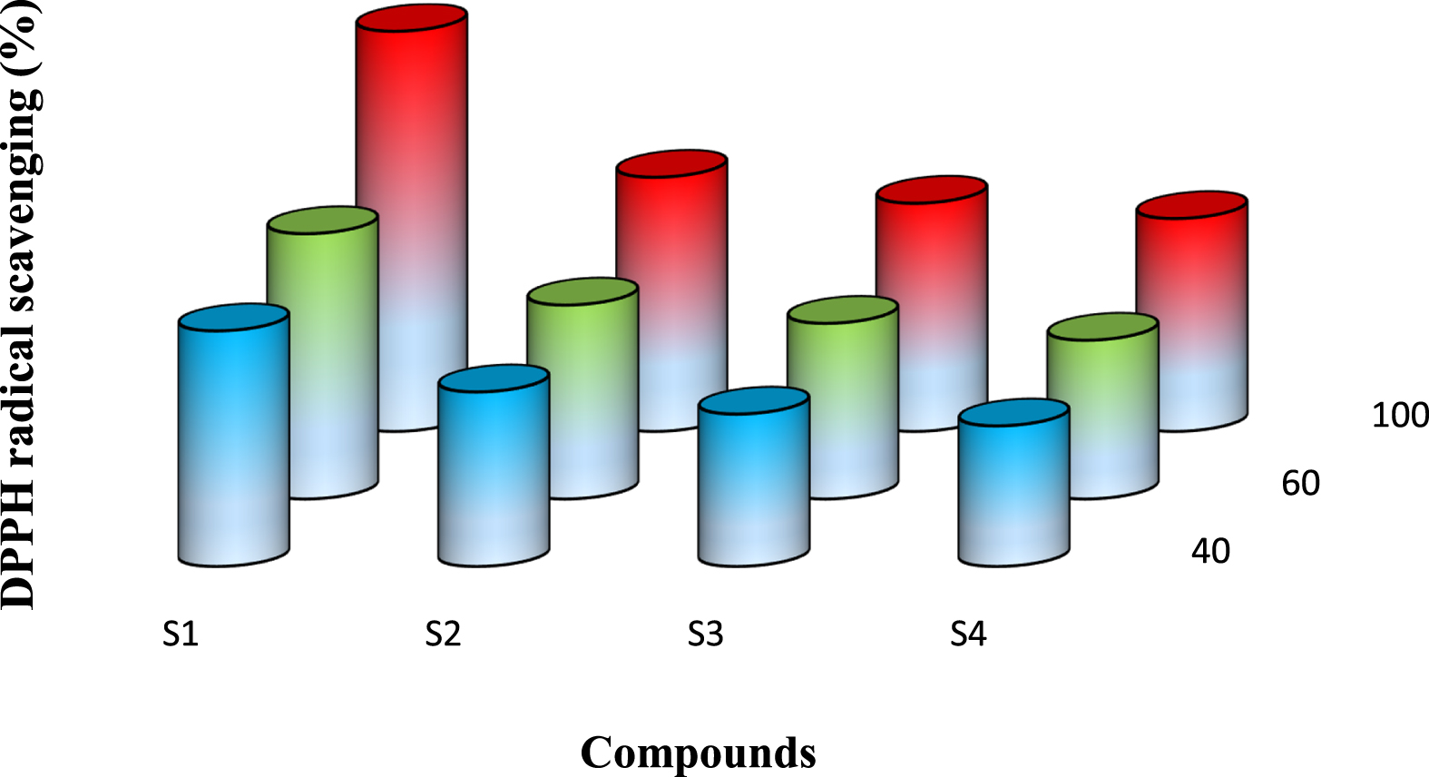
Antioxidant activity results from the DPPH method.
The antioxidant activity of the samples was measured using the FRAP method. The results of the FRAP assay also revealed that thymol has stronger antioxidant properties than its derivatives and that the hydroxyl functional group is largely responsible for this (Fig. 8). Results from the two techniques, DPPH and FRAP, demonstrated that thymol had greater antioxidant activity than silyl derivatives. We can also infer that the hydroxyl functional group is responsible for thymol’s antioxidant properties.
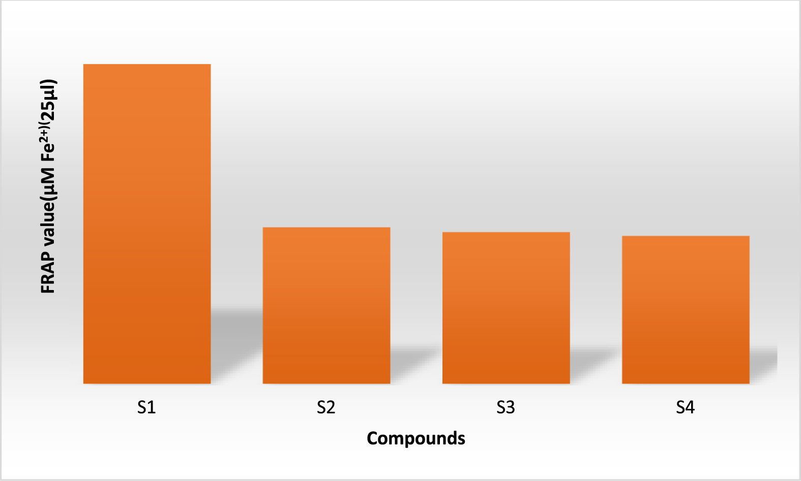
Antioxidant activity results from the FRAP method.
A common protecting group for the functional group of alcohols and phenols is silicon. In this study, the hydroxyl group of thymol was shielded by silicone. Results show that the inhibition zone diameter of native thymol-containing discs is bigger than the inhibition zone diameter of thymol silyl derivatives in all cultures, including gram-positive and gram-negative bacteria (Table 2). The hydroxyl group of thymol is the only difference between it and its derivatives, as can be seen by comparing the structures of the three compounds. Results with several bacteria types demonstrated that the hydroxyl functional group plays a significant role in thymol’s antibacterial action. Eliminating functional group will significantly limit anti-bacterial activity. Thymol affects bacteria by impairing the efficiency of the bacterial cell membrane and external membrane. These findings indicate that thymol interferes with the ATPase enzyme’s ability to function and that its hydroxyl functional group is primarily responsible for its antibacterial effects. It is possible that thymol’s antibacterial properties result from a reaction between its hydroxyl functional group and the two amino acids in the ATPase active site (asparticacid and lysine). Another explanation is that thymol’s ability to enter the ATPase active site and exert its antibacterial effects was diminished when the hydroxyl functional group was replaced with sylil.
Growth inhibitory zone (millimeter) of thymol and silyl derivatives by disk diffusion method
Growth inhibitory zone (millimeter) of thymol and silyl derivatives by disk diffusion method
In this research, thyme essential oil was extracted as one of the main sources of natural thymol with a clevenger, and the identification of this natural compound was done with HPLC. The properties of the silylated compounds increased dramatically, which was confirmed by their lipophilic properties with HPLC, which increased the time of inhibition in these compounds compared to thymol. The antioxidant activity of the thymol and its three derivatives was studied using two methods, DPPH and FRAP, which showed that the activity in the thymol was much higher than its derivatives, which showed the antioxidant activity of the thymol to its hydroxyl group was high. It was dependent on the proclamation of this group’s antioxidant activity. The next part of the work of antibacterial thymol was investigated by its dissemination method, which was also observed in this section regarding the antibacterial activity of thymol against warm bacteria. The Gram-positive is much more than its assistant, which is the result of the very effective role of the thymol hydroxyl group in its antibacterial activity.
Footnotes
Acknowledgments
The authors gratefully acknowledge the research council of Azarbaijan Shahid Madani University for financial support.