
Abstract
Beetroot (Beta vulgaris) has been reported to possess many benefits and medicinal properties. However, the protective effect of beetroot against isoproterenol (ISO) induced myocardial damage has not been clarified sufficiently yet. The present study was aimed to investigate the effects of beetroot on oxidative stress, fibrosis, and myocardial damage in ISO-induced rats. Our investigation revealed that ISO administration markedly increased the oxidative stress parameters, while the level of cellular antioxidants and catalase activities were decreased in ISO-administered rats. Beetroot supplementation and gallic acid treatment to ISO-administered rats prevented the rises of lipid peroxidation products malondialdehyde, nitric oxide and advanced protein oxidation product concentration. Moreover, elevated activities of alanine aminotransferase, aspartate aminotransferase, and creatinine kinase-muscle brain enzymes activities in ISO-administered rats were also lowered by both gallic acid and beetroot supplementation. Furthermore, gallic acid and beetroot supplementation prevented the inflammatory cells infiltration and fibrosis in ISO-induced rats. In conclusion, these results suggest that beetroot supplementation is capable of protecting ISO-administered myocardial infarction in rats probably by preventing oxidative stress.
Introduction
Cardiovascular diseases such as coronary heart disease become the leading causes of death and disability in both developed and developing countries. Growing evidence indicates that reactive oxygen species (ROS) mediated oxidative stress is responsible for the development of cardiovascular diseases (CVD) [1]. In acute myocardial infarction (MI), reactive oxygen species (ROS) are generated in the myocardium and may trigger cell membrane destruction and cellular death. However, ROS may also stimulate signal transduction to elaborate inflammatory cytokines in the ischemic region and surrounding myocardium [2]. Isoproterenol (ISO) has been used to induce infarct-like lesions in the heart of experimental animal species in the laboratory. The ISO-induced myocardial alterations are similar to human myocardial infarction. It is suggested that ISO exerted β-adrenergic stimulatory activity in the heart and increased cardiac oxidative metabolism which lacks oxygen available to the myocyte. The energy imbalance, altered calcium flux, stimulation of the adenylyl cyclase system, aggregation of platelets, and formation of reactive oxygen species appear to contribute to the pathogenesis of myocyte damage [3].
Beetroot or red beet (Beta vulgaris) is grown throughout the Americas, Europe, and Asia with some varieties of edible taproots [4]. Betalains present in the cell vacuoles of beetroot are the reasons behind its’ red color [5]. These betalains are widely used as food colorants in many industries. Beetroot is also a rich source of antioxidants. The presence of caffeic acid, catechin hydrate, 4-hydroxybenzoic acid and epicatechin in the beetroot was detected previously [6]. These antioxidants showed great promise in scavenging free radicals and prevents various degenerative diseases such as cardiovascular diseases [7, 8]. As no extensive report has been found on the pharmacological activities of the beetroot on cardiac remodeling, the rationale of this study was to evaluate the anti-inflammatory and cardio-protective actions of the beetroot powder in ISO administered rats.
Material and methods
Animals
The Animal House of Department of Pharmaceutical Sciences, North South University, Dhaka, Bangladesh produced the Long Evans rats (3-4 months of age, weighing 200–240 g) and were used in this experiment. All animals were kept at constant temperature (22±2°C), humidity (55%), and light-dark conditions (12/12 h light/dark ratio). The animals were provided with standard laboratory chow diet and drinking water ad libitum. Ethics Committee of the Department of Pharmaceutical Sciences, North South University, Dhaka, Bangladesh approved the experimental protocols and the procedure of sacrifice having the approval number AEC-005-2016.
Induction of myocardial infarction
Experimental myocardial infarction was induced by injecting isoproterenol (ISO) hydrochloride (dissolved in physiological solution) subcutaneously to rats. To test the effect of beetroot on ISO-induced cardiac disturbances, the animals were divided into five groups as described below. Group I (control)-was administered saline only with normal food and water. Group II (ISO)-was administered isoproterenol (ISO) (at a dose of 50 mg/kg twice a week for one week). Group III (Control + Beetroot)-control rats, treated with beetroot powder (2.5% of powdered chow food every day for two weeks). Group IV (ISO + Beetroot)-was administered isoproterenol (ISO) (at a dose of 50 mg/kg twice a week for one week) and treated with beetroot powder (2.5% of powdered chow food every day for two weeks). Group V (ISO + gallic acid)-was administered isoproterenol (ISO) (at a dose of 50 mg/kg twice a week for one week) and treated with gallic acid (100 mg/kg every day for two weeks).
All animals were weighed and sacrificed after two weeks using a high dose of pentobarbitone anesthesia. The blood was drawn from the abdominal aorta and kept in citrate buffer containing tubes. Heart, kidney and spleen tissues were also harvested. Immediately after collection of the organs, they were weighed and stored in neutral buffered formalin (pH 7.4) for histological analysis and in the refrigerator at – 20°C for further studies. The collected blood was centrifuged at approximately 3000×g, and the plasma was separated and stored in the refrigerator at – 20°C for further analysis.
Assessment of biochemical parameters
Commercially available kits for the assay of alanine aminotransferase (ALT) and aspartate aminotransferase (AST) enzyme activities were used, which were obtained from DCI Diagnostics (Budapest, Hungary) followed by the manufacturer’s protocol. Creatine kinase-MB (CK-MB) activity was also evaluated in plasma using commercial kit obtained from DCI Diagnostics (Hungary) according to the manufacturer’s protocol.
Preparation of sample tissues for the assessment of oxidative stress markers
Oxidative stress markers were measured in heart tissues and were homogenized in 10 volumes of phosphate buffer (pH 7.4) solution. The homogenates were then centrifuged at 12,000×g for 30 min at 4°C. The supernatant was collected and used for the determination of protein and enzymatic studies as described below.
Lipid peroxidation in tissues was determined as malondialdehyde and we followed a previously described method in our lab [9, 10]. The absorbance of the final solution was read in a spectrophotometer at 532 nm. The content of malondialdehyde (MDA) (nmol/mL) was then calculated, using a standard curve of MDA solution. The concentration of nitric oxide (NO) was determined according to the previously mentioned method as nitrate [10]. The absorbance of the final solutions was taken at 540 nm against the corresponding blank solutions (UV-1800 UV-Vis spectrophotometer, Shimadzu Scientific Instrument). NO level was also measured by using the standard curve and expressed as nmol/mL. The advanced protein oxidation product (APOP) level was assayed by the method described previously [10]. The chloramine-T absorbance was found linear within the range of 0 to 100 nmol/mL at 340 nm; APOP concentrations were expressed as nmol·mL–1 chloramine-T equivalents. CAT activities were also determined using a previously described method [10]. Changes in absorbance of the reaction solution at 240 nm were determined after one minute. One unit of CAT activity was defined as an absorbance change of 0.01 as unit/min. Changes in absorbance of the reaction solution at 240 nm were taken after one minute (UV-1800 UV-Vis spectrophotometer, Shimadzu Scientific Instrument). One unit of CAT activity was defined as an absorbance change of 0.01 as unit/min. SOD activities were also assayed in plasma and tissue homogenates by using previously described method [11]. A 50% inhibition of auto-oxidation of epinephrine was considered as one unit of enzyme activity present in the assay system. In the tissue homogenates, MPO activity was determined followed by a previously described method using dianisidine-H2O2 system [12]. The rate of absorbance change was measured at 460 nm. Results were expressed as units of MPO/mg protein.
Histopathological studies
Myocardial histoarchitecture were examined after processing of the heart followed by routine procedures. The heart tissues were washed with normal saline and fixed in 10% neutral buffered formalin for several days. These tissues were then processed using several consecutive ethanols and xylene treatment. The paraffin-embedded blocks were prepared from the processed tissues. About five μm thick sections were cut by employing a rotary microtome. These sections were stained with Hematoxylin and Eosin using routine procedures. Sirius red staining for fibrosis was also done in heart sections. Sections were then studied and photographed under a light microscope (Zeiss Axio Scope) at 40 magnifications. The slides were examined for morphological changes.
The phenolic content determination in beetroot extract by HPLC-DAD system
The phenolic composition of the ethanol extract of beetroot was determined by HPLC (Dionex UltiMate 3000 system, Thermo Scientific, equipped with quaternary rapid separation pump (LPG-3400RS) and photodiode array detector (DAD-3000RS)), as described previously [12]. Acetonitrile (solvent A), acetic acid solution (pH 3.0, solvent B), and methanol (solvent C) were used as mobile phase. For UV detection, the wavelength program was optimized to monitor phenolic compounds at their respective maximum absorbance wavelengths as follows:λ 280 nm held for 18.0 min, changed to λ 320 nm and held for 6 min, and finally changed to λ 380 nm and held for the rest of the analysis. The DAD was set at an acquisition range from 200 nm to 700 nm. The detection and quantification of gallic acid (GA), catechin hydrate (CH), vanillic acid (VA), caffeic acid (CA), and epi-catechin (EC) was done at 280 nm, of p-coumaric acid (PCA), rutin hydrate (RH), and ellagic acid (EA) at 320 nm, and of myricetin (MC), quercetin (QU), and kaempferol (KF) at 380 nm, respectively.
A stock standard solution (100μg/ml) of each phenolic compound was prepared in methanol by weighing out approximately 5 mg of the analyte into 50 ml volumetric flask. A solution of ethanol extract of beetroot at a concentration of 5 mg/ml was prepared in ethanol by vortex mixing (Branson, USA) for 30 min.
Statistical analysis
The values are expressed as mean±standard error mean (SEM). The results were evaluated by using the one way ANOVA followed by Tukey’s test using Graph Pad Prism Software. Statistical significance was considered p < 0.05 in all cases.
Results
Effect of beetroot supplementation on oxidative stress parameters in plasma and heart tissue homogenates of ISO-administered rats
ISO administration led to a significant increase in the concentration of oxidative stress parameter MDA in plasma and heart tissue homogenates of rats compared to that of control rats (Fig. 1A-B). However, Beetroot supplementation in ISO rats significantly (p < 0.05) prevented the rise of MDA concentration compared to ISO-treated rats (Fig. 1A-B). Beetroot supplementation to control rats did not cause any significant change in MDA concentrations which indicates that normal physiology of rats is not affected by beetroot supplementation (Fig. 1A-B). Gallic acid treatment in ISO-administered rats also exhibited (p < 0.05) decreased MDA level in plasma and heart tissue compared to the ISO-induced rats (Fig. 1A, B).

Effect of beetroot supplementation on oxidative stress parameters in plasma and heart tissue homogenates of ISO administered rats. Data are expressed as mean±SEM, n = 8. Statistical analysis was done by One Way ANOVA followed by Newman-Keul’s post hoc test. Statistical significance was considered as p > 0.05 and marked as asterisk mark.
Moreover, NO concentrations were found significantly (p < 0.05) higher in ISO-treated rats compared to control rats (Fig. 1C-D). Treatment with beetroot significantly (p < 0.05) prevented the rise of NO concentrations (Fig. 1C-D). The level of NO was not changed in control rats supplemented with beetroot (Fig. 1C-D). This response indicates that beetroot itself does not alter the normal physiology of rats. Gallic acid treatment in ISO-administered rats also exhibited significantly (p < 0.05) lower levels of NO concentration as in control rats compared to the ISO-administered rats (Fig. 1C-D).
Furthermore, APOP concentration was increased in plasma and tissue homogenates significantly (p < 0.05) in ISO-administered rats compared to the control rats (Fig. 1E-F). However, beetroot supplementation significantly (p < 0.05) lowered the APOP level in plasma and heart tissue. This effect is also comparable to gallic acid treatment in ISO-administered rats. Gallic acid treatment in ISO-administered rats also showed significant (p < 0.05) protection by lowering the APOP development in plasma and tissues (Fig. 1E-F). Although beetroot supplementation in the control group did not show any significant change in APOP level in plasma and heart compared to the control rats (Fig. 1E-F).
AST and ALT enzyme activities were increased significantly (p < 0.05) in the ISO-treated rats compared to the control rats (Fig. 2A-B). When the ISO-administered rats were treated with the beetroot, AST and ALT enzyme activities were found to be significantly (p < 0.05) normalized as the control group (Fig. 2A-B). In the ISO + Gallic acid group rats, the concentrations of AST and ALT activities were also found to be normalized when compared to diseased group (Fig. 2A-B). Beetroot supplementation alone did not possess any adverse effect on the parameters, suggesting that beetroot did not alter the physiological condition of rats (Fig. 2A-B).
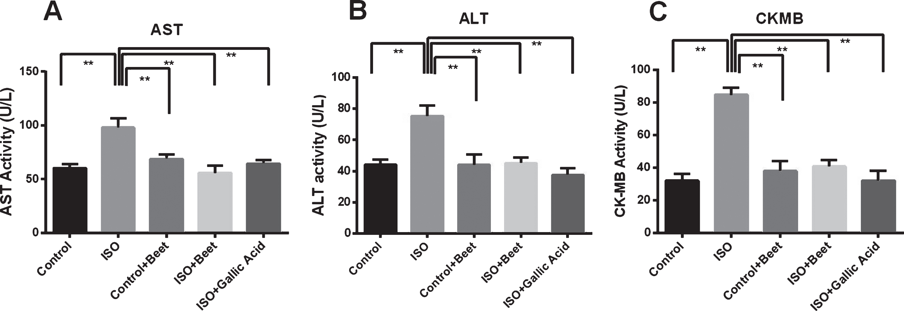
Effect of beet root supplementation on AST, ALT and CK-MB activities in plasma of ISO administered rats. Data are expressed as mean±SEM, n = 7. Statistical analysis was done by One Way ANOVA followed by Newman-Keul’s post hoc test. Statistical significance was considered as p > 0.05 and marked as asterisk mark.
Moreover, CK-MB activities in heart homogenates were increased significantly (p < 0.05) compared to the control rats (Fig. 2C). Beetroot supplementation prevented the rise of CK-MB activities in ISO-administered heart and normalized to near control rats (Fig. 2C). This result is also comparable to gallic acid treatment in ISO-administered rats which showed a reduction of CK-MB activities in ISO-administered rats (Fig. 2C). Also, beetroot supplementation showed significantly (p < 0.05) decreased the level of CK-MB in plasma compared to the ISO-administered rats.
Uric acid and creatinine concentrations were markedly elevated in ISO-administered rats (p < 0.05) compared to control (Fig. 3A-B). Further, uric acid and creatinine concentrations were normalized in ISO-administered rats treated with beetroot compared to the ISO-induced rats (Fig. 3A-B). Gallic acid treatment showed that the concentrations of uric acid and creatinine in plasma were reduced to near normal compared to the ISO-administered rats (Fig. 3A-B). Beetroot treated control rats also showed unaltered concentrations of uric acid and creatinine compared to ISO-administered rats, indicating beetroot had no adverse effect on the normal physiological condition of rats (Fig. 3A-B).
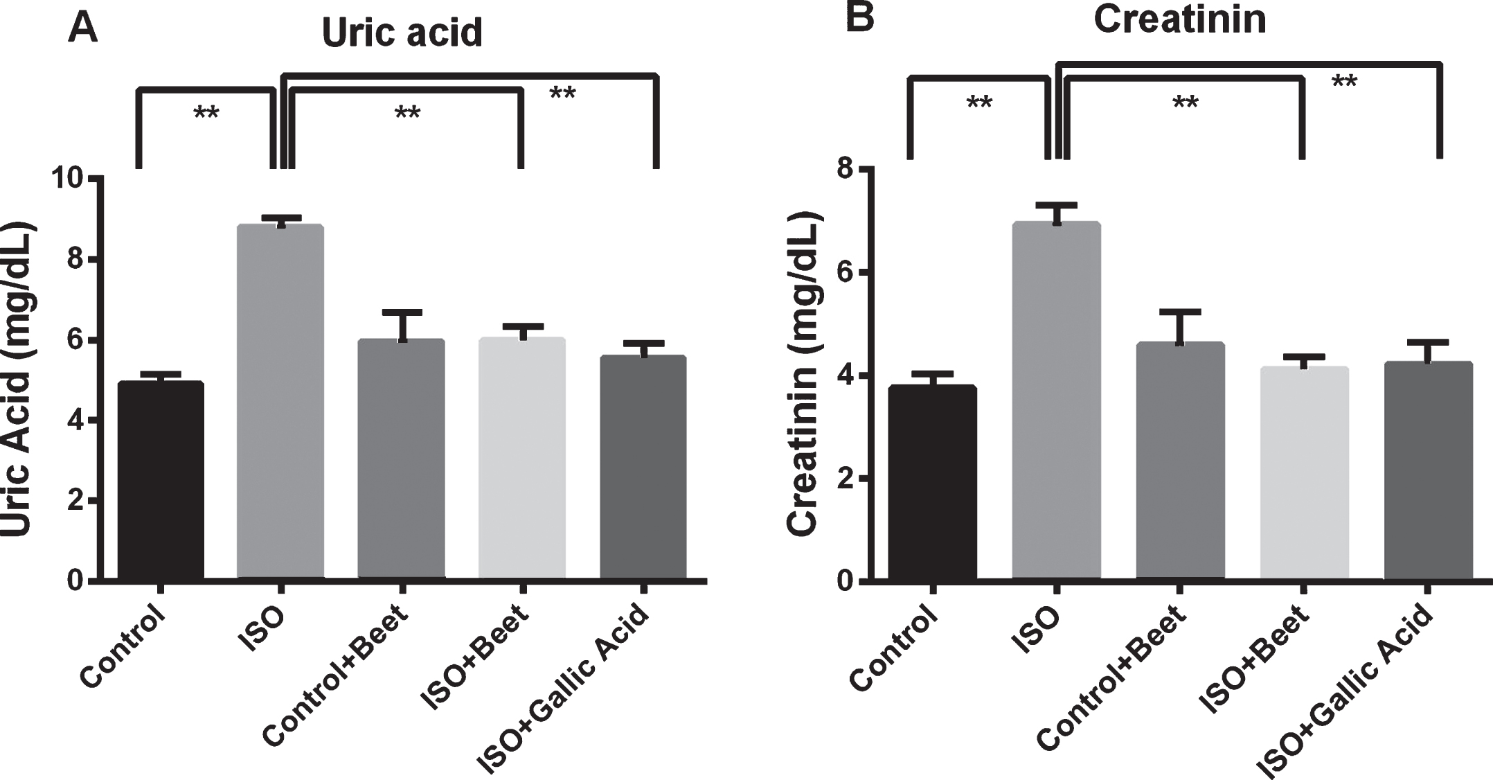
Effect of beet root supplementation on uric acid and creatinine concentration in plasma of ISO administered rats. Data are expressed as mean±SEM, n = 7. Statistical analysis was done by One Way ANOVA followed by Newman-Keul’s post hoc test. Statistical significance was considered as p > 0.05 and marked as asterisk mark.
ISO administration markedly decreased the cellular antioxidant capacities by decreasing catalase activities compared to control rats (Fig. 4A-B). Whereas, ISO-induced rats treated with beetroot showed no significant change in the level of antioxidant enzyme catalase in plasma but significantly (p < 0.05) increased in heart tissue compared to ISO-administered rats (Fig. 4A-B). In the case of ISO-administered rats treated with gallic acid, the catalase activity significantly (p < 0.05) increased (Fig. 4A-B).

Effect of beet root supplementation on antioxidant enzyme activities in plasma and heart of ISO administered rats. Data are expressed as mean±SEM, n = 7. Statistical analysis was done by One Way ANOVA followed by Newman-Keuls post hoc test. Statistical significance was considered as p > 0.05 and marked as asterisk mark.
Moreover, ISO administration significantly (p < 0.05) lowered the activity of SOD in plasma compared to control rats (Fig. 4C). Gallic acid and beetroot treatment normalized the SOD activity in plasma of ISO-administered rats (Fig. 4C). ISO administration also increased the MPO activity significantly (p < 0.05) in rats compared to control it was statistically significant (Fig. 4D). Gallic acid and beetroot treatment were also significantly (p < 0.05) lowered the MPO activity in the heart of ISO administered rats (Fig. 4D).
Hematoxylin and eosin staining (H&E staining)
The heart of the normal control group showed an intact and homogenous histoarchitecture without necrosis, edema and inflammation (Fig. 5A). The heart of the control + beet group also showed an intact and homogenous histoarchitecture without necrosis and inflammation which is similar to the control group (Fig. 5B). Heart of ISO-administered rats showed pivotal necrosis of muscle fibers with inflammatory cell infiltration, and increased extracellular matrix deposition compared to the control group (Fig. 5C). The ISO-administered rats treated with beetroot powder showed protection from myocardial injury evidenced by decreased necrosis as well as inflammatory cell infiltration compared to the ISO group (Fig. 5D). Gallic acid treatment in ISO-administered rats also showed protection from myocardial injury evidenced by decreased necrosis and inflammatory cell infiltration compared to the ISO group (Fig. 5E).

Beet root supplementation prevented mono-nuclear inflammatory cells infiltration in heart of ISO administered rats (H & E staining). A, Control; B, Control + BR; C, ISO; D, ISO + BR and E, ISO + Gal. Magnification 40X.
The image of control rats showed normal collagen distribution and alignments in the left ventricle of the heart (Fig. 6A). Control rats treated with beetroot also showed less collagen deposition in the heart which is similar to the control group (Fig. 6B). ISO administration in rats showed excess collagen deposition and fibrosis compared to the control rats (Fig. 6C). Beetroot and gallic acid treatment significantly prevented the collagen deposition and fibrosis in ISO-administered rats (Fig. 6D-E).
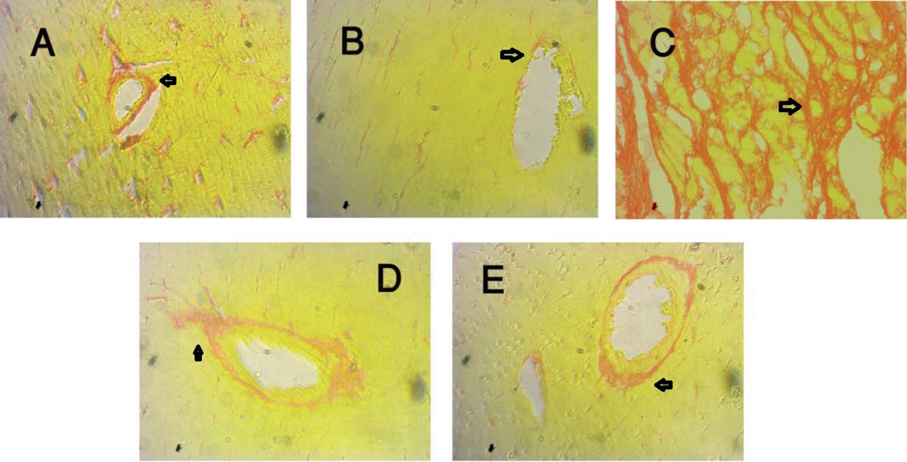
Beet root supplementation prevented fibrosis in heart of ISO administered rats (Sirius red staining). A, Control; B, Control + BR; C, ISO; D, ISO + BR and E, ISO + Gal. Magnification 40X.
Ethanolic extract of beeroot showed the presence of caffeic acid, para-coumaric acid and trans-ferulic acid in the HPLC chromatogram (Fig. 7).
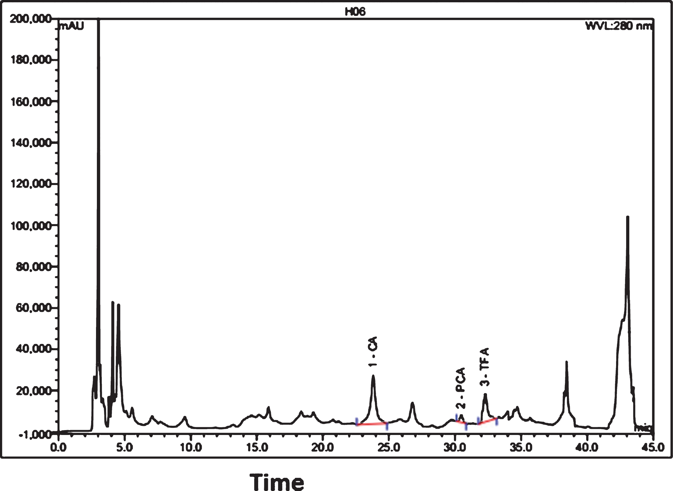
HPLC-DAD analysis of beet root extract.
The current investigation revealed that beetroot powder supplementation ameliorated the oxidative stress and inflammation in the heart of ISO-administered aged rats. A large dose of ISO administration in experimental animal induces morphological and functional alterations in the heart leading to myocardial necrosis. ISO-administration also produces excessive free radicals resulting from the oxidative metabolism of catecholamine. There is increasing evidence that cardiotoxicity of ISO occurs followed by oxidative stress mechanism [13].
Superoxide and hydrogen peroxide are several reactive oxygen species (ROS) produced in large quantities which contributes to myocardial tissue injury during myocardial infarction [14]. ISO may undergo auto-oxidation which results in the excessive formation of free radicals and lipid peroxidation [15]. This free radical mediated peroxidation of membrane phospholipids leads to permeability changes in the myocardial membrane, intracellular calcium overload, and irreversible damage [16]. Previous reports suggest that strawberries and blue berries rich in polyphenol are strong scavengers of free radicals and restores antioxidant defenses [17, 18]. In our study, beetroot powder administration decreases the levels of lipid peroxidation product MDA in ISO-administered rats. This result is also comparable to a potent antioxidant gallic acid which also significantly prevented the lipid peroxidation in plasma and tissues of ISO-administered rats. The presence of antioxidants in beetroot powder is responsible for the prevention of lipid peroxidation and myocardial tissue damage [6]. The previous report suggests that beetroot extract possess 4-hydroxybenzoic acid, caffeic acid, catechin hydrate, and epi-catechin. However, our HPLC-DAD analysis of the beetroot extract revealed the presence of caffeic acid, p-coumaric acid and trans-ferulic acid, all of them are strong scavengers of free radicals [8].
It has been reported that inducible nitric oxide synthase (iNOS) expression and nitric oxide (NO) production increased in the myocardial infarcted heart [19]. Inducible nitric oxide synthase (iNOS) expression by β-Adrenergic stimulation was also reported previously and significantly increased the production of NO [20]. Nitric oxide in the presence of other reactive oxygen species (ROS) such as superoxide and generates the powerful oxidant molecule peroxynitrite (ONOO–) and creates nitrosative stress. Inhibition of superoxide production may be beneficial in peroxynitrite production. In our study, beetroot treatment prevented the rise of nitric oxide level in ISO-treated rats.
Enzyme activities such as ALT and AST, which are present in cardiac muscle, may increase in the bloodstream and released from injured heart tissues [21]. ISO administration induces the leakage of these enzymes as a result of necrosis to the myocardium. Beetroot supplementation in ISO-administered rats prevented the rise of AST and ALT enzyme activities in plasma. In this investigation, creatine kinase-MB (CK-MB), which is a specific marker to the acute myocardial infarction, activity was also raised significantly in plasma of ISO-administered rats compared to control rats. Heart muscle localizes CK-MB predominantly, and it is considered as a valuable diagnostic tool for MI since CK-MB levels found to be elevated in the myocardium during failure [22]. These observations are supported by previous studies done on rats treated with isoproterenol [22, 23]. Beetroot supplementation and gallic acid treatment prevented the increased CK-MB activity in plasma of ISO administered rats.
Tissue antioxidant enzymes and several natural antioxidants constitute the scavenging mechanism of free radicals and prevent free radical-mediated damage in tissues. SOD, CAT, and GPx are several free radical scavenging enzymes and are considered as the first line defense enzymes in cells against oxidative stress and nitrosative stress [24]. Increased lipid peroxidation decreases these enzymes in tissue level [25]. ISO-administered myocardial damage is also associated with decreased activities of superoxide dismutase (SOD) and catalase enzymes which may be impaired structurally and functionally by free radicals [26, 27]. In this study, SOD and CAT activities were lowered in plasma and heart in ISO-administered rats when compared to control rats. Further, gallic acid treatment and beetroot supplementation normalized the catalase activity in ISO-treated rats. Gallic acid induced normalization of catalase activity was also observed in the previous study [28]. A recent report also supported the idea that polyphenols showed increased antioxidant enzyme activities and decreased lipid peroxidation in aging related complications in heart [29].
ISO-administration also causes an increase in the levels of serum uric acid and creatinine concentration in plasma. The increase in blood uric acid, creatinine can accelerate the progression of MI [30]. In our study, significantly higher levels of serum uric acid and creatinine were observed in ISO administered rats when compared to control rats. Gallic acid treatment and beetroot supplementation normalized the elevated serum uric acid and creatinine levels in ISO-treated rats.
To further assess the development of heart failure following isoproterenol injection, we performed distinct morphological studies using different staining. We found that administration of high dosages of ISO primarily resulted in an extensive amount of cardiomyocyte necrosis and deposition of extracellular matrix collagen. This finding is in agreement with the previous report showed massive collagen deposition in the left ventricle of heart due to a high dose of catecholamines [31–33]. Collagen fiber deposition in ISO-administered rats was also accompanied by a lot of mononuclear cells infiltration. Cardiomyocyte necrosis and lipid peroxidation mediated oxidative stress may trigger monocyte migration to scar site of damaged tissue after myocardial infarction [34–36]. Beetroot supplementation in ISO-administered rats prevented the inflammatory cells infiltration and collagen deposition in the heart. Antioxidants present in beetroot powder may be responsible for the cardiomyocytes loss and inflammation in the heart. Potent antioxidants gallic acid also prevented the lipid peroxidation and inflammation in the heart of ISO-administered rats. This result is also supported by the previous report suggests a cardioprotective role of gallic acid in ISO-administered rats [28] and streptozotocin-induced diabetic rats [37].
In conclusion, our study reveals that beetroot powder exerts significant cardioprotective effect against ISO-induced myocardial infarction in aged rats. Also, this study provided experimental evidence that beetroot powder improved the antioxidant enzyme levels in heart tissue and lowered lipid peroxidation levels following exposure to high dose ISO. The protective effect of beetroot supplementation could be linked with the enhancement of the antioxidant defense system and attenuation of inflammation in the myocardium.
Funding
This research did not receive any specific grant from funding agencies in the public, commercial, or not-for-profit sectors.
Conflict of interests
The authors declare that there is no conflict of interests regarding the publication of this paper.
Footnotes
Acknowledgments
The research was conducted in the Department of Pharmaceutical Sciences, North South University, Bangladesh. The authors gratefully acknowledge the logistic support provided by the Department of Pharmaceutical Sciences, North South University Bangladesh.