



Abstract
A flexible piezoelectric thin film sensor has been proposed recently in several studies for detection of muscle movements. The objective of this study was to investigate the ability of this sensor to assess skeletal muscle performance and fatigue under isokinetic contractions. Simultaneous noninvasive measurements of muscles activity were done using surface electromyography (EMG) electrodes and two thin film piezoelectric sensors. Measurements were taken from the biceps during slow and fast elbow flexion with and without strong grip, during different weight lifting and from the gastrocnemius during treadmill marching at speeds of 4 and of 10 kph. The results shows correlation between the onset of EMG and the piezoelectric sensors (Piezo) signals during muscle contraction. Increasing contraction intensity increase significantly both EMG and Piezo signals. Higher contractions velocity increased Piezo signal. Opposite linear relation was found between the average maximal EMG envelope amplitudes and the average maximal Piezo peaks with increasing loads. The significant decrease in the maximal Piezo peaks with time of all 3 subjects during elbow flexion while holding weight suggests the ability of piezoelectric thin film sensor to track muscle fatigue during isokinetic contractions.
Introduction
Several methods are being used to assess skeletal muscle activity as electromyography (EMG), mechanomyography (MMG) and ultrasound imaging [1, 2, 3]. Surface EMG is common and reliable technique that detects superimposed motor unit action potentials from fibers in the area of the detecting electrodes [1]. EMG has been used extensively for assessing skeletal muscle activity, to detect human movements and muscle fatigue [4, 5, 6]. Sonomygraphy and MMG are other techniques for the evaluation of skeletal muscles function [7, 8, 9]. Sonomygraphy uses ultrasound to measure muscle morphological changes during static and dynamic contraction [10]. MMG signal is being measured at the skin surface using an accelerometer sensor, piezoelectric contact sensor or condenser microphone [8, 11] to detect the dimensional changes of the active muscle fibers. MMG measurements were done to detect neuromuscular disorders [12, 13, 14, 15], evaluate muscle mechanical activity [16], assess muscle fatigue [17, 18, 19, 20, 21], muscle pain [22] and for prosthetic control [23, 24, 25].
A flexible piezoelectric thin film sensor has been proposed in several studies for detection of muscle movements [2, 26, 27]. This sensor detect changes in the circumferential length of the skin during muscle contraction [27]. Its flexibility, high sensitivity and the low cost are major advantages for wearable devices that can be used for monitoring skeletal muscle performance and fatigue. The objective of this study was, therefore, to further investigate its ability to assess skeletal muscle performance and fatigue under different dynamics motion.
Methods
Simultaneous measurements of surface EMG and piezoelectric (Piezo) signals during different tasks were taken from three healthy subjects mean age 29
Experimental setup and protocol
The experimental system was designed for noninvasive measurements of muscle activity using EMG amplifiers (BIPAC-EMG100C), disposable EL503 vinyl surface electrodes and DT piezoelectric sensors. The signals were sampled at 1 kHz and were analyzed using MATLAB software. Two surface electrodes and two piezoelectric thin film sensors were attached to the same measured muscle. The EMG and Piezo signals were measured: from the biceps during slow and fast elbow flexion with and without strong grip, during different mass lifting (3, 5 and 12.5 kg), during mass lifting of 1 kg for 150 sec and from the gastrocnemius muscle during marching at speeds of 4 and of 10 kph.
Data analysis
The raw EMG signals were fully rectified to yield the absolute value of the surface EMG signal and filtered with a low pass filter (e.g., Butterworth with a cut-off of 10 Hz) to yield the signal envelopes (IEMG). The root mean square (RMS) of the EMG signal (EMG_RMS) were computed for successive segments of 1 second. For each muscle, the instantaneous mean of EMG_RMS values was calculated over 8 seconds intervals (Fig. 1a–c).
An example of (a) raw EMG, (b) the EMG_RMS values of each second, (c) the instantaneous mean of EMG_RMS over 8 seconds, (d) raw Piezo and (e) the negative Piezo peaks during biceps flexion.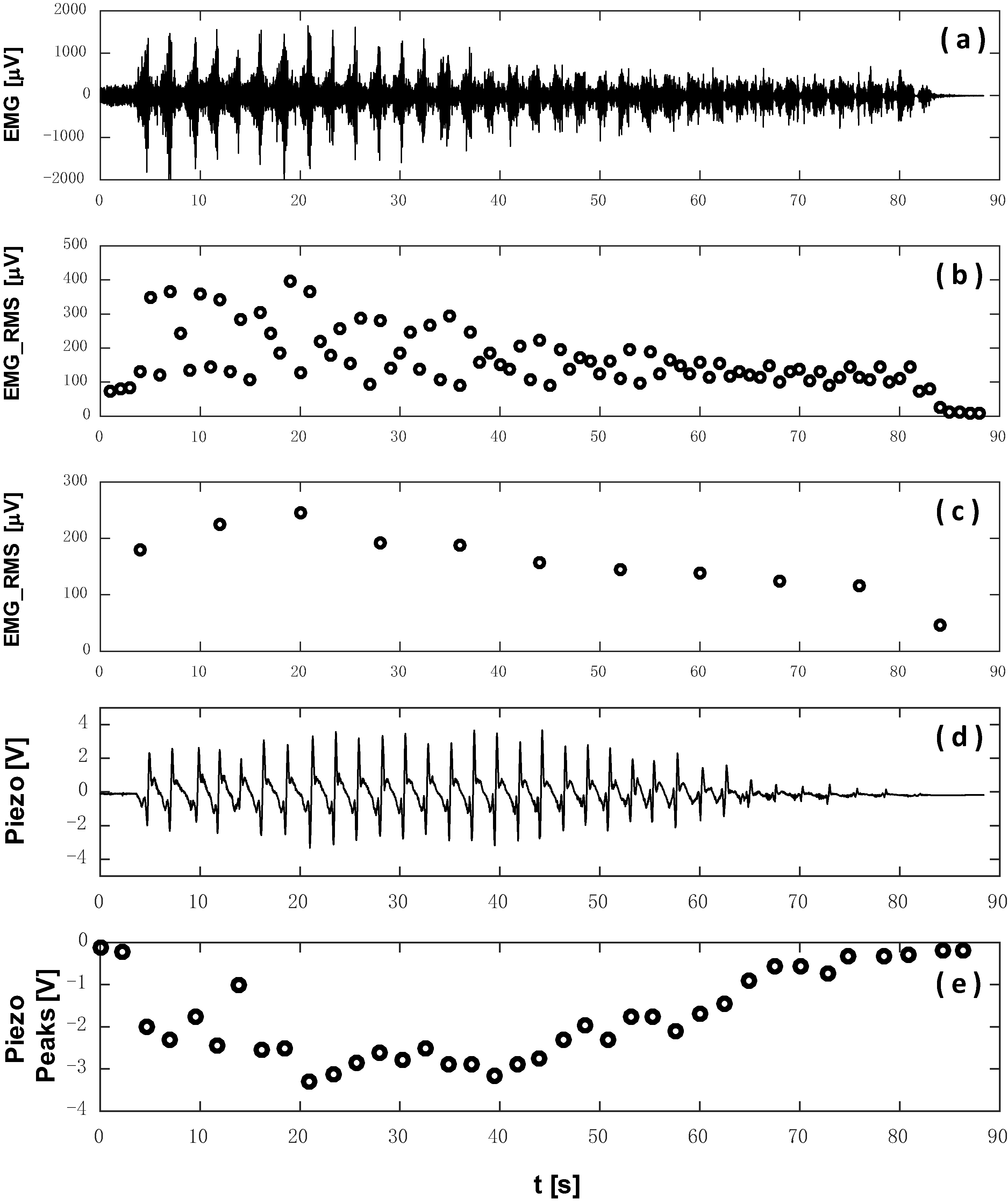
In order to assess muscle fatigue during mass lifting of 1 kg for 150 sec, the EMG_RMS of EMG signal was divided into four equal time segments. The EMG_RMS values were normalized with respect to the maximal value (EMG_RMSn) and then the means
The negative values of the raw Piezo signal correlates with muscle contraction. Therefore, only the negative peaks were derived (Fig. 1d and e). The Piezo signal, during the fatigue assessment was divided, as the EMG signal, into the same four equal time segments. The Piezo signal peaks were normalized with respect to their maximal value (Piezon) and then the means
Analysis of variance (One way ANOVA) and Tukey honest significant difference were applied to compare the distributions of the average EMG_RMSn and the average Piezon across the time segments and to compare the IEMG amplitudes and the Piezo peaks between the different maneuvers. Value of alpha set at 0.05 and P-values less than 0.05 (
Increased intensity and load
Increasing contraction intensity during slow and fast elbow flexion increase significantly (
IEMG envelope (a–d) and Piezo signals (e–h) measured from the biceps during elbow flexion while lifting mass of 0 kg (a&e), 3 kg (b&f), 5 kg (c&g) and 12.5 kg (d&h). The correlation between the average Piezo peaks and the average IEMG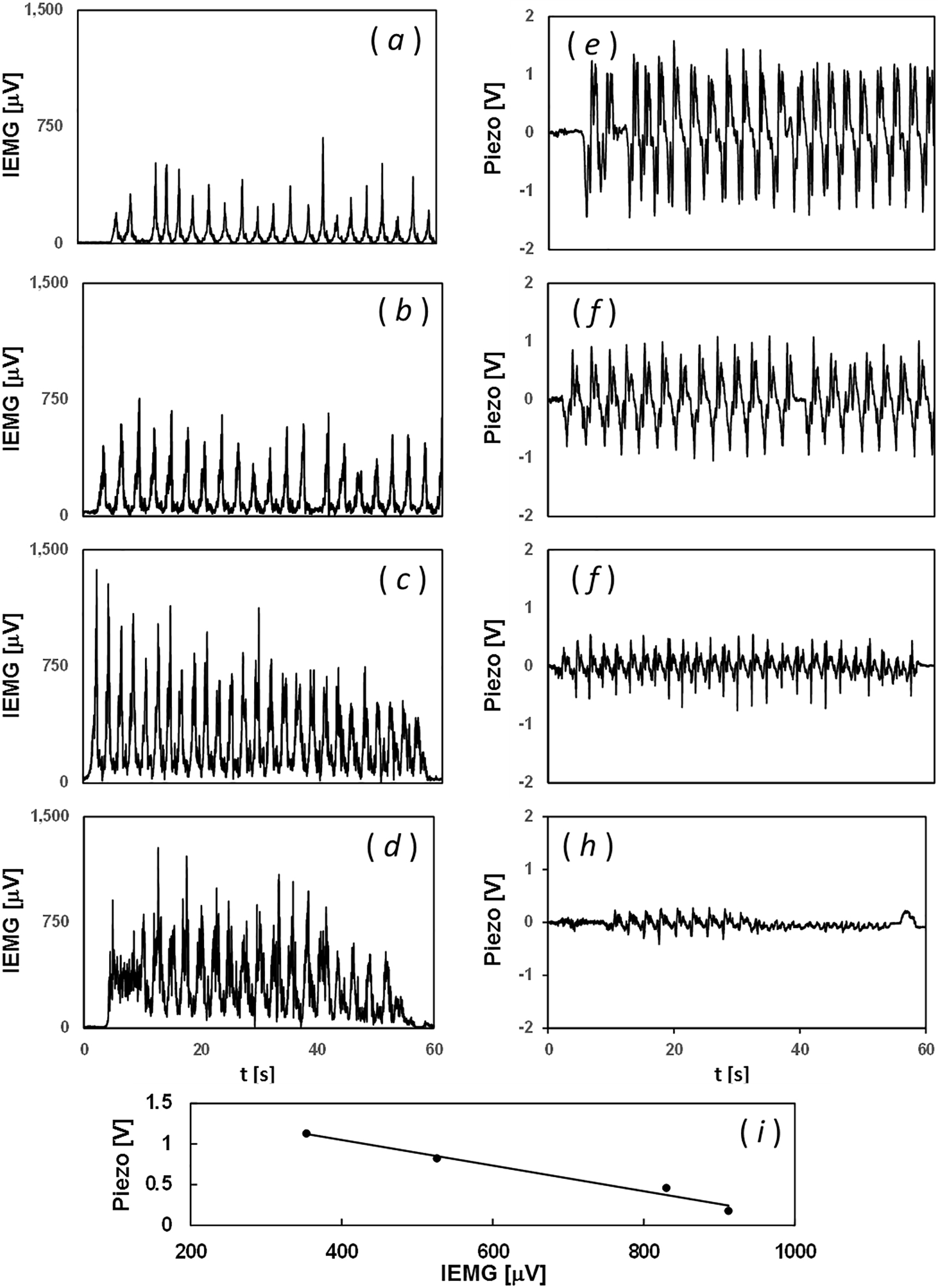
During different mass lifting (0 kg, 3 kg, 5 kg, and 12.5 kg), in all 3 subjects, the IEMG signal increased while the Piezo peaks decreased with the increased mass (Fig. 2). The average IEMG
Significant increase in the average EMG_RMSn and significant decrease in the average Piezon of all subjects was found between time segment 1 or 2 to time segment 3 or 4 and between time segments 3 to 4 (Fig. 3). Average values of the EMG_RMSn and Piezon of all subjects increased from 0.54
The average EMG_RMSn (a,c) and the average Piezon (b,d) at each time segment of one subject (a–b) and the average of all subjects (c–d). The correlation between the average EMG_RMSn and the average Piezon with the increase in the time segments (e).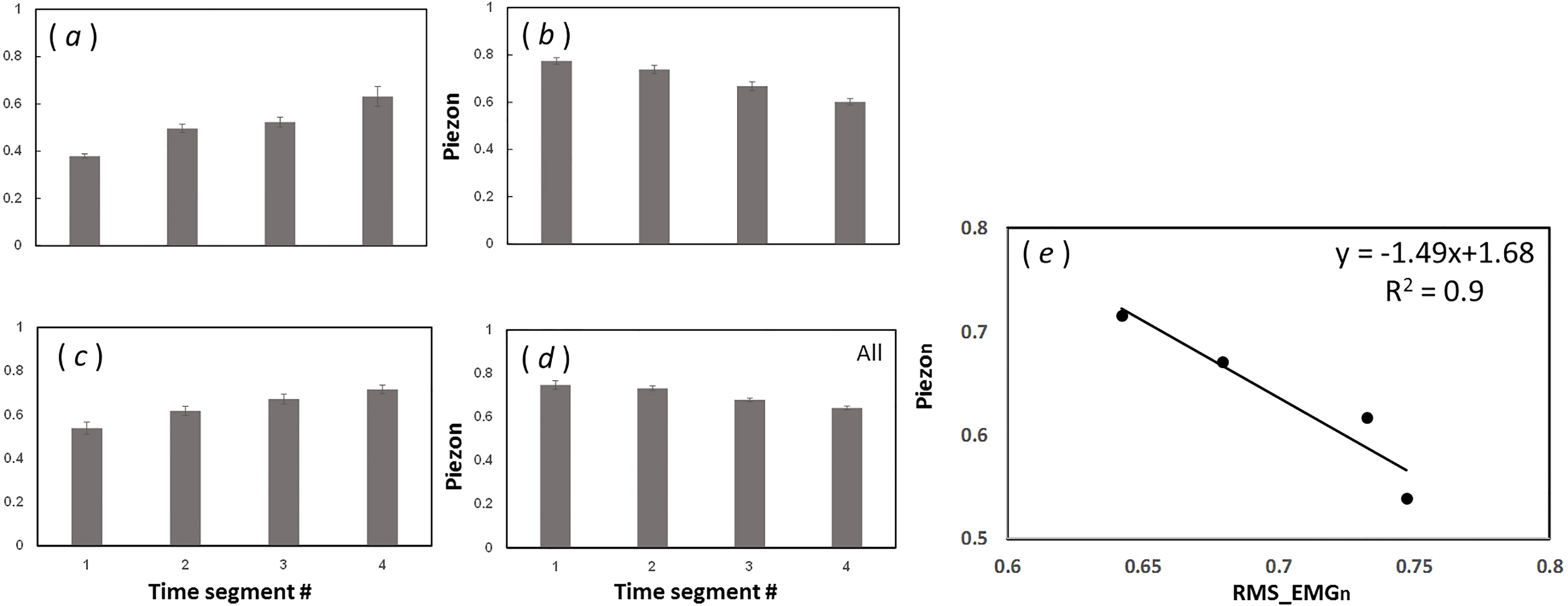
As was expected, correlation was found between the onset and the duration of the biceps EMG and Piezo signals. Other studies found that the time of Piezo peaks (both positive and negative) provide a good indicator of the starts and ends of contractions [2, 26].
Increasing the biceps contractions velocity increase significantly (
Elbow flexion with grip increase significantly (
A significant increase with time in the average EMG_RMSn of the biceps muscle was seen in all subjects during the fatigue maneuver as was expected [34, 35, 36]. The average Piezon, on the contrary, decreased significantly in all subjects, and an opposite linear relation (
Conclusion
Increasing contraction intensity and velocity increase significantly both IEMG and Piezo peaks. Opposite linear relation was found between the average IEMG
Footnotes
Conflict of interest
None to report.