
Abstract
BACKGROUND:
Symmetry of gait is an important component of rehabilitation in stroke patients. Insufficient weight-bearing causes gait asymmetry.
OBJECTIVE:
This study aimed to identify the immediate effects of sufficient weight-bearing on the forefoot during the stance phase using visual feedback.
METHODS:
Twenty-seven individuals with stroke enrolled in this study. All patients were evaluated for gait parameters with and without visual feedback. Visual feedback was provided through a smart application and a beam projector screen that showed a weight shift as a change in color. Spatiotemporal gait parameters were evaluated, resulting in values for a calculated symmetry index, in addition to heel % and toe % temporal values.
RESULTS:
Velocity and cadence were significantly decreased when visual feedback was provided (
CONCLUSION:
This study suggests that visual feedback aids in the improvement of gait symmetry, forefoot weight-bearing on the affected side, and spatiotemporal parameters.
Introduction
Hemiplegic gait in stroke patients is characterized by reduced symmetry, reduced step length, reduced stance time on the affected side, reduced range of motion in swing phase, and reduced balance [1]. Symmetrical and functional gait can improve gait quality in stroke patients. Muscle weakness and reduced sensation on the affected side causes asymmetrical gait due to insufficient weight-bearing during stance phase [2]. Asymmetrical gait results in musculoskeletal problems on the less affected side while it causes decreased bone density [3], decreased gait speed [4], and inefficient energy consumption on the affected side [5]. Furthermore, stroke patients exhibit limited weight-shifting from the heel towards the toe during the stance phase on the paralyzed side and anterior-posterior force on the affected side is decreased at the push-off phase [6]. Insufficient weight shifting to the forefoot on the affected side causes asymmetrical gait, which progresses to decreased function. Instability due to loss of sensation, fear of falling and low balance confidence are the main contributors to decreased function [7].
In order to improve gait ability in stroke patients, visual [8], auditory [9], electromyographic [10], and vibrotactile [11] feedback have been used. A study by Walker et al. [12] reported that visual feedback could potentially prevent falls in stroke patients by providing important information on the walking environment, which leads to improved dynamic balance regulation. Furthermore, visual perception could be improved by practicing various tasks including directional discrimination [13]. A systematic review [14] showed that biofeedback has more positive effects than traditional/placebo therapy on improvement of lower extremity activities such as sitting, sit to stand, standing and walking. Moreover, these effects were reported to remain in effect for several months after the intervention. Feedback shares information on movement errors and methods of correction, which either improves the skill level or speeds up the learning process. Hence, feedback provides fundamental information to maintain motivation during learning [15].
Moreover, an observational study by Talvitie [16] reported that visual and verbal cues are the most effective means of enabling patients to understand their movements and objectives. Byl et al. [17] performed gait training in chronic stroke and Parkinson’s disease patients using visual kinematic biofeedback, and reported that visual feedback was similar to verbal feedback from a therapist. The positive effects were significantly greater than in a control group and included improved gait speed, step length, endurance, gait quality, balance, range of motion, and strength [17]. In a study of nine healthy elderly people and one stroke patient, visual feedback for toe clearance during pre-swing phase effectively reduced the risk of tripping [18]. However, most visual feedback was through center of pressure (COP) movement during gait [19] or provided graphs of kinetic data, such as timing, location, and amplitude of ground reaction forces [8, 17].
Commonly used devices that display weight distribution on the soles as colors are mostly fixed to the ground and the data are frequently provided as feedback for maintaining balance rather than for gait. When the devices are fixed to the ground, the steps are restricted to specific locations during gait. Therefore, these can cause unnatural gait and make it difficult to continuously provide visual feedback during ambulation. To regulate head and trunk movement during gait, stability is required [20]. Vision plays an important role in regulating postural stability [21]. As stability of the head and trunk decreases after stroke, the quality of visual information decreases, resulting in decreased balance [22]. Due to fear of falling and instability, most stroke patients look downwards when walking for visual compensation. Therefore, this study provided real-time visual feedback in front of the patient at eye level to improve visual stability during gait. With weight-shifting during gait, the pressure of the affected lower extremity was displayed in different colors based on intensity to emphasize anterior shifting of the COP during stance phase. Accordingly, the changes in weight-shifting during gait were provided as real-time visual feedback and the effects on spatiotemporal variables of ambulation and symmetry were investigated. The hypothesis of this study is that gait training using visual feedback will increase the forward movement of body weight on the forefoot and increase the symmetry of gait.
Methods
Participants
For this study, 72 stroke patients at the “H” rehabilitation hospital in Jungnang-gu, Seoul, whose symptoms occurred within the two prior years using brochures and oral public activities promotion were recruited. Selection criteria included stroke symptoms for at least six months, mini-mental status examination-Korea (MMSE-K) score of
Procedures
Training with visual feedback.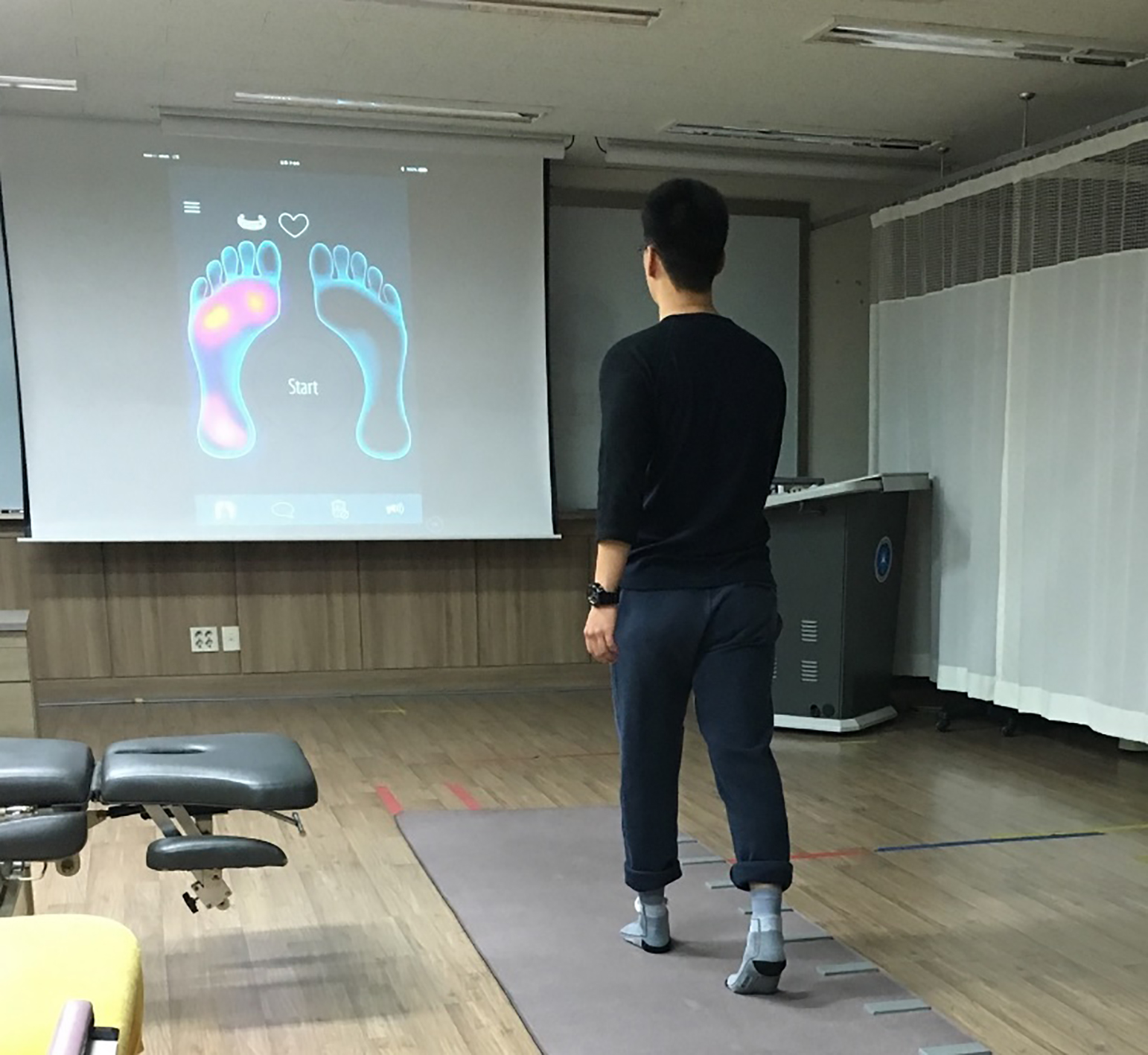
This was a cross sectional study. One participant withdrew for personal reasons during the study. The remaining nine participants were asked to walk on a GAITRite
Screen showing the weight of the left forefoot during gait.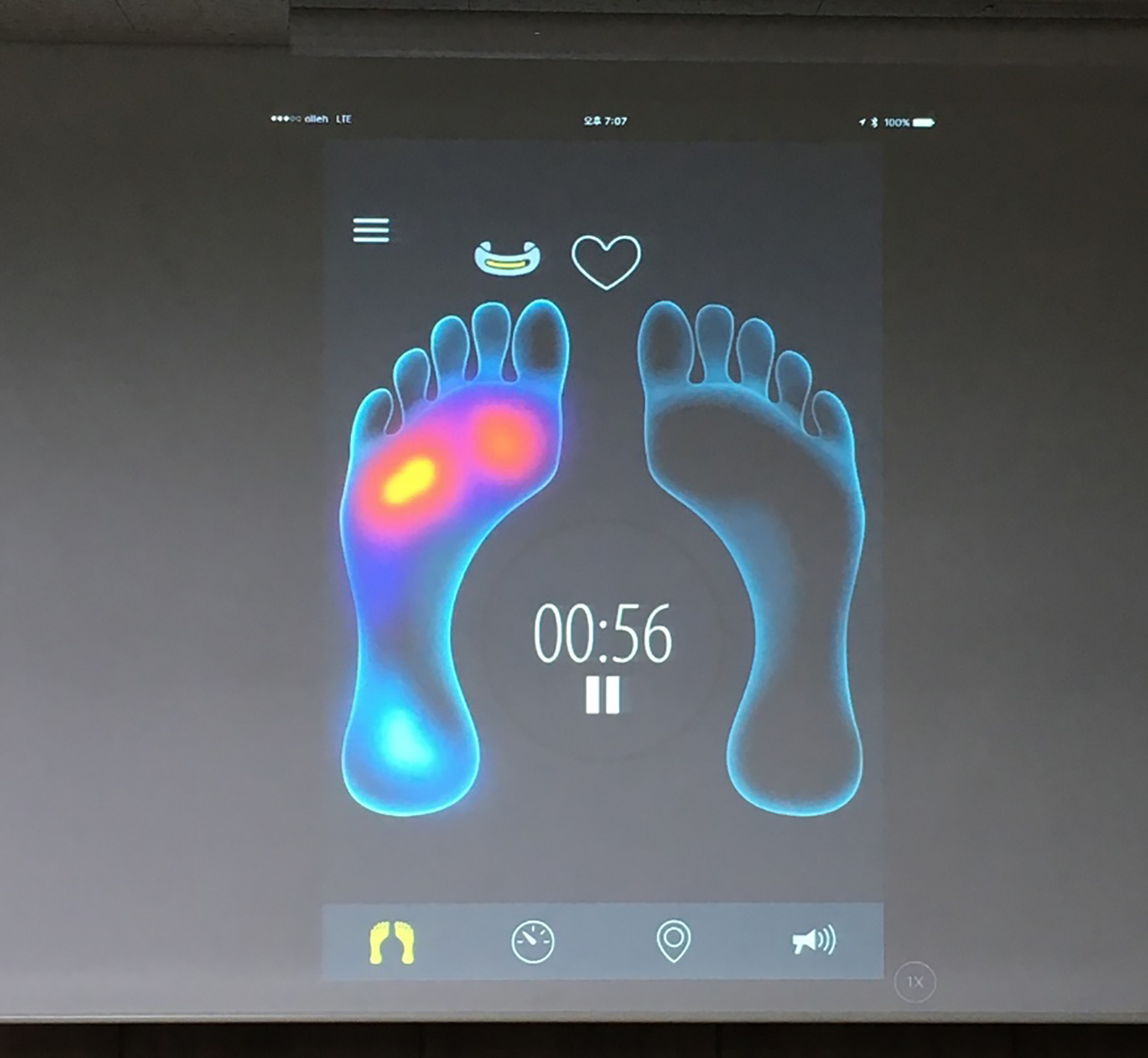
Temporal and spatial gait parameters
This study used a GAITRite
Heel-on time and toe-on time values
Heel-on time and toe-on time values expressed heel only and toe only [27]. Heel only (heel-on time) was defined as the time when only the heel was on the floor. Toe only (toe-on time) was defined as the time when only the toe was on the floor. These values represented two-limb support through a gait cycle. The calculation method is as follows Eqs (1) and (2). Mid on and mid off mean that the middle foot is on and off.
The symmetry index (SI) was calculated using the following Eq. (3), where X is the stance phase time (s) of the gait cycle. As it approaches zero, the SI reflects increasing symmetry.
Data were analyzed using SPSS version 19.0 software for Windows. The Mann-Whitney U test was used for analysis of differences in general characteristics including age, height, weight, MMSE-K score, and post-stroke duration in months. The Wilcoxon signed-rank test was used for nonparametric data since the sample size in this study was small and the data were not normally distributed by the Shapiro-Wilk test. The Wilcoxon signed-rank test was performed to identify differences within paired scores based on pre-test and post-test results for each condition. For all tests, statistical significance was set as 0.05.
Results
The characteristics of the nine participants are listed in Table 1.
General characteristics of the participants
General characteristics of the participants
MAS: Modified Ashworth Scale. MMSE-K: mini-mental status examination-Korea.
The gait speed and cadence were significantly reduced when real-time visual feedback was provided (
Change in spatiotemporal variables with real-time visual feedback for gait
Change in spatiotemporal variables with real-time visual feedback for gait
Change in symmetry index with real-time visual feedback for gait
Change in heel and toe % temporal values
The SI was significantly increased when real-time visual feedback was provided (
Discussion
In this study, real-time visual feedback was performed using smart socks, and gait symmetry and spatiotemporal variables were improved by focusing on weight shifting on the affected forefoot. Gait analysis was performed based on the time of contact between the heel and the floor surface, and between the forefoot and the floor surface using spatiotemporal variables. Unlike graphing of bilateral step length using two bars as proposed in a previous study [28], or determination of COP movement using measured pressure on each plane (frontal, sagittal, transverse plane) on a Wii Fit board [29], the real-time visual feedback method in this study used changes in color based on the magnitude of weight shifting and weight bearing on the soles. The participants were intrigued by the feedback during ambulation. Kim and Krebs [30] reported that real-time visual feedback motivates participation in walking training and may induce gait adaptation, hence providing implicit motor learning. Yang et al. [29] used center of posture for visual feedback in stroke patients and reported decreased severity of pusher syndrome and improved lower extremity motor regulation. Real-time posture feedback improved concentration and promoted awareness of posture and subsequent self-adjustment [29]. The real-time visual feedback used in this study also induced immediate regulation of weight-bearing and improved the quality of gait.
In this study stance phase time was used to investigate gait symmetry because real-time visual feedback focused on weight shifting on the affected foot. Previous studies on gait symmetry reported significant improvement in the step length symmetry when swing resistance was applied using robots on the affected lower limb in stroke patients [31]. The intent was to increase muscle strength in the lower extremity to overcome resistance; however, the real-time feedback in the present study can be applied without resistance as long as visual ability is not restricted. A case study by Lewek et al. [32] also reported that visual feedback effectively improved gait symmetry. Symmetrical changes in gait can be explained as improved ability to correct gait when an error occurs during training [32].
Khallaf et al. [33] used E-med
In this study, real-time visual feedback was provided using a large frontal screen for gait. The advantage of this type of intervention is that stroke patients can perform gait by fixing their gaze to the front rather than looking at the feet or walking with a smartphone application based on walking. However, when using a large screen during walking, it is difficult to provide continuous visual feedback when changing directions. Limitations of this study include the small number of participants, which impedes generalization of the results. This study was limited by space constraints in the ability to provide real-time visual feedback. In addition, different sizes of socks were needed to accommodate different foot sizes. Although the stroke participants in this study (mean age of 50.67 years) reported that the visual color display was very good, objective satisfaction was not studied. In further studies, the participant satisfaction should be confirmed using objective indicators and changes in other gait variables other than symmetry should be investigated for a longer period.
Conclusion
In conclusion, real-time visual feedback was used to induce weight-bearing on the forefoot, and significantly improved gait symmetry and quality in stroke patients. In addition to quantitative changes, qualitative improvement in gait is very important for satisfaction in stroke patients, with the goal of early discharge and return to society.
Footnotes
Acknowledgments
This study was supported by Sahmyook University.
Conflict of interest
None to report.