




Abstract
OBJECTIVE:
To cure atrial fibrillation, the maximum ablation depth (
METHODS:
The finite element model of the endocardial microwave ablation of pulmonary vein was simulated by electromagnetic thermal flow coupling. The ablation power was 30 W and the ablation time was within 30 s. The blood flow in the coupling of fluid mechanics equation and heat transfer equation results in the heat damage. Furthermore, the cause of the different lesion dimensions is the blood flow velocity. The flow velocities were set as 0, 0.02, 0.05, 0.07, 0.12, 0.16, 0.20, 0.25 and 0.30 m/s.
RESULTS:
When the flow velocities were 0, 0.02, 0.05, 0.07, 0.12, 0.16, 0.20, 0.25 and 0.30 m/s, the maximum ablation depth were 6.00, 5.56, 5.16, 5.12, 5.04, 5.01, 4.98, 4.96 and 4.94 mm, respectively; the maximum ablation width were 12.53, 9.63, 9.23, 9.16, 9.07, 9.05, 8.94, 8.91 and 8.90 mm, respectively; the maximum ablation length were 12.00, 11.61, 8.98, 8.59, 8.37, 8.23, 8.16, 8.06 and 8.04 mm respectively. To achieve transmural ablation, the time was 3, 3, 3, 3, 3, 4, 4, 4, 4 s, respectively when the myocardial thickness was 2 mm; the time was 7, 8, 8, 8, 9, 9, 9, 9, 9 s, respectively when 3 mm; the time was 15, 16, 18, 19, 19, 20, 20, 20, 20 s, respectively when 4 mm.
CONCLUSIONS:
When the velocity increases from 0 m/s to 0.3 m/s, the microwave lesion depth decreases by 1.06 mm. To achieve transmural ablation, when the myocardial thickness is 2 mm, 3 and 4 s should be taken when the velocity is 0–0.12 and 0.12–0.30 m/s, respectively; when the myocardial thickness is 3 mm, 7, 8 and 9 s should be taken when 0, 0–0.07 and 0.07–0.30 m/s respectively; when the myocardial thickness is 4 mm, 15, 16, 18, 19, 20 s should be taken when 0, 0–0.02, 0.02–0.05, 0.05–0.12, 0.12 m/s–0.30 m/s.
Introduction
Atrial fibrillation (AF) is the most common sustained tachyarrhythmia. When AF occurs, regular and orderly electrical activity of the pulmonary vein is lost and replaced by the rapid and disordered fibrillation wave. AF is the most serious disorder of atrial electrical activity [1]. It can cause a series of clinical symptoms, such as palpitation, chest distress, dizziness, syncope. It will reduce the quality of life of patients, even threaten the life of patients and increase mortality [2].
The pulmonary vein is an important source of spontaneous electrical activity in the mechanism of AF [3]. Pulmonary vein is not only involved in the occurrence of AF, but also plays an important role in the maintenance of AF. Therefore, pulmonary vein isolation is an effective method to treat AF [4]. Catheter ablation is the most commonly used method to eliminate abnormal electrical signal conduction in pulmonary vein [5]. This method can cause irreversible damage to endocardium and epicardium, block abnormal electrical signal conduction to achieve transmural ablation and finally treat AF. Catheter ablation methods include endocardial ablation and epicardial ablation. The epicardial ablation was performed through the right thoracic wall incision. After the cannula was inserted, the ablation device was guided to the epicardium for ablation [6]. The endocardial ablation was performed through the right femoral vein puncture to establish a venous approach. The atrial septal puncture to the left atrium designated location for ablation after entering the right atrium. This method is commonly used in clinic [7].
At present, radiofrequency ablation is the dominant ablation method. However, due to the presence of the ground pads, there will be complications related to skin burns during radiofrequency ablation [8]. Because of its different heating mechanism, microwave ablation (MWA) will not cause skin burns. In addition, microwave power tends to be deposited in the whole biological medium. The principle of MWA is that the conductive medium absorbs electromagnetic energy. When the electromagnetic wave penetrates, it will be converted into thermal energy [9], making the target tissue temperature reach above 50
In the process of ablation, intracardiac blood flow also plays an important role in ablation effect except to the ablation time and microwave power [14]. Pérez et al. [15] used numerical simulation to analyze the thermal impact of intramyocardial capillary blood flow on radiofrequency cardiac ablation, and proved that all calculation depths excluding blood perfusion terms were larger than those including blood perfusion terms. Lai et al. [16] analyzed the lesion dimensions of endocardial radiofrequency ablation using different convective heat transfer coefficients instead of blood flow. Their results showed that the lesion dimensions of high blood flow were smaller than that of low blood flow. Although some studies have investigated the influence of blood flow on radiofrequency ablation, most studies have estimated the different blood flow velocities by using the convective heat transfer coefficient. However, the values are different in different studies [17]. In this study, the flow rate measured in clinic was directly used to simulation. The effect of heat dissipation caused by pulmonary vein was evaluated by coupling the fluid flow model (Navier-Stokes equation) with the bioelectromagnetic and bioheat transfer model of thermal ablation. This method provides more accurate lesion dimensions. If the ablation is not complete, it is easy to cause recurrence of AF. At present, there is no report on the effect of MWA of pulmonary vein in different blood flow velocities and ablation parameters to realize transmural lesions. It has become a key problem to use appropriate ablation parameters (power and ablation time) to achieve transmural ablation.
The study aims to investigate the effect of different blood flow on MWA of pulmonary vein, and to obtain ablation parameters (ablation power and time) for different myocardial thickness. Only the endocardial MWA was considered in this study. The temperature field distribution of the myocardium and the lesion dimensions of endocardial MWA of pulmonary vein were simulated by finite element method.
Methods
Numerical models
COMSOL Multiphysics software is used to divide mesh and simulation. We established an ideal pulmonary vein model composed of microwave antenna, the myocardium and blood. To simulate the real ablation situation during the operation, the antenna was placed vertically on the endocardium and pressed into the myocardium by 0.5 mm [18]. According to the CT scan results, the diameter was 12 mm in this study [19]. In addition, the thickness of left lower pulmonary vein opening was measured according to the anatomy, the myocardial thickness was 6 mm [20]. The model simulated a similar state of real pulmonary vein orifice isolation and performed MWA at the pulmonary vein orifice [21]. To observe the lesion dimensions, the pulmonary vein orifice was lengthened properly (Fig. 1). The microwave antenna was proposed by Bernardi et al. [22] with frequency of 2450 MHz. The antenna was made of copper, and the inner and outer conductors were made of Teflon. The antenna structure and parameters of each part are shown in Fig. 2.
Myocardial model.
Geometric model of MWA antenna.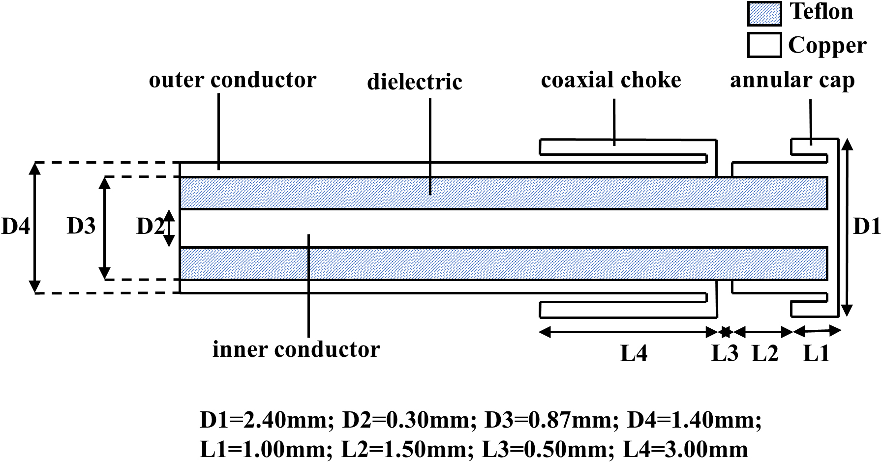
The ablation range and temperature field distribution were obtained by the coupling calculation of electromagnetic field, temperature field and flow field.
The electromagnetic used in the research was considered to be plane shear wave. The wave equation of magnetic strength was solved as follows [23]:
In the formula,
The control equation of temperature field was biothermal equation, which was solved by classical Pennes equation [24]. Considering the flow field, the heat loss caused by the blood flow was added to the right side of the equation. as follows:
In the formula,
The
Among them,
The maximum flow rate was 28.5 ml/s, which was measured by 4D flow MRI [26]. According to the conversion formula of flow rate and velocity:
The ablation time was within 30 s. The lesion dimensions were analyzed under different ablation times. The ablation power was set at 30 W. The ablation parameters set in the study were consistent with those set in conventional MWA treatment [29]. Shortening the ablation time can effectively shorten the operation time. In the simulation, the thermophysical parameters of the myocardium are shown in Table 1 [22].
Material parameters
Boundary conditions of the model.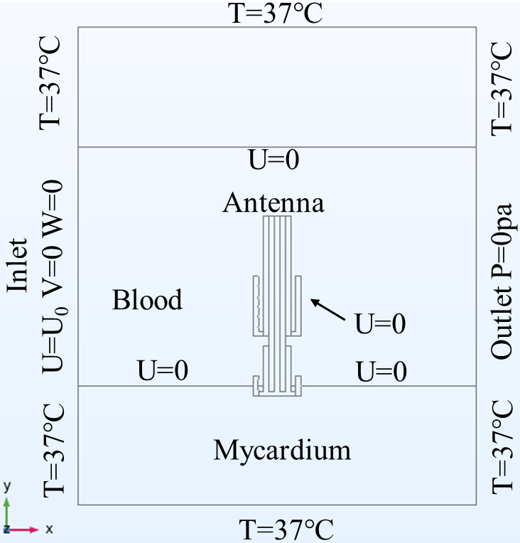
The mesh was automatically divided by COMSOL software. The grid independence study of the model was conducted. Several number of grid cells were explored, including 41,778, 50,259, 90,440, 147,782 and 247,814 at the initial time
(a) Grid convergence results and (b) Models after gridding.
Many scholars use 50
Schematic view of the lesion shape created and nomenclature used to characterize the lesion geometry. MLD: Maximum lesion depth; MLW: maximum lesion width; MLL: Maximum lesion length.
Location of five points (0.5, 1.5, 2.5, 3.5 and 4.5 mm directly below the microwave antenna).
Temperature distribution after 30 s in different velocities, considering (a) lateral and (b) frontal. The solid black line corresponds to the 50
Temperature distribution of five points (0.5, 1.5, 2.5, 3.5 and 4.5 mm directly below the microwave antenna after (a) 10 s, (b) 15 s, (c) 20 s, (d) 25 s and (e) 30 s).
Temperature comparison of MWA in different flow velocities
Figure 7 shows the lateral and frontal temperature distribution of the myocardium after 30 s in different velocities. When the flow velocity is 0 m/s, the maximum myocardial temperature of the myocardium is located on the surface. When there is blood flow, the maximum temperature lies in the interior of the myocardium, and a layer of hypothermia is formed on the surface of endocardium. Due to blood flow, the lesion dimensions become asymmetric and extends to the outflow side. In addition, when the flow rate is 0.02 m/s, the lesion dimensions extend to the outflow side most obviously. When the flow is 0.16–0.30 m/s, the shape of lesion dimensions on both sides is basically symmetrical. The maximum temperature does not appear in the myocardium but the antenna at the flow velocity of 0, 0.02, 0.05, 0.07, 0.12 and 0.16 m/s. The results show that the temperature decreases significantly when there is blood flow instead of no blood flow. The increase in blood flow velocity will reduce the maximum temperature, but the change is not significant when the flow is greater than 0.05 m/s. The influence of flow velocity on MLL is significant compared with the influence on MLD and MLW. The increase in blood flow velocity will decrease the maximum myocardial temperature, but the change of MLL is not significant when 0.16–0.30 m/s.
Figure 8 shows the temperature distribution in different myocardial depth under different velocities. When the ablation time was 10 s, the temperature at 0.5 mm was higher than that at 1.5 mm. When the ablation time was 30 s, the temperature at 1.5 mm at high velocity (0.07–0.3 m/s) was higher than that at 0.5 mm.
Comparison of lesion dimensions in different velocities
The change of lesion dimensions in different blood velocities are studied. And then investigate the appropriate ablation parameters of different myocardial thicknesses in different velocities.
Lesion dimensions in different velocities. (a) MLD. (b) MLL. (c) MLW.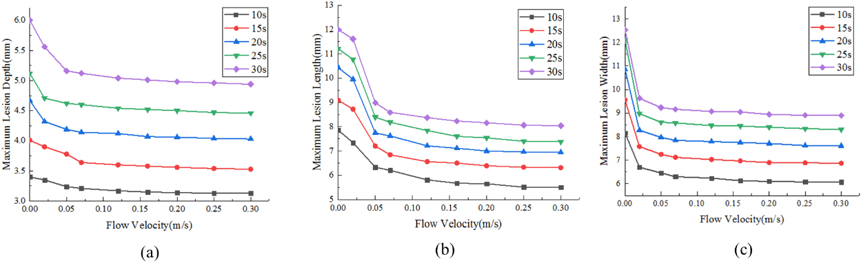
The lesion dimensions with 30 s ablation time in different flow velocities.
Figure 9 shows the lesion dimensions (MLD, MLW, MLL) in three velocities. The results show that MLD, MLL and MLW decreased with the increase in flow velocity. The blood flow in the blood vessel can reduce the heat energy, which leads to incomplete ablation. This effect is called the heat-sink effect. The faster the flow velocity is, the more obvious the heat-sink effect is, and the lower the MLD, MLL and MLW are. However, compared with different ablation times, the results show that different ablation times have more influence on lesion dimensions than the flow velocity. Figure 10 shows the lesion dimensions (MLD, MLW, MLL) in different flow velocities at 30 s. The lesion dimensions decrease with the increase of blood flow. The change is obvious at 0 m/s–0.05 m/s. The trend of change is similar as myocardium temperature.
The ablation time and power of required myocardial thickness in different pulmonary vein velocities
It can be seen that the cooling effect of blood flow will reduce lesion dimensions. However, the decline rate is different. MWA can cause transmural injury to myocardial thickness in different velocities. Table 2 gives the recommended ablation parameters for different myocardial thicknesses (2, 3, 4 mm) in different blood flow velocities of pulmonary vein. When the myocardial thickness is 3 mm and the velocity is 0.07 m/s, 30 W and 8 s can achieve transmural ablation. The corresponding MLW is 5.81 mm, MLL is 5.81 mm and the maximum myocardial temperature is 74.9
The numerical simulation in this study shows that the myocardium temperature decreases with increasing flow velocity. The cooling effect of blood flow will affect the therapeutic effect of MWA of AF. At 0 m/s, the maximum myocardial temperature was located on the surface of the myocardium and the lesion dimensions were around the endocardium. However, the lesion dimensions were semicircular with the increase in flow velocity. Blood flow forms a layer of hypothermia, which significantly reduces the lesion dimensions and the temperature of the antenna. In addition, the maximum myocardial temperature point will move down with the increase of flow velocity. Compared with the temperature of 0.5 and 1.5 mm below the microwave antenna, the temperature at 2.5, 3.5 and 4.5 mm below the microwave antenna is less affected by the flow velocity. It indicates that the cooling effect of blood flow on the surface of myocardium (
Lower flow velocity can lead to larger lesion dimensions. The blood flow leads to the temperature asymmetry of the antenna and the shift of MLL to the blood outflow side. At present, there are few studies using MWA for treating pulmonary vein. The results are compared with those on radiofrequency ablation of AF. The blood flow leads to the asymmetry of the lesion dimensions, which is consistent with González et al. [37]. When Yu et al. [38] increased the velocity from 0.067 to 0.1 m/s, the myocardial temperature did not decrease significantly, which was consistent with the results in this study. Jain et al. [39] found that with the increase in flow velocity, the shift of the lesion dimensions to the outflow side will be weakened. In the study, the antenna pressed the myocardium vertically and the material properties were set uniformly. Therefore, the cause of uneven temperature distribution is blood flow. When the flow velocity increases, the electrode temperature distribution and MLL are more symmetrical. However, the antenna temperature distribution and MLL are symmetrical when the blood is still (0 m/s). It shows that the asymmetry between the antenna temperature distribution and MLL achieves the maximum value between 0–0.07 m/s, and then gradually becomes symmetrical with the increase of flow velocity. The maximum temperature does not appear in the myocardium but the antenna at the flow velocity of 0, 0.02, 0.05, 0.07, 0.12, 0.16 m/s. In the actual situation, the antenna temperature will change because water cooling is set in the microwave antenna, so the maximum temperature at this time will not be taken.
The MLD, MLW and MLL decreased with the increase in flow velocity. However, the myocardial temperature is not reduced by equal amplitude with the increase in flow velocity. Table 2 shows the ablation parameters (MLD, MLL, MLW and maximum temperature) of different flow velocities to achieve transmural ablation for different myocardial thickness. To achieve therapeutic effect, the ablation time should be at least 4, 9 and 20 s when the myocardial thickness is 2, 3 and 4 mm. Although the maximum myocardial temperature at 0 and 0.02 m/s is higher than 100
There are also some limitations in this study. First, the change of myocardial thermophysical properties with temperature is not considered. Second, the ablation parameters at different flow velocities were summarized when the myocardial thickness was 2, 3, 4 mm. More myocardial thickness and ablation parameters will be considered in the future. The in vitro experiments will be added to verify the experimental results in the future.
Conclusion
In this study, the effect of blood flow on MWA of AF was investigated. These results indicate that the cooling effect of blood flow makes the maximum myocardial temperature moves to the interior of the myocardium. The temperature around the antenna and the lesion dimensions is asymmetric when the flow velocity is less than 0.16 m/s. When the flow velocity increased from 0 to 0.16 m/s, the lesion dimensions change obviously. The ablation parameters in different flow velocities are given, which can provide a reference for future research.
Footnotes
Acknowledgments
This research is supported by the National Natural Science Foundation of China (31771021, 11832003), and Beijing International Science and Technology Cooperation Base for Intelligent Physiological Measurement and Clinical Transformation.
Conflict of interest
None to report.