



Abstract
BACKGROUND:
Artificial intelligence (AI) technology is a promising diagnostic adjunct in fracture detection. However, few studies describe the improvement of clinicians’ diagnostic accuracy for nasal bone fractures with the aid of AI technology.
OBJECTIVE:
This study aims to determine the value of the AI model in improving the diagnostic accuracy for nasal bone fractures compared with manual reading.
METHODS:
A total of 252 consecutive patients who had undergone facial computed tomography (CT) between January 2020 and January 2021 were enrolled in this study. The presence or absence of a nasal bone fracture was determined by two experienced radiologists. An AI algorithm based on the deep-learning algorithm was engineered, trained and validated to detect fractures on CT images. Twenty readers with various experience were invited to read CT images with or without AI. The accuracy, sensitivity and specificity with the aid of the AI model were calculated by the readers.
RESULTS:
The deep-learning AI model had 84.78% sensitivity, 86.67% specificity, 0.857 area under the curve (AUC) and a 0.714 Youden index in identifying nasal bone fractures. For all readers, regardless of experience, AI-aided reading had higher sensitivity ([94.00
CONCLUSION:
The AI model might aid less experienced physicians and radiologists in improving their diagnostic performance for the localisation of nasal bone fractures on CT images.
Introduction
The nasal bone, which is a prominent part of the facial skeleton, is vulnerable to traumatic fractures. A nasal bone fracture is the most common injury in the otolaryngology emergency department and can be caused by even relatively weak forces [1, 2]. Moreover, a nasal bone fracture requires an early diagnosis and accurate initial treatment. Improper management could result in deformity and functional discomfort [3]. A facial computed tomography (CT) analysis is an effective tool for diagnosing a nasal bone fracture. However, using a CT analysis to diagnose nasal bone fractures requires time-consuming and repetitive work by radiologists [4]. As the diagnosis of nasal fractures requires rapid and accurate determination to prevent nasal deformity, a new rapid diagnostic technique is needed.
Artificial intelligence (AI) has been applied to various medical technologies. Currently, an increasing number of studies attempt to use AI techniques to detect fractures as an adjunct to a clinical diagnosis. For example, Olczak et al. [5] demonstrated that AI technology with a deep-learning approach could be used to diagnose ankle and wrist fractures on plain X-rays. Oka et al. [6] developed an AI system that can diagnose distal radius fractures using bi-planar X-ray images and found that the AI system has a high diagnostic accuracy with a diagnostic rate of 98%. A multicentre study conducted by Sato et al. [7] developed a new AI system for hip fractures on plain X-rays and found that the diagnostic accuracy of hip fractures could be improved using this system. A recently published meta-analysis revealed that the diagnostic performance in fracture detection was not significantly different between the clinician and AI performances [8]. These studies suggest that AI technology is a promising diagnostic adjunct in fracture detection.
However, few studies describe the improvement of clinicians’ diagnostic accuracy for nasal bone fractures with the aid of AI technology. Therefore, the aim of this study is to determine the value of the AI system in improving the diagnostic accuracy for nasal bone fractures.
Methods
Patient inclusion
This retrospective study was approved by the Ethics Committee of our hospital in accordance with the Declaration of Helsinki. All participants provided written informed consent for publishing the data.
A total of 252 consecutive patients who had undergone facial CT between January 2020 and January 2021 were enrolled in this study. Patients with image artifacts or who did not meet the diagnostic requirements were excluded. All CT images were evaluated by two radiologists, with 20 years of experience, for the presence or absence of a nasal bone fracture. If the results differed, a decision was made after a discussion. Consensus was achieved after consulting a third senior orthopaedist with 25 years of orthopaedic professional experience. A total of 152 patients with confirmed fractures by the expert radiologists were included in the fracture group, and 100 patients without fractures were included in the non-fracture group.
The enrolled cases were randomly selected for training (
Model building
Multislice CT scans were performed with 120 kVp tube potential and 180 mAs tube current using GE Optima CT 660 CT (General Electric Healthcare, Chicago, IL, USA). The CT protocols were as follows: spiral scan mode; pitch, 0.516; layer thickness, 0.625 mm; and interval, 0.312 mm. The scan ranged from orbit to palatum durum. The bone algorithm reconstruction was used to observe the bone with a CT window width of 4,000 Hu and window level of 700 HU using the GE AW4.6 workstation. The CT images of these patients were downloaded from the Picture Archiving and Communication System in Digital Imaging and Communications in Medicine format. After masking the patients’ personal information, the CT images were uploaded to the AI workstation for analysis.
The AI algorithm based on the deep-learning algorithm of the Feature Pyramid Network (FPN) was engineered, trained, optimised and validated to detect and localise the fractures [9]. Input data were obtained by cropping the nasal bone region from the facial CT images, and the entire nasal bone image was implemented in a three-dimensional isotropic voxel form (64 px
The training set was used to train the AI model. The validation data were used to validate the model.
Readers and readings
After the AI model was developed, the CT images of 252 patients in the fracture and non-fracture groups were independently reviewed by 20 readers (10 radiologists and 10 physicians) with various levels of experience (range: 1–15 years). The readers were blinded to the clinical data and experts’ judgments. Before the study, the readers were trained to use the software and perform the required task. All readers were randomly presented with the radiographic images twice – once with the assistance of AI software and once without assistance, with a minimum washout period of 3 months. Each of these readers provided their informed consent to participate in this study.
The accuracy, sensitivity and specificity with and without the aid of the AI system among readers with different experience were calculated and compared.
Statistical analysis
All data in this study were analysed using the Statistical Package for the Social Sciences (version 22.0; IBM Corp. Armonk, NY, USA) and MedCalc statistical package (version 19.0.5; MedCalc Software Ltd., Ostend, Belgium). The categorical data were described as numbers and percentages and compared using the Chi-square test. The quantitative data were described as mean
Results
A total of 252 patients with a mean age of 45.65
Patient characteristics in the non-fracture and fracture groups
Patient characteristics in the non-fracture and fracture groups
Note: BMI, Body Mass Index.
In the validation set, there were four false negative, seven false positive, 26 true negative and 39 true positive results for the AI model using the expert’s readings as the reference standard for detecting nasal bone fractures. According to these results, the AI model was calculated as having 84.78% sensitivity (95% confidence interval [CI]: 71.10, 93.70) and 86.67% specificity (95% CI: 69.30%, 96.20%) when distinguishing between normal and fractured nasal bone. The Youden index was 0.714. The AI model showed a diagnostic performance with an AUC of 0.857 (95% CI: 0.758, 0.928) for differentiating nasal bone fractures from non-fracture cases (Fig. 1). The representative CT images of the AI model’s diagnoses are shown in Fig. 2.
The diagnostic performance of AI model in diagnosing the nasal bone fractures.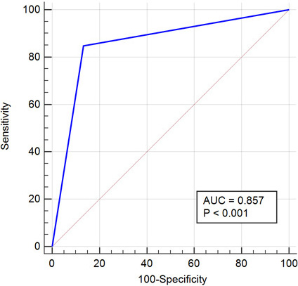
The representative CT images that were diagnosed by AI models. (A) A case of true positive by the AI model; (B) A case of false negative by the AI model; (C) A case of false positive by the AI model. The white arrow indicated the fractures detected by 2 expert radiologists (ground truth). The blue box indicated the fractures detected by the AI model.
A total of 20 readers (10 radiologists and 10 physicians) with various levels of experience were included to determine the value of the AI model in improving diagnostic accuracy for nasal bone fractures. Among them, 10 readers (50%) had experience of 1–5 years, six (30%) had experience of 6–10 years and four (20%) had experience of 11–15 years. The characteristics of the readers are shown in Table 2.
The characteristics of readers
Diagnostic performance of 20 readers for fracture detection with and without AI assistance
Note: Numbers in parentheses are 95% CIs. AI, artificial intelligence; AUC, area under curve.
For all readers, as shown in Table 3, the sensitivity was estimated at 83.52%
Improvement in diagnostic performance of fracture detection with AI Assistance: Subgroup analysis
Note: Numbers in parentheses are 95% CIs, and numbers in brackets are
The subgroup analyses are shown in Table 4. With the aid of AI, 10 physicians had an average of 11.65% (95% CI: 4.57, 18.73;
With the aid of AI, the sensitivity, specificity and AUC were significantly improved in readers with 1–5 years or 6–10 years of experience (all
In this study, we developed an AI model based on the deep-learning algorithm for the detection of nasal bone fractures and validated its diagnostic performance in a splitting dataset. The main findings can be summarised as follows: 1) the AI model had an acceptable performance in distinguishing between patients with nasal bone fractures and those without; 2) the AI-aided reading significantly improved the sensitivity, specificity and accuracy of fractured nasal bone detection by radiologists and physicians; and 3) the improvement of the diagnostic performance in the AI model was mainly for less experienced readers. Our study suggests that the AI model might be a useful tool for clinicians for the precise diagnosis of nasal bone fracture, which has not been fully addressed before.
Several studies have investigated the efficacy of AI technology for fractures. Oka et al. [6] developed an AI system that could diagnose distal radius fractures using bi-planar X-ray images and found that the AI system had a high diagnostic accuracy with a diagnostic rate of 98%. Tomita et al. [10] developed a convolutional neural network algorithm to detect osteoporotic vertebral fractures in CT images, and the detection accuracy was 89.2%, which was equivalent to that of experienced radiologists. Moreover, the study by Gan et al. [11] demonstrated that AI technology exhibited a diagnostic ability for detecting distal radius fractures like that of orthopaedists. Sato et al. [7] developed a new AI system with a deep-learning algorithm and found that the AI system achieved a high diagnostic performance for hip fractures, regardless of the experience of the doctors. Ozkaya et al. [12] found that AI technology based on convolutional neural networks was useful for scaphoid fracture diagnoses, particularly in the absence of an experienced orthopaedist or hand surgeon. Similarly, Liu et al. [13] found that the AI algorithm was an efficient method for the clinical diagnosis of tibial plateau fractures and could be a useful assistant for orthopaedic physicians. In addition, the AI algorithm has been widely used for the diagnosis of other diseases. Based on the patients’ lung function and symptoms, Badnjevic et al. [14] developed an expert diagnostic system to distinguish between patients with asthma, chronic obstructive pulmonary disease (COPD) or normal lung function, which could correctly identify patients with asthma and COPD with a sensitivity of 96.45% and a specificity of 98.71%. Additionally, an artificial neural network-based expert diagnostic system was reported to have excellent diagnostic efficiency for five aneuploidy syndromes [15]. Similarly, the AI-based model could achieve high accuracy (95.14%) for the real-time classification of epileptic seizures [16]. Compared to these studies, our model may be simpler and more practical because it can help less experienced physicians to diagnose nasal fractures using only CT images. The evidence suggests that AI algorithms will probably play a more important role in aiding diagnosis than as fully autonomous replacements for radiologists.
Different from the limb long and flat bones, the anatomical structure of nasal bone and its adjacent blood vessels is complex, which creates a challenge for the construction of AI models. The study by Seol et al. [17] developed an AI-aided diagnosis system to determine nasal bone fractures based on a three-dimensional-convolutional neural network. The AI model was determined to have higher accuracy and reliability in the automatic classification of nasal fractures [17]. In the present study, we developed an AI model for fractured nasal bone detection based on the deep-learning algorithm of FPN. The deep-learning AI model had a sensitivity of 84.78%, specificity of 86.67%, AUC of 0.857 and a Youden index of 0.714 when identifying nasal bone fractures. Previously, the sensitivity of conventional radiography (such as CT) in detecting the fracture line of the nasal bone was approximately 79% [18, 19]. Our AI model had higher sensitivity and accuracy.
We invited 20 readers with various experience to read CT images with or without AI to determine the value of the AI system in improving the diagnostic accuracy for nasal bone fractures. As expected, we found that our AI model significantly improved the diagnostic accuracy of radiologists or physicians in fractured nasal bone detection. For all readers, regardless of their experience, AI-aided reading had higher sensitivity, specificity and AUC compared with reading without AI. The subgroup analyses showed that less experienced readers were more likely to benefit from AI-aided reading, which is consistent with other studies [20, 21]. With the aid of AI, the sensitivity, specificity and AUC were significantly improved in readers with experience of 1–5 and 6–10 years. For experienced readers (with experience of 11–15 years), we found no evidence that AI could improve sensitivity and AUC because the experienced readers already had high accuracy regarding diagnosing fractures without AI. Therefore, AI-aided reading has the potential to minimise the gap between less experienced readers and experienced readers for fractured nasal bone detection.
The present study has several limitations. First, this is a retrospective, single-centre study with a relatively small sample size. The results might be susceptible to selection bias because the AI model in this study was developed using a small amount of learning data. Therefore, further large, multicentre studies are needed to examine the diagnostic accuracy of the AI model. Second, the reference standard for nasal fractures in this study was based on the readings by senior radiologists. Although their diagnostic accuracy is high, this detection method may still misdiagnose cases. Third, evaluating lateral nasal wall injuries on conventional radiographs is more difficult than the midline of nasal bone [18, 22]. Therefore, the detection of nasal wall injuries may have a higher misdiagnosis rate. However, in this study, we did not conduct a subgroup analysis to evaluate the efficacy of AI-aided reading in different types of nasal bone fractures. This problem should be determined in the future. Finally, in this study, we did not record the reading time when readers reviewed the CT images with the aid of AI. A shorter reading time is one of the potential benefits of AI. Even if it is quicker by only a few seconds per radiographic examination, a meaningful amount of time will be saved for radiologists who may read 200–300 radiographs per day [23]. Therefore, further studies are needed to determine whether AI-aided reading could reduce the reading time of nasal CT images.
Conclusion
We developed an AI model based on the deep-learning algorithm and confirmed that the AI model had an acceptable performance when identifying patients with nasal bone fractures. Our results suggested that the AI model might aid less experienced physicians and radiologists by improving their diagnostic ability for the localisation of nasal bone fractures on CT images.
Funding
The study was supported by the S&T Program of Hebei (No. 20377733D) and Youth Science and Technology Project of Hebei Provincial Health and Family Planning Commission (No. 20210815).
Ethics statement
This study was conducted in accordance with the Declaration of Helsinki and received approval from the Ethics Committee of the Second Hospital of Hebei Medical University. Written informed consent was obtained from all participants.
Availability of data and materials
All data generated or analyzed during this study are included in this article.
Footnotes
Acknowledgments
None to report.
Conflict of interest
None of the authors has any personal, financial, commercial, or academic conflict of interest to report.