
Abstract
BACKGROUND:
Due to the unfavourable neck-shoulder muscle loads caused by poor posture, the people who use the laptop for a long time may face the risk of neck and shoulder injuries.
OBJECTIVE:
The purpose of this study investigates the impact of the screen height on the muscle activation of head flexion, neck and shoulder, and the cervical spine torque to provide the favorite screen height for laptop user.
METHODS:
Twelve healthy young participants completed a 15-minute task of the reading at the four different screen heights. sEMG signals of the splenius capitis (SC) and upper trapezius (UT) were measured and calculated the root mean square (RMS) and mean power frequency (MPF) to determine muscle fatigue. The different height of laptop users was simulated and the forces on the spine of users at different screen heights were analyzed by Jack.
RESULTS:
Adjusting the height of the laptop screen can effectively reduce head flexion and muscle activity of SC and UT, and has a positive effect on reducing fatigue of SC, but has no significant effect on UT.
CONCLUSIONS:
Adjusting the height of the laptop screen can delay the occurrence of SC muscle fatigue to a certain extent. The joint analysis of sEMG spectrum and amplitude reports that the screen heights of D15 and D45 have the highest and the lowest frequency of fatigue, respectively. At the same time, the moment of spineT1/T2 and spineL4/L5 decrease with the increase of screen height.
Introduction
Nowadays, using computers for office work has become a common thing in life and is taken for granted by the student population [1, 2]. The majority of individuals, particularly students, maintain a stationary posture for prolonged periods while using electronic devices like laptops and tablet computers to work or study [3, 4]. According to a study, the majority of students utilize computers for more than eight hours per day, and a significant number of female university students reported experiencing musculoskeletal discomfort during or after computer usage [5]. Prolonged laptop usage results in extended tension in the posterior neck muscles, significantly heightening the chance of developing musculoskeletal disorders [6–9].
According to recent findings, over 53.4% of students have encountered computer-related musculoskeletal symptoms in their upper limbs [10], with the neck being one of the most susceptible sites for musculoskeletal disorders (MSD) in computer users [11], and the incidence of musculoskeletal disorders of the neck has been reported to be 65% –75% [12], with a tendency to be at a younger age. There has been a notable rise in the prevalence of neck musculoskeletal disorders (MSD) among individuals under 30 years, with jumping from 26% in 1996 to 37% in 2016 [13]. MSD pose not only physical challenges [14], but also result in substantial economic costs [15, 16].
Therefore, investigating the potential risk factors for neck and shoulder discomfort associated with laptop could be effective in preventing and reducing the occurrence of associated musculoskeletal disorders. Studies have shown that working with laptops is more likely to result in neck hyperflexion compared to desktop computers [17–19]. This flexion may be caused by the low position of the laptop screen, which requires the user to look down at the screen. This poor working posture and discomfort [18, 20] may be due to the interconnected screen and keyboard of laptop computers, which are designed in such a way as to improve the compactness and portability of the laptop at the expense of the user’s comfort and health while using it [21] and the screen height setting of the laptop computers is also well below the recommended working height [22, 23].
Some studies have attempted to improve the discomfort caused by working on a laptop from an ergonomic perspective by changing the screen height. Berkhout et al. [24] found that using an assistive device to increase the height of the laptop screen can reduce neck discomfort and mechanical load. Asundi et al. [22] compared the head and neck posture of using a laptop with a non-tilt bracket, a 12° tilt bracket, and a 25° tilt bracket. The results showed that using a tilt bracket can reduce head forward tilt and neck bending. Straker et al. [25] compared the 3D head, neck, and upper limb postures of 18 young men and 18 young women working at different screen heights, and found that lower screen heights increased the degree of head and neck bending. Therefore, increasing the height of the laptop screen can improve the posture of the head and neck.
Some studies have also used electromyography, a biometric technique, to analyze the neck and shoulder muscle activity of laptop users at different screen heights, and objectively evaluate the effect of changing the screen height on muscle activity. Yadegaripour et al. [26] compared 8 minutes of work on a traditional laptop screen height and a laptop with a higher screen height, and found that adjusting the laptop screen height can effectively reduce the cervical (–4.53°) and thoracic (–3.14°) flexion angles, as well as the muscle activity of the cervical pinch muscle (–10.31%) and multifidus muscle (–15.57%). Lee and Liu [27] collected the activities of the sternocleidomastoid muscle, trapezius muscle, clavicular muscle, and erector spinae muscle after watching the screen for 1 minute at 5 viewing angles (+40°, +20°, 0°, –20°, –40°). The results showed that the neck and chest flexion angles increased with the increase of the viewing angle, increased activity of trapezius and erector spinae at higher or lower screen heights, and the activities of the cervical pinch muscle decreased with the increase of the viewing angle. Maria et al. [28] collected the electromyogram of neck and shoulder muscles of 10 healthy subjects while they performed mouse-driven interactive tasks at screen heights of 80, 100, and 120 cm respectively. They found that with the increase of screen height, the neck became significantly more upright, and the muscle activity of the cervical muscles was significantly reduced. The subjects showed a more backward-tilted trunk position, and the trapezius muscle activity of some subjects gradually decreased.
However, the above study only showed that increasing the height of the laptop screen could improve the posture of the head and neck and reduce the muscle activity of the cervical muscles, but did not further explore the impact of changing the screen height on neck and shoulder muscle fatigue by monitoring the muscle state of neck and shoulder and not given the favorable height of laptop screen for alleviation of the muscle fatigue. In addition, laptop users are of different heights, and their sitting posture and eye height are also different. Each laptop user should have their own reasonable screen height. Therefore, this study investigated the impact of changing the screen height on neck and shoulder muscle fatigue through electromyography, and provided reasonable screen height recommendations for laptop users of different heights.
Experimental methods
The hybrid system for neck and shoulder comfort evaluation mainly includes both real and virtual experimental environments, and the flow is shown in Fig. 1. Among them, the real environment consists of surface electromyographic (sEMG) signal acquisition, through which real muscle response results can be obtained from the real experimental environment. The virtual experimental environment is analyzed by Jack to determine the forces on the thoracic and lumbar vertebrae of users of different heights at different screen heights.

Hybrid system flow for neck and shoulder comfort evaluation.
Participant
The subjects are 12 postgraduate students, including 8 males and 4 females. The mean age was 25.6±1.3 years, height 175.1±6.3 cm, and weight 65.7±12.1 kg (Table 1). Subjects with any chronic pain or injuries in the back or neck within the last 12 months were excluded, and those who had not participated in strenuous exercise within 72 hours prior to the experiment were included to ensure good muscle condition. All subjects were mentally fit and had no signs of sleep deprivation or depression. The computer mouse was operated by the subjects’ right hand during daily use.
General information of subjects
General information of subjects
A 12-channel sEMG device (Core Future, Wuxi, Jiangsu, China) was used to measure the sEMG signals of the left and right splenius capitis (SC) and the left and right upper trapezius (UT), and the sampling rate of the sEMG device was 1000 Hz. Silver-silver chloride (Ag-AgCl) electrodes were used for this experiment, and the distance between the center of the electrodes was 20 mm. Use Jack to analyze the forces on the thoracic and lumbar vertebrae of users of different heights at different screen heights.
Experimental design
The four screen height settings were used in the experiment i.e. 15 cm, 25 cm, 35 cm, 45 cm (D15, D25, D35, D45), the screen height is the distance from the table top to the center of the screen, and the screen height is varied in increments of 10 cm. The table has a height of 75 cm, and the chair featured a backrest and a seat height of 45 cm, as shown in Fig. 2. Participants completed a 15-minute task reading literature at the four different screen heights, with the order of heights randomized. An external keyboard and mouse served as the input devices for the experiment, and participants rested for at least 20 minutes between tests to allow their neck and shoulder muscles to return to baseline.
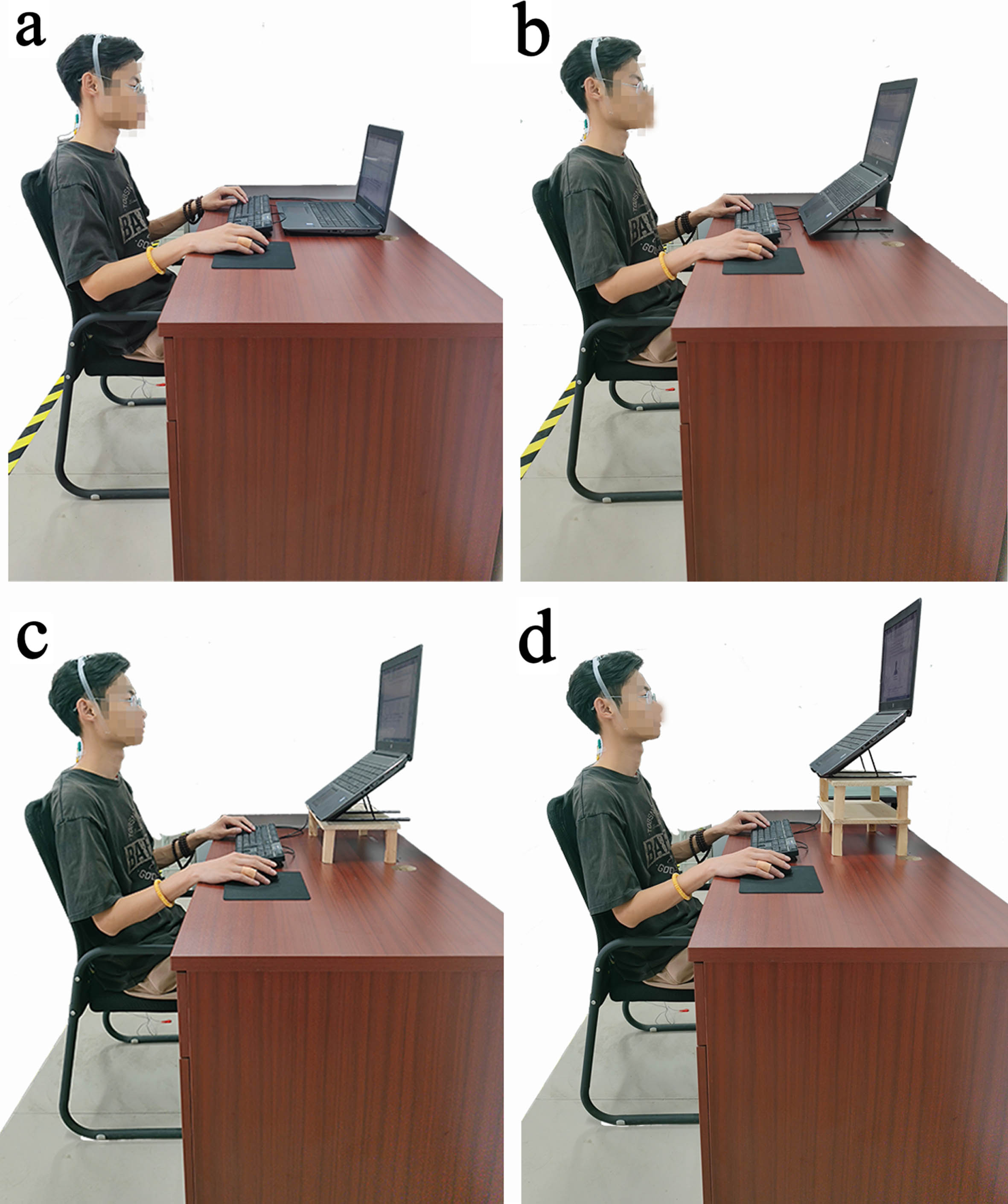
Laptop workstation setup (with external keyboard and external mouse). (a): Conventional setup, laptop flat on the desktop with a screen height of 15 cm (D15); (b): Screen height raised to 25 cm (D25); (c): Screen raised to 35 cm (D35); (d): Screen raised to 45 cm (D45).
Then, the laptop user model and office scenario model were established in the Jack software. According to different screen heights, the digital human was analyzed using the Static Strength Prediction module. The variation trends of the moment between the first and second thoracic vertebrae (spineT1/T2 M) and the moment between the fourth and fifth lumbar vertebrae (spineL4/L5 M) were obtained.
Participants were shown the experimental tasks and then positioned in front of the testing apparatus, as given in Fig. 2, and freely become familiar with the experimental setting. The 75% concentration medical alcohol were used to repeatedly wipe the subjects’ neck and shoulder where the electrode sheet was pasted and eliminate any sweat stains, dead skin, and cuticle from the skin surface. If required, a razor was employed to remove surface hair. After the alcohol on the skin surface had dried, the disposable electrode sheet was placed along the direction of muscle fibers for the measurement of the sEMG signals of the SC and UT [29]. The electrodes were positioned parallel to the spine on both sides of 1 cm apart from the C5 spine to measure the SC. At the same time, the electrodes were placed at the midpoint between C7 and the acromion to measure the UT, as given in Fig. 3(a). The electrodes were secured using medical tape [26]. To measure head posture, a postural measurement sensor was positioned at the top of the head where the coronal suture meets the sagittal suture, as well as on the C7 spinous process, recording the angle of head bend in the sagittal plane.
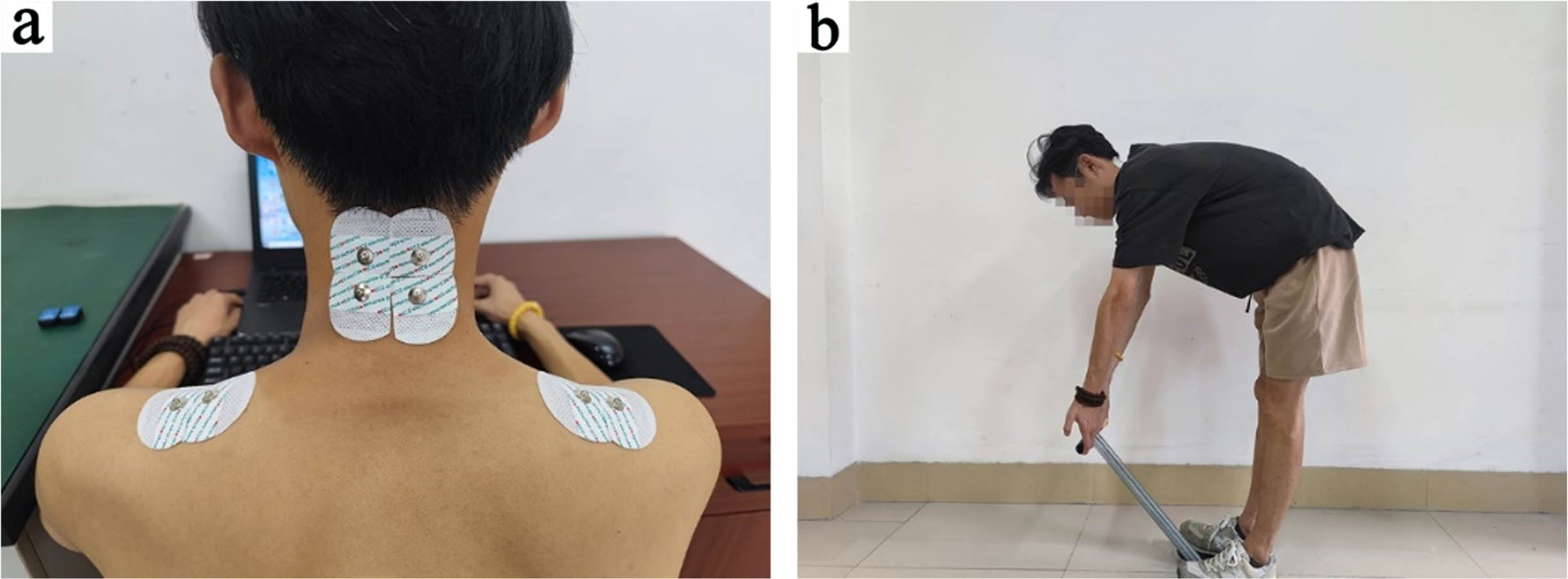
Schematic diagram of patch position and maximal voluntary muscle contraction test. (a) Schematic diagram of electrode positions for the SC and UT; (b) Schematic diagram of movements for the maximal voluntary contraction test of the SC and UT.
During the experiment, the subjects maintained a seated position and engaged in a literature reading task for 15 minutes at a randomly assigned screen height. sEMG signals from the SC and UT of the subjects at 51st-60th s per minute were collected using an sEMG device, which yielded 15 segments of sEMG data. Furthermore, participants were instructed to refrain from any unnecessary actions during the experiment, including talking, nodding, body movements, and leg swinging.
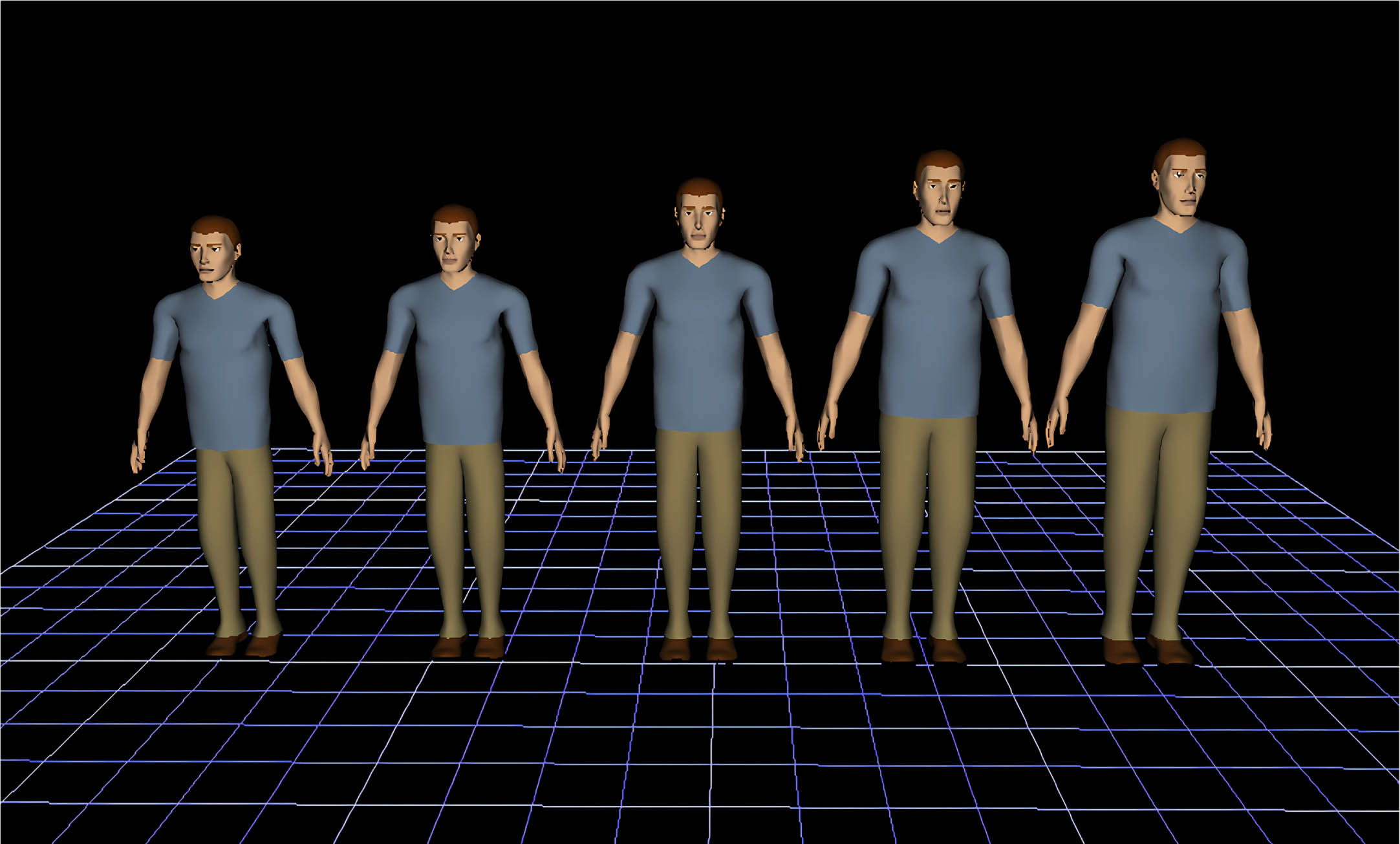
The human body models of male P1, P5, P50, P95 and P99 laptop users were established in Jack software, as shown in Fig. 4. The heights of laptop users are different. As shown in Table 2, 50% of females are taller than 154 cm. Therefore, the human body models of male P1, P5, P50, P95 and P99 were selected in this model, which can cover the height dimensions of more than 50% of female office workers and 99% of male office workers (human body dimensions are from GB/T 1000-1988 Chinese Adult Human Body Dimensions).
Mannequin in Jack.
Human size parameters at different percentiles
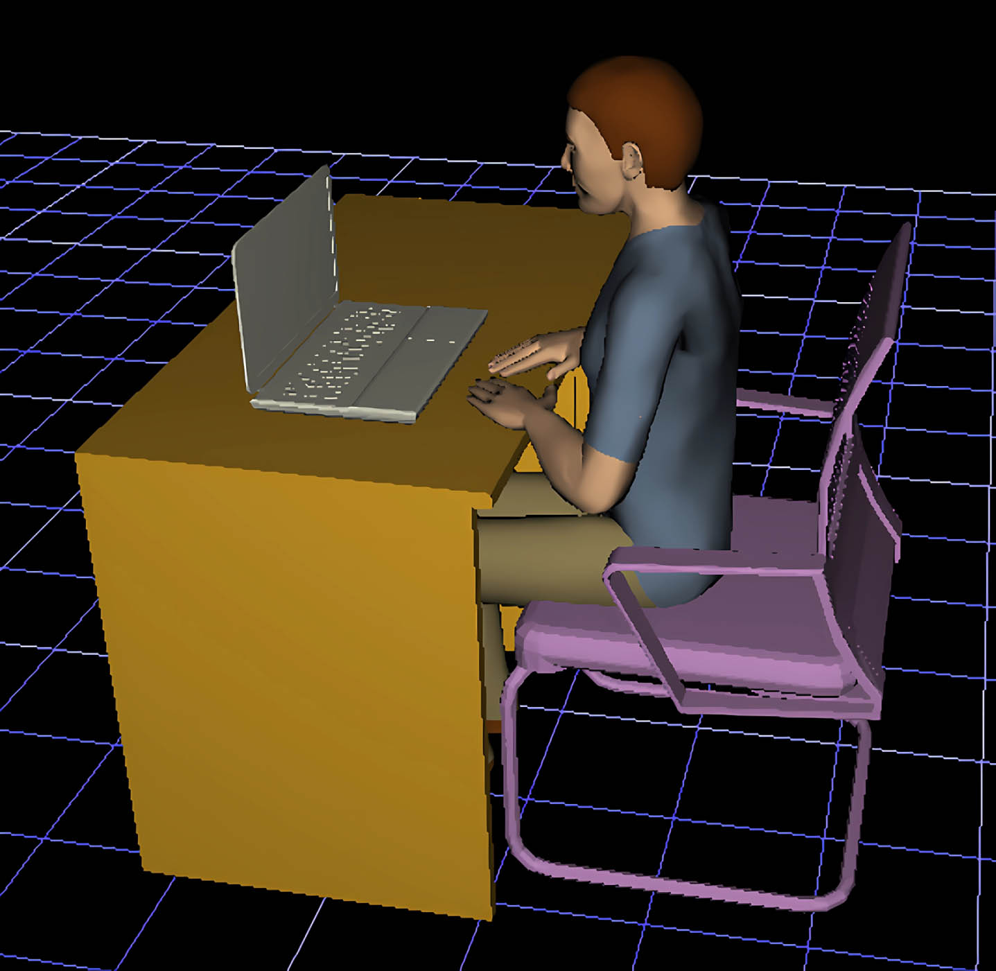
During the experiment, the digital human was placed on a 45 cm high chair, with the upper body always kept upright, the sternum 15 cm away from the edge of the table, and the hands flat on the 75 cm high table as shown in Fig. 5. In order to simulate the force on the human spine when the laptop was placed at different heights, the digital humans’ eyes were set to follow the change of the center point position of the screen in the Jack software, thus realizing the change of the torque of the spinal joint. The vertical height (h) and horizontal distance (d) of the center height of the laptop screen in the experiment are shown in Table 3.
Simulation model of office scene.
The vertical height and horizontal distance of the center of the laptop screen
Each subject was tested 4 times, each test lasted 15 min to obtain 15 segments of sEMG signals, and the duration of each segment of sEMG signals was 10s. Each segment of the acquired sEMG signals was pre-processed using custom written code in MATLAB. A trap filter was used to remove contamination from 50 Hz industrial frequency noise, and a fourth order Butterworth filter was used to bandpass filter the acquired sEMG signals at 15–350 Hz to remove ECG noise and motion artefacts. The maximum voluntary contraction (MVC) of the SC and UT was obtained by performing the three tests of the specified action as shown in Fig. 3(b), and the sEMG signals obtained from the MVC test were pre-processed in the same way, and the average was taken as the MVC of the SC and UT. The MVC was used to normalized the muscle activity of each subject in order to reduce the variability between replicates of a single subject or the variability between experimental results of multiple subjects [30, 31]. The time and frequency domain features were extracted for muscle fatigue assessment, and the pre-processed data were then smoothed by full-wave rectification and low-pass filtering at 6 Hz to obtain the root mean square (RMS) value of the time-domain eigenvalues, and the frequency-domain eigenvalues were used to calculate the mean power frequency (MPF) by performing a fast Fourier transform of the pre-processed data. The time-domain eigenvalue RMS and the frequency-domain eigenvalue MPF were linearly fitted and the respective slopes were obtained, and the joint analysis of sEMG spectrum and amplitude (JASA) proposed by Hägg et al. [32] was used to investigate the fatigue state of the muscles. The main relationships between sEMG amplitude and spectrum and muscle condition are then plotted in an intuitive four-quadrant plot [33], JASA can effectively discriminate the changes in the sEMG signals, such as determining whether muscle strength is increasing, decreasing or muscle fatigue is occurring.
A one-way repeated measures ANOVA was used to determine the effect of screen height on head flexion, muscle force, and joint torque. Paired t-tests were performed on the left and right sEMG signals to find if there was a significant difference between the left and right sEMG signals. IBM SPSS Statistics 26 was employed for performing statistical analyses. Results with p < 0.05 were treated as statistically significant.
Results
Experimental results
Head posture
Figure 6 illustrates the angles of flexion of the head and viewing at different screen heights. It is evident that the neck is more upright with increased screen height, and there is a consequential decrease in head flexion. Table 4 is a significant relationship between screen height and head flexion change (p < 0.001). Figure 6(a) is the head flexion at the four screen heights. The head reaches its maximum flexion state (25.5±1.1°) at D15 and its minimum flexion state (17.0±0.5°) at D45. The head’s flexion falls between 21.1±1.4° and 18.6±0.6° under D25 and D35, respectively. In comparison with D15, the D25, D35 and D45 condition decrease 4.1°, 6.6° and 8.1° in head flexion, respectively. The alteration in screen height also has a significant effect on the alteration in the viewing angle (p < 0.001) (Table 4). Figure 6(b) is the alteration in the viewing angle with an increase in height, the viewing angle gradually descending as the screen height increased, from 27.8±2.5° in D15 to –1.2±2.9° in D45 a negative angle indicates that the viewing angle is above the horizontal plane). D15 resulted in a greater increase in head forward and viewing angle when compared to other screen heights.

Head flexion angle and viewing angle at different screen heights. (a) Variation of head flexion with height; (b) Variation of viewing angle with height.
One-way repeated measures ANOVA and pairwise comparison results for head flexion, muscle activity and C3-C7 joint torques
*Indicates a significant difference at p < 0.05; **Indicates a significant difference at p < 0.01.
Figure 7 displays the normolized sEMG activity of the splenius capitis (SC) and upper trapezius (UT) for four different screen heights. The results showed a significant effect of screen height on the muscle activities of the left splenius capitis (LSC) and right splenius capitis (RSC) (p = 0.005, p = 0.006) in Table 4. However, there was no significant effect of screen height on the muscle activity of the left upper trapezius (LUT) and right upper trapezius (RUT) (p = 0.603, p = 0.064). It can be found from Fig. 7 that the muscle activity of RSC and RUT is higher than that of LSC and LUT. However, a paired t-test on both left and right sides muscle activity of SC and UT indicated that this difference was not statistically significant (P > 0.05). As shown in Fig. 7(a), the muscle activity of the LSC and RSC gradually declines with the increase of the screen height of the laptop from 15 cm to 45 cm. These findings corroborate those of Lee and Liu [27]. In particular, the greatest muscle activity of the SC (LSC 12.4% MVC, RSC 13.0% MVC) was observed in the D15 condition, while the lowest muscle activity (LSC 9.19% MVC, RSC 10.15% MVC) was recorded for the D45 condition. The muscle activity of LSC decreases by 1.08%, 1.66%, and 3.24% MVC for the D25, D35, and D45 conditions, respectively, when compared to D15. Similarly, the RSC exhibited a decrease in muscle activity by 0.98%, 1.69%, and 2.89% MVC for the D25, D35, and D45 conditions, respectively. The decrease in muscle activity of the left and right upper trapezius (LUT and RUT) as the height of the laptop screen increases is also found from Fig. 7(b). The decline in muscle activity of the UT was notably less than that of the SC.

Muscle activity diagram. (a) Muscle activity of LSC and RSC; (b) Muscle activity of LUT and RUT. % MVC is percentage maximum voluntary contraction.
Results of the joint analysis of sEMG spectrum and amplitude are given in Fig. 8. The point which lies in the fourth quadrant implies that the muscle is in a fatigued state. It is found from Fig. 8(a) that the regression coefficients of the paired values of RMS and MPF for the SC are mainly in the second quadrant, which indicates that the muscle is in a normal state. The highest number of fatigue occurs in the SC in the D15 condition, and the lowest number of fatigue occurs in the SC in the D45 condition. At the same time, the regression coefficients for the paired values of RMS and MPF for the UT are also predominantly distributed in the second quadrant, with a small part in the fourth quadrant from Fig. 8(b). The D15 condition had the highest number of UT fatigue occurrences, while the D25 and D45 conditions had the lowest number of occurrences (Fig. 8(b)).

The JASA results for SC and UT. (a) The JASA diagram for SC; (b) The JASA diagram for UT. The blue symbol represents in the first quadrant (force increase); the black symbol represents in the second quadrant (recovery); the green symbol represents in the third quadrant (force decrease); finally, the red symbol represents in the fourth quadrant(fatigue). KRMS= the slope of the regression line of the RMS vs time. KMPF= the slope of the regression line of the MPF vs time.
Figure 9 shows the changes of spineT1/T2 M and spineL4/L5 M with the same horizontal distance of the laptop and only changing the screen height. It can be seen from Fig. 9 that the spineT1/T2 M and spineL4/L5 M of different heights decrease with the increase of screen height, and the decrease trend of each digital person is divided into two parts, the first part is greater than the second part, and the decrease trend is basically linear. The decrease trend of spineT1/T2 M and spineL4/L5 M of P95 and P99 changes at h = 40 cm, the decrease trend of spineT1/T2 M and spineL4/L5 M of P50 changes at h = 35 cm, and the decrease trend of spineT1/T2 M and spineL4/L5 M of P1 and P5 changes at h = 30 cm. The larger the percentile is, the greater the moment of spineT1/T2 and spineL4/L5 is, and the greater the rate of decrease is. This shows that when using a computer at the same screen height, the higher the height of the person, the greater the moment of the cervical spine and lumbar spine is.

Changes of spineT1/T2 M and spineL4/L5 M at different vertical distances. (a) Changes in spineT1/T2 M; (b) Changes in spineL4/L5 M.
The display height was gradually increased from the conventional screen height setting to 45 cm with a gradient of 10 cm, resulting in the head flexion and angle of view changed by 8.1° and 29.0° respectively. The head flexion and angle of view were the largest under the conventional laptop screen height setting (D15), and the head flexion and angle of view were the smallest when the screen height was 45 cm (D45). The relationship between Laptop placement, head flexion and viewing angle is consistent with previous findings, that is, with the increase of screen height, head flexion and viewing angle gradually decrease [25, 34]. Lee and Liu’s research has proved that there is a strong relationship between the angle of view and the angle of head bending, and the change of the angle of view is adjusted by the change of the angle of head bending [27]. The results of this study conform to this rule, and the change trend of head flexion is consistent with the change trend of visual angle.
Villanueva et al. [35] reported that increasing screen height during mouse work resulted in the decrease of neck and shoulder muscle activity and a high correlation between the degree of neck flexion and SC muscle activity. Excessive head flexion causes to increase SC stretching, resulting in the highest SC muscle activity at the normal laptop screen height. In comparison to the conventional screen height (D15), SC muscle activity decreased by 1.03% MVC when raised by 10 cm (D25), decreased by 1.68% MVC when raised by 20 cm (D35), and decreased by 3.07% MVC when raised by 30 cm (D45). We were surprised to find no significant differences in UT muscle activity at varying display heights (LUT p = 0.603, RUT p = 0.064), and SC muscle activity was only slightly reduced by 0.89% in the natural head posture, compared to the standard screen height setting. It is worth noting that many authors have reported no significant difference in UT muscle activity at different display heights [35–37]. The study revealed that muscle activity in the RSC and RUT was higher than that in the LSC and LUT at varying display heights. This may be attributed to the exclusively right-handed participants who used the right hand for mouse movements, resulting in greater muscle activity in the right muscles compared to the left muscles.
There are some concerns about the increase in muscle activity in SC, especially when the screen is placed low, because the continuous muscle contraction may lead to musculoskeletal discomfort or injury [36]. In traditional settings, working on a laptop for a long time seems to further increase the risk of neck diseases, as users experience more neck bending and an increase in mechanical load on the neck [38]. According to the results of SC and UT fatigue analysis, the regression coefficients of RMS and MPF pairs of SC are mainly distributed in the second quadrant, indicating that the muscle is in a normal state, and only a small part is distributed in the fourth quadrant representing fatigue. In addition, with the increase of screen height, the occurrence of SC fatigue gradually decreases. The occurrence of SC fatigue is the most frequent under the traditional screen height setting, so increasing the screen height can slow down the occurrence of SC fatigue to a certain extent. As expected, UT activity does not change significantly under different screen heights, and the number of UT fatigue does not change significantly either. Therefore, the impact of changing screen height on UT fatigue seems to be not obvious. Relevant investigations indicate that prolonged head bowing can result in excessive pressure on the cervical spine, thus leading to neck disorders and symptoms such as soreness of the neck muscles accompanied by damage to soft tissues surrounding the neck [26]. Ding et al. [39] recorded the sEMG activity of the upper trapezius and latissimus dorsi during 2 hours of seated work, and found that the muscles were most susceptible to fatigue at 40–50 minutes. Waongenngarm et al. [30] reported a statistically significant rise in discomfort in the neck, shoulders, upper back, lower back, and buttocks after one hour of sitting regardless of posture. Therefore, while working for extended periods with computers, modifying screen height may postpone the onset of fatigue.
The simulation results show that the higher the height of a person, the greater the torque on the thoracic and lumbar vertebrae when using a laptop in a seated position. According to biomechanical principles, an upright neck position could potentially lessen the mechanical burden on the neck. Sun et al. [40] and Silva et al. [41] constructed cervical spine model for finite element analysis and found that the vertebral body rotates forwards during flexion (positive torque) and backwards during extension (negative torque), and that the intervertebral joint torque decreases as the vertebral flexion angle decreases. Consistent with the expected results, with the increase of screen height, the torque of spineT1/T2 and spineL4/L5 gradually decreases. In addition, the results also show that the decrease trend is divided into two stages, and the decrease rate of the first stage is greater than that of the second stage. Therefore, it is recommended that the height of the computer screen center should not be less than the height value at the inflection point, as shown in Fig. 9 (a) and (b). That is, the lowest center height of the computer screen should belong to 30 cm for people below 158.3 cm in height; the lowest center height of the computer screen should belong to 30–35 cm for people between 158.3 cm and 167.8 cm in height; the lowest center height of the computer screen should belong to 35–40 cm for people between 167.8 cm and 177.5 cm in height; and the lowest center height of the computer screen should belong to 40 cm for people taller than 177.5 cm in height. Based on the simulation results, a mathematical model is built for the minimum screen height of laptop users between 154 cm and 181 cm in height when using laptops in a sitting position, as follows:
Where y is the minimum screen height (cm), x is the sitting eye height (cm), a is the table height (cm), and b is the height of chair seat (cm). Users can substitute their own sitting eye height, table height, and height chair seat into the formula to get the minimum screen height suitable for them. Of course, users can also make appropriate adjustments on this basis. By adjusting the height of the laptop screen properly, it can greatly relieve the fatigue and pain symptoms of the neck, lower back and waist, and reduce the damage to the body.
Finally, it should be noted that although this study revealed some effects of laptop screen height on head posture angle and muscle fatigue, and provided recommendations for the minimum screen height for laptop users of different heights, the subjects were healthy Asian laptop users, which may weaken and limit the generalizability of the results and cannot be generalized to other symptomatic groups.
Conclusion
sEMG signals of the SC and UT were scaled by twelve healthy young participants performing a15-minute task of the reading at the four different screen heights and gained its the root mean square and mean power frequency to determine muscle fatigue. The findings show that adjusting the height of the laptop screen can significantly reduce the cervical flexion angle and the activity level of SC. The following results were obtained: There is a significant relationship between screen height and head flexion change. The head has its maximum flexion state (25.5±1.1°) and its minimum flexion state (17.0±0.5°) at D15 and D45, respectively. The joint analysis of sEMG spectrum and amplitude shows that the highest and the lowest frequency of fatigue occurs in the SC on the condition of D15 and D45. It can be concluded from the simulation results with Jack that the spineT1/T2 moment and spineL4/L5 moment decrease with the increase of screen height, especially, the moment is the sharp decline at the first stage. The lowest center height of the computer screen should belong to 30 cm, 30–35 cm, 35–40 cm, 40 cm for people with the height of below158.3 cm, 158.3 cm–167.8 cm, 167.8–177.5 and above 177.5, respectively.
Ethical approval
Not applicable.
Informed consent
All participants agreed to participate in the study and signed a consent form.
Conflict of interest
No potential conflict of interest was reported by the authors.
Footnotes
Acknowledgments
We would like to thank the students who participated in this study and all those who helped us during this study.
Funding
This work was supported by Agricultural Key Applied Project of China (No. SD2019NJ015) and National Natural Science Foundation of China (projects No. 52075275).