
Abstract
BACKGROUND:
Streak artifacts in computed tomography (CT) images caused by metallic objects limit the wider use of CT imaging technologies. There have been various attempts to improve CT images containing streak artifacts; however, most of them generate additional artifacts or do not completely eradicate existing artifacts.
OBJECTIVE:
In this paper, we propose a novel algorithm which reduces streak artifacts in CT images.
METHODS:
Using CT numbers reconstructed from a given sinogram, we extract the metal part M and the surrounding area C with similar CT numbers. By filling in the area C ∪ M with the evaluated average CT number of C, we obtain a modified CT image. Using forward projection of the modified CT image, we generate a sinogram containing information about the anatomical structure. We undertake sinogram surgery to remove the metallic effects from the sinogram, after which we repeat the procedure.
RESULTS:
We perform numerical experiments using various simulated phantoms and patient images. For a quantitative analysis, we use the relative l ∞ error and the relative l 2 error. In simulated phantom experiments, all l ∞ errors and l 2 errors approach 10% and 1% of the initial errors, respectively. Moreover, for the patient image simulations, all l ∞ errors are decreased by a factor of 20 while the l 2 errors are decreased less than 5%. We observe that the proposed algorithm effectively reduces the metal artifacts.
CONCLUSIONS:
In this paper, we propose a metal artifact reduction algorithm based on sinogram surgery to reduce metal artifacts without additional artifacts. We also provide empirical convergence of our algorithm.
Introduction
X-ray CT is one of the most popular nondestructive techniques for visualizing anatomical structures in the human body. X-ray CT uses radiation from X-rays to produce an internal image of the body. When the X-rays pass through the body, the energy of the X-rays is absorbed according to the X-ray attenuation coefficients of the tissues on their path [7]. Given that the X-ray attenuation coefficients differ depending on the tissues involve, the measured X-ray energy data taken from the different angles around the body reflect the distributional information of the tissues in the body.
The quality of an X-ray CT image is degraded by several artifacts. These artifacts are classified into four categories based on the causes [2]: (i) physics-based artifacts, which result from the physical processes involved during the acquisition of the CT data; (ii) patient-based artifacts, which are caused by such factors as patient movement or the presence of metallic materials in or on the patient; (iii) scanner-based artifacts, which result from imperfections in certain scanner functions; and (iv) helical and multisection artifacts, which are produced by the image reconstruction process. Meanwhile, artifacts can be grouped by their shapes. One of the major shapes of CT artifacts is a streak. Streaks are caused by metallic objects such as dental implants and surgical clips, and are referred to as streak artifacts. They can result from multiple mechanisms, including beam hardening, scatter effects, and Poisson noise [4]. Given that metal artifacts are spread over the entire image as a bright and shadow crown shape in the region of interest and degrade the quality of CT images, they prevent accurate diagnoses in the medical and dental fields. For this reason, the importance of metal artifact reduction (MAR) technique increases as CT imaging becomes more popular.
Several studies have attempted to understand metal artifacts, and many approaches have been proposed to reduce them. Existing MAR methods can be roughly classified into the three categories: methods based on sinogram interpolation, those based on iterative reconstruction, and others. With methods based on sinogram interpolation, the sinogram data are corrected by various types of inpainting techniques, such as polynomial interpolation, wavelets, Euler’s elastica models, and interpolation using neighboring pixel values [1 , 12]. However, these methods generate additional artifacts in the reconstructed CT image due to inconsistencies in the boundary of the projected region of metallic objects after interpolation. These extra artifacts worsen the quality of X-ray CT images. In iterative reconstruction methods, both the image and sinogram data are updated in a feed-back manner to complete the corrupted parts due to metallic objects [5, 6]. Iterative reconstruction methods achieve better image quality than interpolation based methods; however, the high computational cost makes it difficult to implement these methods practically. Park et al. [16] proposed a method which interpolates the missing part in the CT image directly using the Poisson equation. However, this method has limitations when used with cases with multiple metallic objects. In addition, they reveal metal artifacts in X-ray CT which are characterized using the wave front set and artifacts are corrected mathematically [17]. Prell et al. [19] proposed a projection-based metal artifact reduction method for flat-detector computed tomography. They used three-tissue-class segmentation to replace the corrupted part of a sinogram. The first clinical application of the iterative MAR algorithm was achieved by Philips Health Care [18] though it was applied only to cases of orthopedic implants. The algorithm used by Phillips Health Care was based on work by Timmer and Koehler [13, 20], whose methods were investigated experimentally only for a simply shaped phantom, with the result showing that residual artifacts still existed. Meyer et al. [14] introduced what was termed a normalized metal artifact reduction (NMAR) algorithm, which is one of the most up-to-date MAR algorithms. It uses a generalized normalization technique to inpaint the corrupted part of the sinogram. Kano and Koseki [10] developed an iterative metal artifact reduction algorithm based on a deteriorated CT image. Their method only requires reconstructed images and the projection conditions.
In this paper, we propose a novel algorithm which properly reduces metal artifacts. Even when a CT image reconstructed from a corrupted sinogram contains severe artifacts near metallic objects, we can obtain structural information apart from the metallic region. Unlike other interpolation based MAR methods, the proposed algorithm uses this structural information to complete the missing part of the sinogram. The sinogram completion process iteratively proceeds using the basic principles of CT image reconstruction. We apply our method to simulated phantoms of various shapes, in this case a simple circular phantom and a Shepp-Logan phantom and to patient images obtained from clinical CT. Note that in these numerical experiments, only the beam hardening effect is considered. Numerical experiments show that our algorithm reduces metal artifacts effectively by filling in the missing sinogram data properly. We analyze the simulation results quantitatively and qualitatively.
We also demonstrate the empirical convergence of our algorithm.
Methods
The proposed algorithm consists of two steps: a preprocessing step and an iterative reconstruction step. In the preprocessing step, we extract the metal part from the given CT image and determine its corresponding sinogram region by forward projecting it using the Radon transform. In the iterative reconstruction step, we undertake the processes of average fill-in, sinogram surgery, and reconstruction from the updated sinogram to moderate metal artifacts. Detailed descriptions of each of these steps are given below.
Preprocessing step
Metal extraction: To manage streak artifacts, we find the metal region M which can be extracted by simple thresholding based on the fact that metallic objects have much higher attenuation coefficients than normal human tissue. Surgery region designation: Once the metal region M has been extracted, it is forward-projected using the Radon transform
Iterative reconstruction step
The iterative reconstruction step has three steps: average fill-in, sinogram surgery, and reconstruction of the updated sinogram. Average fill-in: Let f
(n-1) represent the reconstructed CT image from the previous step. We segment a connected region C surrounding the metallic object region M by means of CT number thresholding. The shape and size of C differ depending on the distribution of the CT numbers of f
(n-1). We evaluate v
(n-1), the average of the attenuation coefficients f
(n-1) in region C, and fill in the region C ∪ M with v
(n-1). We then have
Projection and sinogram surgery: After the average fill-in step, we obtain a new sinogram Because This approach enables us to use all of the metal-effect-free part of the initially given sinogram without missing data in order to complement the corrupted sinogram using the available structural information of the CT image. CT image reconstruction: Using the updated sinogram, we reconstruct the new CT image
This results in a CT image with less streak artifacts.
As we repeat the iterative reconstruction step, streak artifacts are reduced gradually because the missing data is complimentarily replaced for both the sinogram and the reconstructed CT image. The iterative reconstruction step is terminated when the relative difference between the sinogram data becomes less than the tolerance level. We demonstrate the convergence of our algorithm empirically in the Appendix.
Remark. When we have a priori information about the anatomical structures, we consider m connected regions {C
i
} in f
(n-1) according to the anatomical structures. In this case, we adopt a modified average fill-in step; we evaluate
Algorithm 1
Pseudo code of the proposed algorithm.
Algorithm 1 shows the pseudo code of the proposed algorithm and Fig. 1 provides a schematic diagram of the proposed algorithm.

Diagram of the proposed algorithm: A CT image f
(0) is reconstructed from the given sinogram S
(0). The metal part M is extracted from f
(0) and it is projected to obtain the metal mask
Simulation conditions
The performance of the proposed algorithm is tested on simulated phantoms (Fig. 2), specifically a simple circular phantom and on a Shepp-Logan phantom. All images are 512 × 512 in size, and the field of view is 200 mm. We embedded metallic objects of different sizes into the simulated phantoms at different positions, and the inserted metallic objects were assumed to be iron. To generate the polychromatic sinogram, we modeled the parallel beam with 512 channels per detector and 1800 views per half rotation. We defined seven discrete energy bins (10, 20, 30, 40, 60, 80, 100 keV) (Table 1), and all X-ray coefficients were obtained from the National Institutes of Standards and Technology (NIST) database [15].
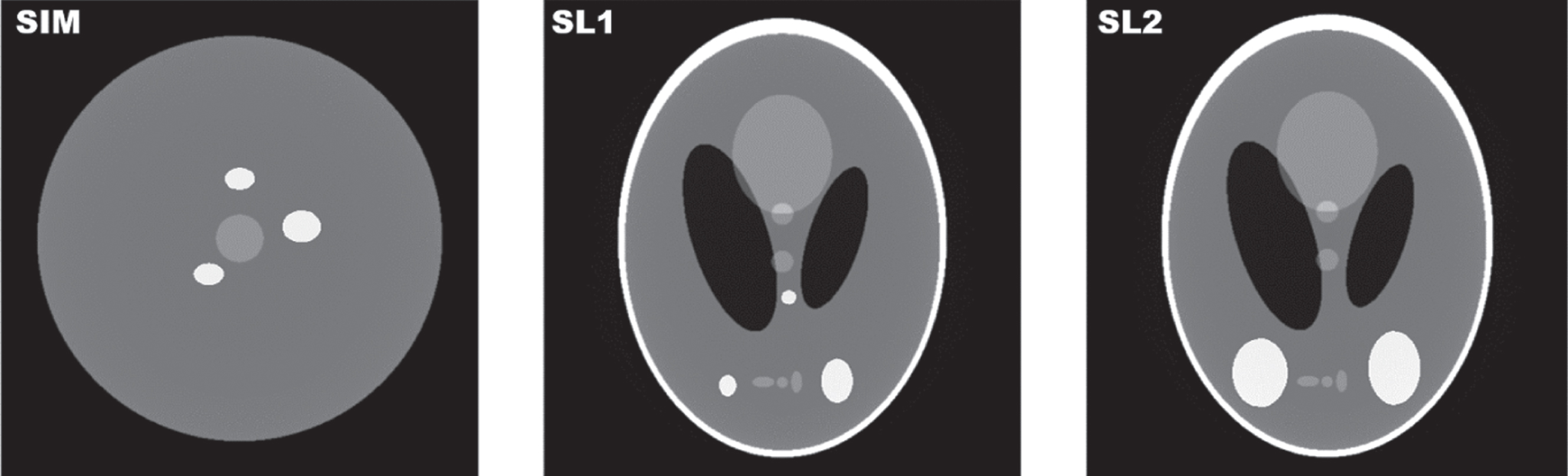
The simulated phantoms.
X-ray intensity and nominal Hounsfield units (HU), and linear attenuation coefficients for materials used in phantom simulation
For a quantitative analysis, we generated metal-effect-free sinograms using the phantoms before the metallic objects are inserted. From the generated sinograms, we reconstructed the metal-effect-free CT images f
⋄ as references and measure the performance of MAR algorithms with two error measurements: the relative l
2 error and the relative l
∞ error. We used a simple notation for the relative error between a CT image f and the reference CT image f
⋄,
In all phantom images and reconstructed images, we use the window level with a width of 1000 centered at 0 (C/W=0/1000 (HU)).
To analyze the performance of the proposed algorithm, we compared the performance of the proposed algorithm with that of NMAR [14], one of the best MAR algorithms currently available. Figure 3 shows the results of NMAR and of the proposed algorithm with the uncorrected CT images, and Table 2 shows the corresponding relative errors.

A comparison between NMAR and the proposed algorithm for SIM, SL1, and SL2: C/W=0/1000 (HU).
Performance comparison between NMAR and the proposed algorithm for the simulated phantom experiments. The number of iterations is denoted by n.
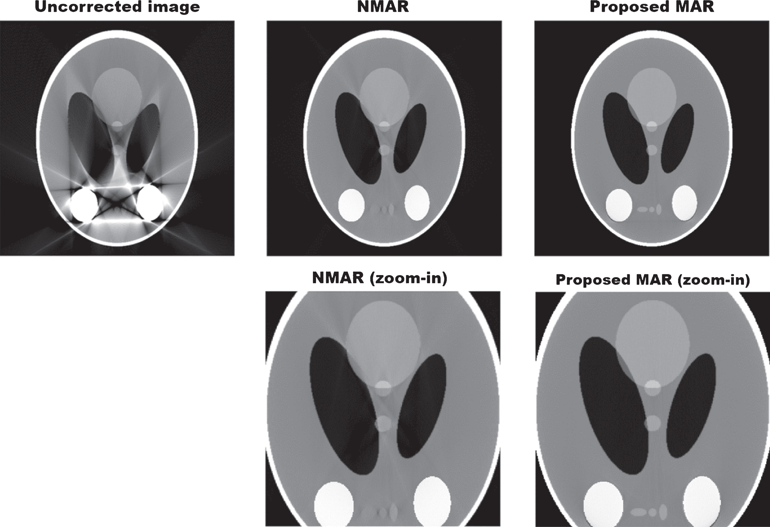
As shown in Fig. 3, most of the streak artifacts are reduced effectively by both NMAR and the proposed algorithm. For SIM, the relative l ∞ and l 2 errors in the NMAR result are 0.1476 and 0.0151; both are decreased by a factor of ten from the initial relative l ∞ and l 2 errors of 1.7205 and 0.1464, respectively. The proposed algorithm has smaller relative l ∞ and relative l 2 errors than NMAR (Table 2), at 0.0725 and 0.0064, respectively. Moreover, as shown in Fig. 4, the proposed algorithm restores the central circular shape more visibly than NMAR. For SL1 and SL2, the proposed algorithm also outperforms. Compared to NMAR, the proposed algorithm shows clearly outstanding results in terms of how it preserves the anatomical structures, as shown in Figs. 5 and 6. While NMAR blurs the anatomical structures and leaves some streak artifacts in the resulting images, the proposed algorithm produces a clean image without blurring the anatomical structures and with less remaining streak artifacts. However, the proposed algorithm has a larger relative l ∞ error, at 0.1154 (NMAR: 0.0981), a smaller relative l 2 error, at 0.0143 (NMAR: 0.0191), and it produces a cleaner image than NMAR for the SL1 case. SL2 represents a case in which large pieces of metallic objects are present, possibly leading to photon starvation. For the SL2 case, the relative l ∞ and l 2 errors are dropped by a factor of six and a factor of nine, at 0.0796 and 0.0135 (Initial: 0.4873 and 0.1133), respectively. For all cases, the proposed algorithm diminishes streak artifacts effectively.
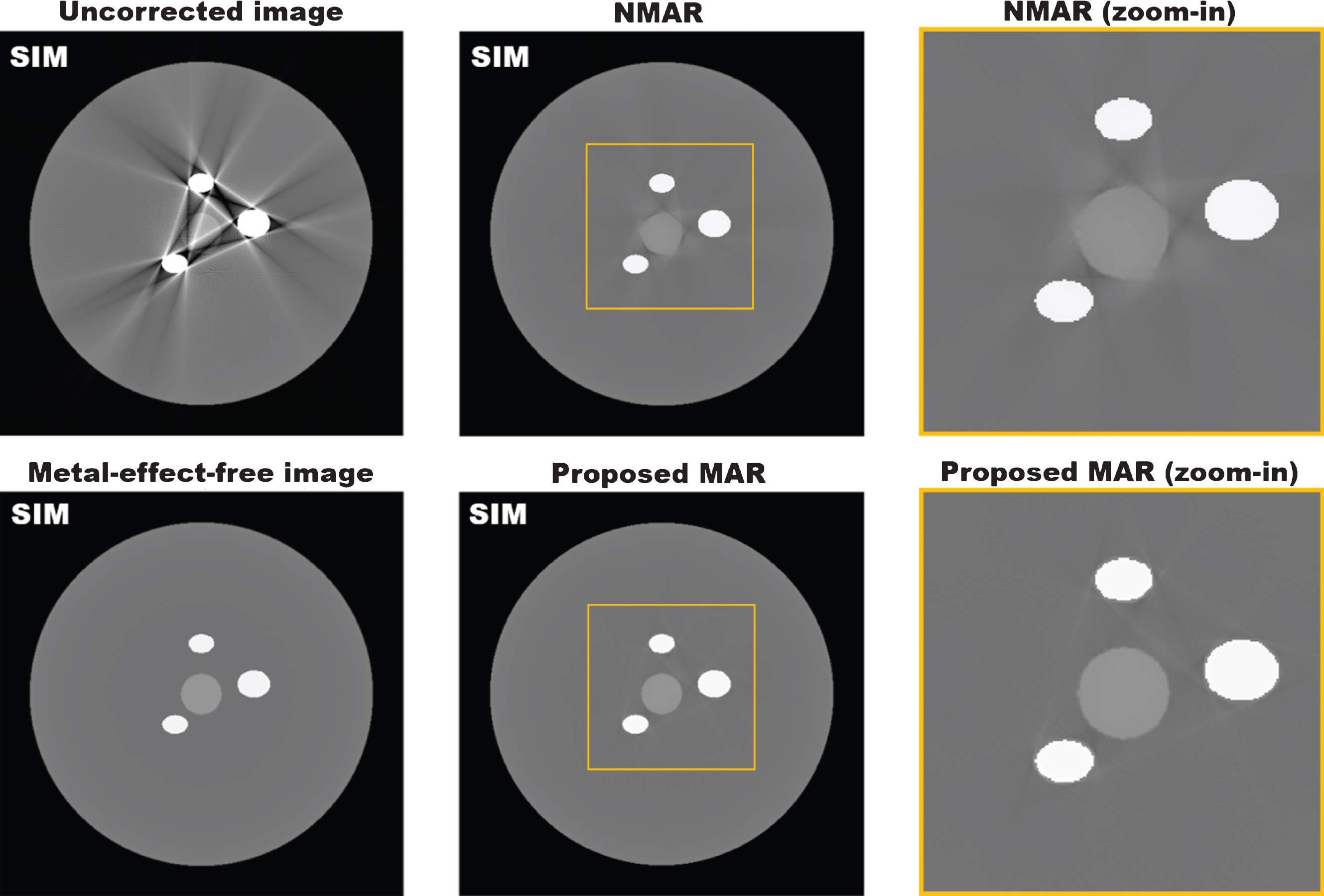
Resulting images of NMAR and the proposed algorithm for SIM: C/W=0/1000 (HU).
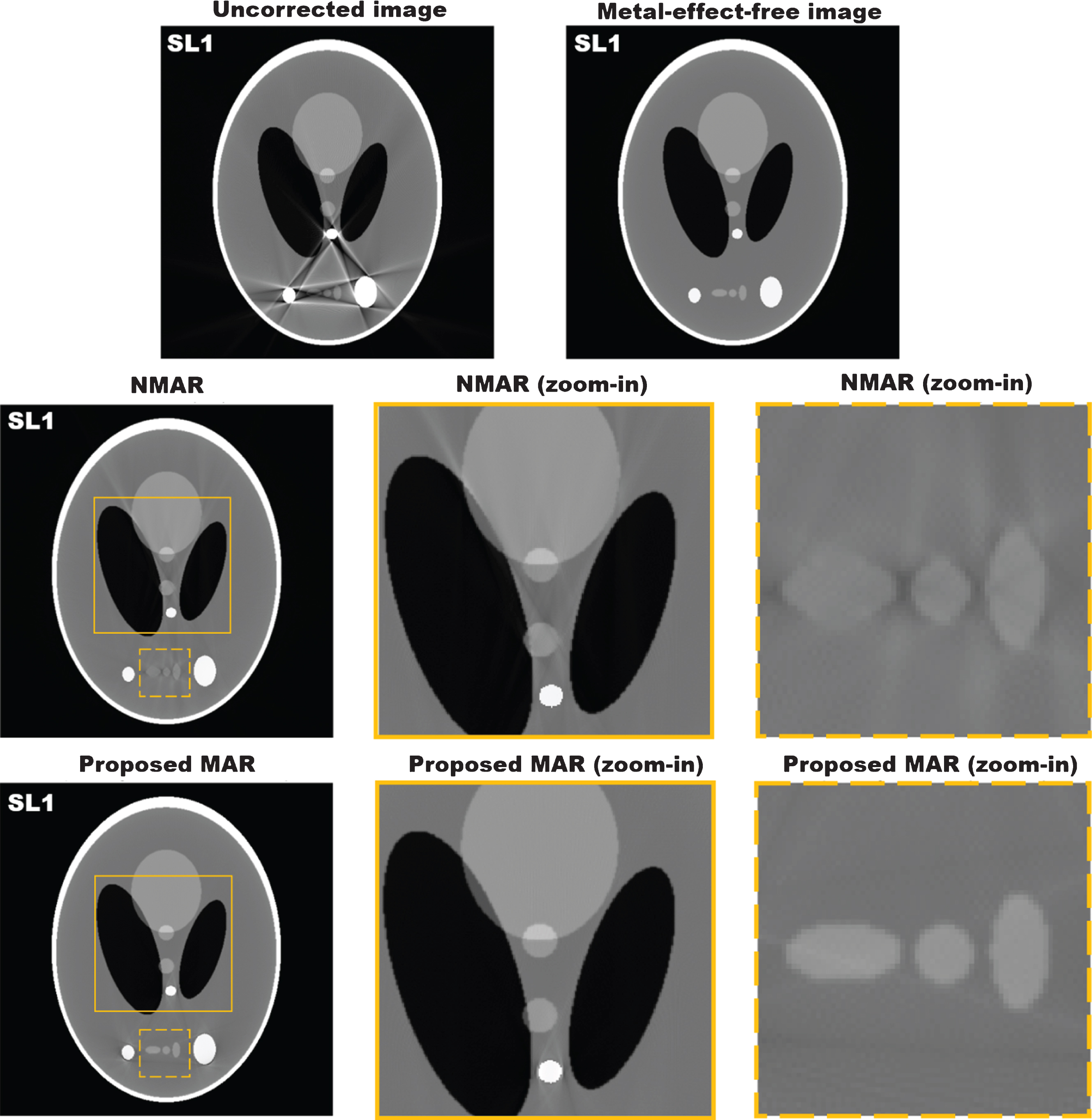
Resulting images of the proposed algorithm result for SL1: C/W=0/1000 (HU).

Resulting images of the proposed algorithm result for SL2: C/W=0/1000 (HU).
Noise was also simulated using a random Poisson model. The total intensity of the incident X-ray is set as shown in Table 1. The randomly generated Poisson noise was added to a pre-logarithm of the simulated raw data before the reconstruction of the SL2 case. As shown in Fig. 7, the quality levels of the initially reconstructed CT images are worse than in the case of the noise-free simulations. Although the streak artifacts severely damage the anatomical structure, the proposed algorithm successfully reduces the streak artifacts in this case as well.
Resulting images of the noise test: C/W=0/1000 (HU).
We also tested the proposed algorithm on patient images (Fig. 8). Three cross-sectional images (pelvis, chest, and dental) are selected from a CT dataset acquired in a dosimetry study of 68Ga-NOTA-RGD PET/CT [11]. All study procedures were approved by the Institutional Review Board of Seoul National University Hospital, Seoul, Korea. We inserted simulated metallic objects into the patient images while assuming that the metallic objects are titanium (Table 1). We use the same X-ray energy spectrum, as shown in Table 1. Because it is difficult to assign all X-ray attenuation coefficients corresponding to each tissue type in patient images, we assume that only the X-ray attenuation coefficient of the metallic objects depends on the X-ray energy level.
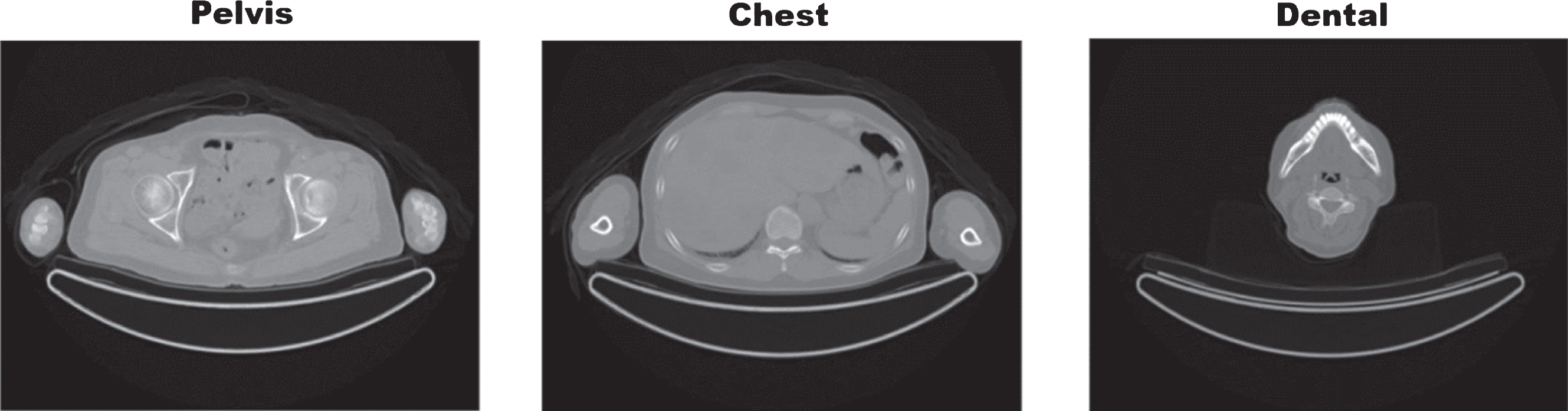
Patient images (pelvis, chest, and dental).
Figure 9 shows the simulation result of the pelvis of a patient with metallic hips. In the reconstructed CT image, there are streak artifacts between the metallic hips, and they corrupt the anatomical structure. NMAR reduces most of the streak artifacts; however, the resulting image contains bright and dark artifacts which blur the anatomical structure. In comparison, the proposed algorithm reduces streak artifacts effectively without generating such bright and dark artifacts. As a result, we obtain a clean CT image, and the textures are also preserved well.
As shown in Table 3, the initial relative l ∞ and l 2 errors, 5.0085 and 0.7044, are decreased by nearly half to 3.5728 and 0.2341, respectively, for NMAR. The proposed algorithm drops the errors more significantly, with the resulting relative l ∞ and l 2 errors becoming 0.2293 and 0.0269, respectively, the values which are decreased by a factor of twenty from the initial levels.
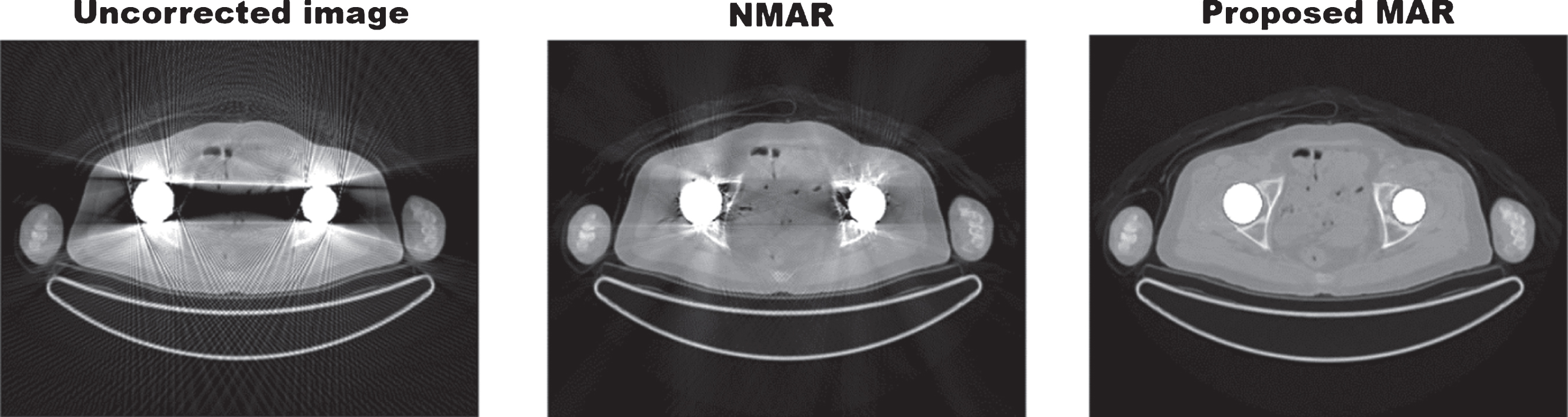
Patient pelvis experiment. Uncorrected CT image (left), the result of the NMAR (middle), and the result of the proposed algorithm (right): C/W=0/1000 (HU).
The performance comparison between NMAR and proposed algorithm for the patient image simulations. The number of iterations is denoted by n.
Figure 10 presents the experimental results of the chest of a patient. Two metallic screws inserted into the spine generate streak artifacts, which severely damage the anatomical structure near the spine. While NMAR does reduce the major part of the streak artifacts, it generates additional artifacts near the metallic objects, resulting in bright and dark patterns. These newly generated artifacts appear near the metallic objects and thus corrupt the anatomical structure. Moreover, in the NMAR result, the metallic objects are thicker than the original metallic objects, whereas the proposed algorithm improves the image quality without generating additional artifacts, so that we can successfully distinguish the anatomical structures near the spine. As shown in Table 3, the initial relative l ∞ and l 2 errors, 12.1957 and 0.9187, are decreased in the NMAR result to 7.8182 and 0.3100, respectively. The resulting relative l ∞ and l 2 errors of the proposed algorithm are 0.5878 and 0.0314, respectively, values which are lower by a factor of twenty from the initial values.
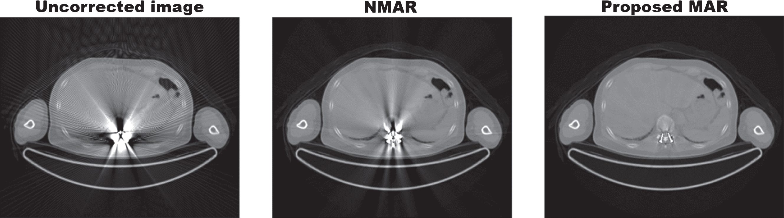
Patient chest experiment. Uncorrected CT image (left), the result of the NMAR (middle), and the result of the proposed algorithm (right): C/W=0/1000 (HU).
In the dental image simulations, streak artifacts appear to connect three metallic objects, as shown in Fig. 11. As shown in the zoomed images of the dotted boxes, both NMAR and the proposed algorithm reduce the streak artifacts. However, NMAR produces the shadow effects even in the region near the teeth and shows undulated artifacts across the entire image domain. Even in a region far from the inserted metallic objects, undulated artifacts appear, as shown in the zoomed images in the solid boxes, and they degrade the image quality. As shown in Table 3, the initial relative l ∞ and l 2 errors, 11.9255 and 1.7471, are decreased for NMAR to 3.4632 and 0.4378, respectively. The resulting relative l ∞ and l 2 errors for the proposed algorithm are 0.3734 and 0.0476, respectively, showing decreases by a factor of thirty from the initial levels.
Unlike NMAR, the proposed algorithm does not generate undulated artifacts in an image; therefore, the result shows a clear image. In the patient image simulations, the proposed algorithm shows visibly better performance than NMAR.
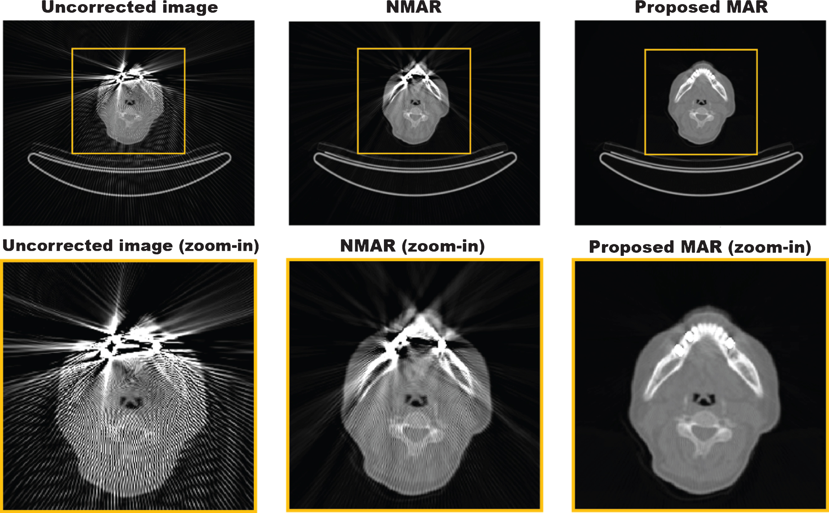
Patient dental experiment. Uncorrected CT image (left), the result of the NMAR (middle), and the result of the proposed algorithm (right): C/W=0/1000 (HU).
The proposed algorithm was applied to various simulated phantoms and to patient images. In the simulated phantom experiments, we observed outstanding performance qualitatively and quantitatively. As in other MAR approaches based on tissue classification, it is essential for the proposed algorithm to find a good tissue classification. The advantage of our algorithm stems from this point. Because the proposed algorithm tends to converge to a moderate value of the image intensity by filling in the classified region with the average values, we obtained a resulting image with less streak artifacts. We then used this image as new input data for the next iteration. Therefore, we managed to obtain better tissue classification as we repeat the iterations. In the patient image simulations, the proposed algorithm also effectively reduced the streak artifacts. Although there were slight shadow effects between the anatomical structures in a case involving the chest, this could have been improved if we had used a more sophisticating segmentation method rather than simple segmentation based on the CT number. Another issue is the complexity of the shape of the metallic object. It is not easy to recover a dented region such as that which arises when a screw is inserted into the spine using the proposed algorithm. Because the two neighboring convex structures create a blind spot between them, the value of this region must be reconstructed from limited sinogram data. Despite the fact that the quality of the image is not severely degraded in the chest case, it can be a problem in cases with more such blind spots.
Conclusion
In this paper, we proposed a novel algorithm for reducing streak artifacts in CT images. We undertook surgery on the corrupted part in the sinogram iteratively to inpaint the region properly considering the basic principles of CT. We compared the performance of our algorithm with that of NMAR, one of the best currently available MAR algorithms, both qualitatively and quantitatively and observed that our algorithm outperforms with simulated phantoms. In addition, we tested the proposed algorithm using patient images. Through numerical experiments including phantom simulations, noise tests, and patient data simulations, we demonstrated that our algorithm reduces metal artifacts effectively without a loss of the anatomical structures. We also demonstrated the empirical convergence of our algorithm, given in the Appendix.
Footnotes
J. B. Fraleigh, Calculus with analytic geometry 3rd edn. (Addison-Wesley Publishing Company, 1990).
Appendix A
Acknowledgement
This work was supported by the NRF grant funded by the MSIT (NRF-2017R1A2B4011627). We also acknowledge the Institutional Review Board of Seoul National University Hospital for providing the patient CT image set.