



Abstract
BACKGROUND:
Medical imaging plays a crucial role in modern medicine. In order to provide fast and accurate medical diagnosis, computed tomography (CT) is a commonly used tool in radiological examinations, and 640-slice CT is the most advanced CT imaging modality.
OBJECTIVE:
To evaluate the radiation dose and the risk under 640-slice abdominal CT examination.
METHODS:
Examinations were performed using a 640-slice CT scanner on an Alderson-Rando anthropomorphic phantom. The used scanning acquisition parameters were the same as those used on abdominal examination without contrast medium injection in clinical practice. To measure the absorbed doses, optically stimulated luminescence dosimeters (OSLDs) were put into liver, stomach, bladder, gonads, colon, small intestine, bone marrow, and skin.
RESULTS:
According to the 1990 Recommendations of the International Commission on Radiological Protection (ICRP Publication 60), the calculated effective doses received from this examination were 0.90 mSv in males and 0.89 mSv in females. According to the 2007 Recommendations of the International Commission on Radiological Protection (ICRP Publication 103), the calculated effective dose received from this examination was 0.83 mSv in both sexes.
CONCLUSIONS:
Radiation doses obtained from the abdominal 640-slice CT examination are lower than the yearly cumulative doses received from natural radiation, revealing there is no deterministic effect and radiation risk is relatively low; therefore, this CT examination is considered safe.
Introduction
Computed Tomography (CT) is a non-invasive diagnostic tool which combines ionizing radiation and computer technology. In recent years, the rapid development of computer technology and the performance of electronic equipment have promoted rapid evolution of CT, from single-slice computed tomography (SSCT) to multi-slice computed tomography (MSCT), with 640-slice CT being the most advanced medical MSCT, providing real-time clinical condition of patients in a timely manner. Two-dimensional or three-dimensional images of any section of the scanned body can also be reconstructed by computer software. According to Reports No. 160 and No. 184 issued by the National Council on Radiation Protection and Measurements (NCRP) in 2009 and 2019 [1, 2], CT cumulative dose as a percentage of the contribution to the “total medical radiation dose” increased from 50% to 62% from 2006 to 2016, so while CT has become increasingly used as a diagnostic tool, its radiation dose has also become of great concern to the public.
According to ICRP Publication 60 and Publication 103 [3, 4], the liver, stomach, lung, gonads (testicles and ovaries), brain, salivary glands, thyroid, breast, esophagus, red bone marrow, bladder, colon, bone surface, skin, and remainder tissues are listed as the radiation-sensitive organs. In clinical routine scans, the whole abdominal CT scan, which includes the whole abdomen and pelvis, covers these most radiation-sensitive organs, and as gonads are also included in its scan range, performing such holistic scan has become a concern to many patients in consideration of explicit risks.
CT has become widely used as a diagnostic tool and how to minimize its radiation risk has become of great concern [5]. In this study, we investigated 640-slice CT that can provide real-time clinical condition of patients quickly and accurately. Overall, the objective of this study intended to evaluate radiation dose and risk under 640-slice abdominal CT examination using optically stimulated luminescence dosimeter (OSLD) in expectation of further abating patient anxiety and developing more optimal CT scanning protocols.
Materials and methods
640-slice Computed Tomography
All the study was performed on a 640-slice CT scanner (AQUILION ONE, Canon Medical Systems, Otawara-shi, Tochigi, Japan) at the Division of Radiology and Nuclear Medicine, Zuoying Branch of Kaohsiung Armed Forces General Hospital. There are 320 rows of detectors in the CT scanner. The CT scanner can produce 640 slices at 0.500 mm thickness (0.250 mm reconstruction interval) per rotation of 0.175 seconds. An alternating focal spot that allows 16-cm z-axis coverage to be sampled twice, thus generating 640 slices in one rotation. The computer software, Adaptive Iterative Dose Reduction 3D (AIDR 3D), was used for reconstruction. The tube voltage range was between 80 and 135 kVp, and the tube current (mA) could be set to fixed or automatic if the tube current modulation system (TCMS) was turned on. The scanning time of each tube rotation could be selected between 0.275 and 1.5 seconds with the scanning mode selected either as helical scan or volume scan. The clinical preset scanning acquisition parameters of the adult abdominal CT examination are shown in Table 1.
Routine adult abdominal scanning parameters on the 640-slice CT
Routine adult abdominal scanning parameters on the 640-slice CT
*FOV: Field of view.
Solid-state detectors, such as the thermo-luminescent dosimeter (TLD) and optically stimulated luminescent dosimeter (OSLD), are commonly used to measure radiation doses in a wide variety of situations. OSLDs are gaining popularity over TLDs because they offer significant advantages in cost-effectiveness and are relatively easy to operate [6]. The fast and multiple readout features coupled with optical erasing choice has increased the degree of research greatly to develop different types of OSL-sensitive phosphor materials for exploring the possibility of their application to passive radiation dosimetry. These developments build an effective and reliable OSL dosimetry application for individuals or environmental monitoring of large-scale radiation workers throughout the world [7]. The optically stimulated luminescence (OSL) technique is now well-established and widely used in various dosimetry applications, particularly in luminescence dating and personal dosimetry [8].
OSLDs have now been used for monitoring occupational radiation doses for more than a decade, and OSLDs can also be utilized to measure the organ doses in diagnostic radiology [9]. The material of the OSLD (Landauer Inc., Glenwood, IL) used in this study was aluminum oxide doped carbon as the activator (Al2O3:C), and the effective atomic number of this OSLD is 11.28. The OSLD nanodot is 0.3 mm thick, 4.0 mm across the diameter, and was placed in a black square plastic case to block external light sources. The volume of the case was 10×10×2 mm3. Each OSLD has its own QR code and serial number on the case (Fig. 1). OSLDs were read using a microStar Reader (Landauer Inc., Glenwood, IL) which has an array of green light-emitting diodes (LEDs) as a high intensity stimulating source (Fig. 2). The theory of OSL phenomenon is well understood and has been described thoroughly in various papers. OSLDs exhibited a high level of batch homogeneity (< 5%), reproducibility (3.3%), as well as a linear response. In addition, there was no statistically significant difference between OSLDs and TLDs in CT measurements (p > 0.1) [10].
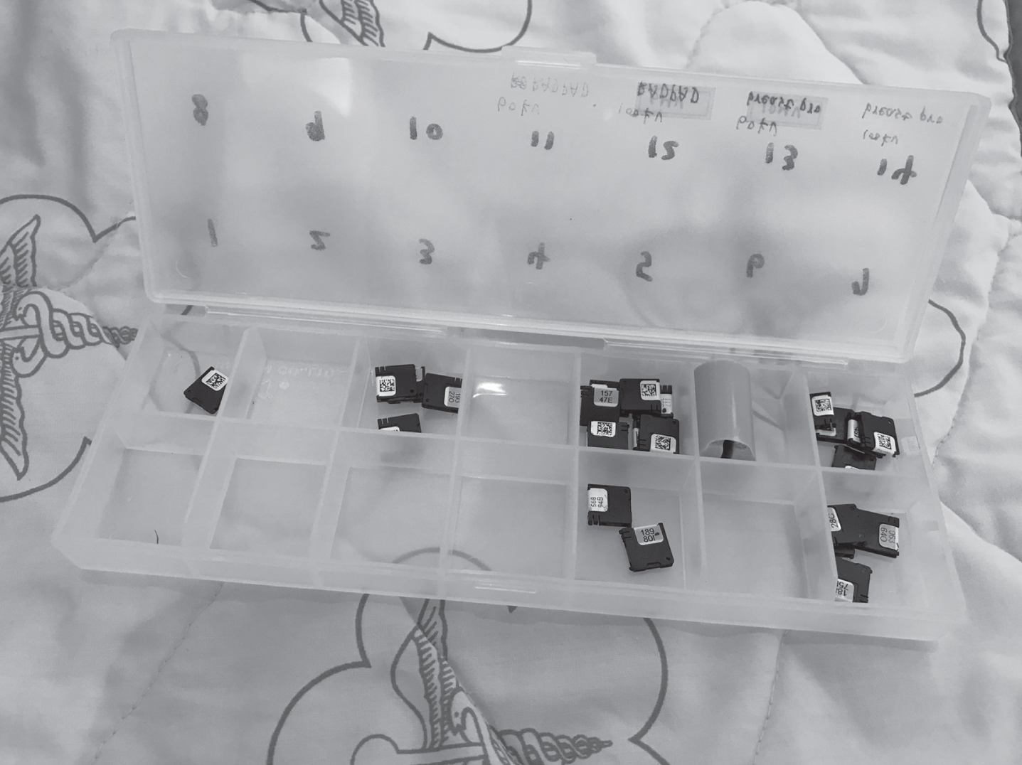
OSLDs black square plastic case with its own QR code and serial number.

OSLDs reading system (including laptop connecting QR code scanner and microStar Reader).
An ATOM-701 anthropomorphic adult phantom (CIRS, Norfolk, VA, USA) with height of 173 cm, weight of 73 kg, and chest width of 23 cm×32 cm was used (Fig. 3). The phantom is made of material equivalent to human tissue. From the top of the skull to the entire pelvic cavity, the anthropomorphic phantom comprised 39 slabs, and each slab is 2.5 cm thick with several cylinders to place the OSLD chips. We placed OSLD in the cylinders (Fig. 4, curved arrow) and then placed the cylinders back into the corresponding position of the human tissues or organs in the anthropomorphic phantom (Fig. 4, straight arrow), and then the position number and the OSLD serial number were noted.

Anthropomorphic phantom with multiple slabs for radiation dose measurement.

Procedures of the OSLD placement into the anthropomorphic phantom.
OSLD can be irradiated inside a CT head or body phantom and later read in a laboratory to obtain the dose profiles for specific CT parameters [8]. Examinations were performed using a 640-slice CT on an Alderson-Rando anthropomorphic phantom simulated as a supine patient (Fig. 5). The radiation-sensitive organs listed in the ICRP Publication 60 and Publication 103 included in the scanning range were selected to place OSLDs. For male, the selected tissues or organs include gonads (testicles), bone marrow, large intestine (colon), stomach, bladder, liver, skin, and small intestine (remainder tissues or organs). For female, the selected tissues or organs include gonads (ovaries), bone marrow, large intestine (colon), stomach, bladder, liver, skin, and small intestine (remainder tissues or organs). The effective doses were obtained by multiplying organ doses with radiation weighting factors and tissue weighting factors according to ICRP publication 60 or ICRP publication 103.

Anthropomorphic phantom simulated as supine patient on the CT examination couch. Arrows represent the alignment lasers.
Routine adult abdominal scanning parameters were used. The tube voltage was 120 kVp, same as used in clinical practice to scan different sections of human body (i.e., from chest to pelvic cavity). Automatic tube current modulation (ATCM) system was adopted in mA to reduce the radiation dose. ATCM system uses the attenuation values on anteroposterior and lateral scanogram to select the optimal tube current in an automated manner so that the user-chosen contrast-to-noise ratio (CNR) of each patient is maintained [11]. The scanning range was between the upper edge of the diaphragm and the end of the whole pelvic cavity, and the scanning direction was from head to feet. The above-mentioned CT scan included one-time anteroposterior topogram, lateral topogram, and helical scan. The measurement results represented the radiation dose received from abdominal CT examination without contrast medium injection.
It is important to ensure that the reproducibility of the phantom placement on the CT examination couch was strictly controlled, and the deviations caused by the phantom placement were minimized, so in each group of experiments, the height of the CT examination couch and scanning range must be confirmed as the same, and there was no rotation or skew in the phantom placement.
Because of the multi-readout feature of OSLD, each scanned OSLD was read in triplicate to obtain the average measurement value. After subtracting the background value, the net count values could be obtained so that the average net count value and standard deviation as the absorbed doses received from this examination could be calculated according to net count values measured within the prior three groups of experiments.
The absorbed doses of each tissue or organ, i.e. liver, stomach, bladder, gonads, colon, small intestine, bone marrow, and skin, measured in the phantom were multiplied with the weighting factor of photons to obtain the equivalent dose of each tissue or organ. After summing the product of the equivalent dose of each tissue or organ and its corresponding tissue weighting factor, the effective dose was obtained and then multiplied with the risk coefficient for stochastic effects to obtain the risk of stochastic effects caused by this examination. The radiation weighting factors, tissue weighting factor, and risk coefficients for stochastic effects were issued by ICRP Publication 60 and Publication 103 according to the collected information from the survivors of atomic bombs. While collecting more information, the suggested weighting factors might be changed. ICRP Publication 60 was published in 1991, and ICRP Publication 103 was published in 2007. These revisions reflected the progress in knowledge about the radiation sensitivity of various organs and tissues [12].
The OSLD readout values and the calculated effective doses are shown in Table 2 and Table 3 respectively. According to ICRP Publication 60, the calculated effective doses received from the examination were 0.90 mSv and 0.89 mSv for adult males and females respectively. The risk of stochastic effect was 5.040×10⁻⁵ and 4.984×10⁻⁵ for adult males and females respectively. According to ICRP Publication 103, the calculated effective dose received from this examination was 0.83 mSv for both adult males and females. The risk of stochastic effect was 3.486×10⁻⁵ for both adult males and females (Table 4). The risk of stochastic effects for male was obtained by multiplying effective doses for male with risk coefficients for stochastic effects for male according to ICRP publications 60 or 103. Same as female. The risk coefficients for stochastic effects for adults (Sv–1) is the possibility of stochastic effects might happen for adults while receiving effective dose per Sievert according to ICRP publication 60 or 103. The yearly cumulative dose received from the natural radiation is about 3 mSv [1]. The effective doses received from this CT examination were less than the yearly cumulative doses.
The equivalent dose of different organs from abdomen CT scan
The equivalent dose of different organs from abdomen CT scan
Effective dose estimation based on ICRP Publication 60 and Publication 103
*WR: Radiation weighting factor. WR of photons for all energies are identical in ICRP Publication 60 and Publication 103. *WT: Tissue weighting factor.
Risk estimation of stochastic effects based on ICRP Publication 60 and Publication 103
CT can provide fast and accurate medical diagnosis and has become a commonly used modality in radiology departments. In this study the radiation doses and the risk for stochastic effects under 640-slice abdominal CT examinations were investigated. The effective doses estimated according to ICRP Publication 60 were 0.90 mSv and 0.89 mSv for male and female adults respectively. The risk of stochastic effects was 5.040×10⁻⁵ and 4.984×10⁻⁵ for male and female adults respectively, while the effective doses estimated based on ICRP Publication 103 were 0.83 mSv for both male and female adults, while the risk of stochastic effect was 3.486×10⁻⁵ for both male and female adults.
Table 2 shows the readout values obtained from three groups of experiments. The readout values showed that there were some deviations of the measured tissue or organ doses between each group of experiments except for the skin dose. The reasons for the deviations between each group of experiments might be attributed to the unstable dose output of the CT, the uncalibrated condition of OSLDs, and the deviations of phantom placement, etc. [13–15].
The same CT scanning parameters were used among each group of experiments. Operations for quality assurance were regularly executed to make sure that the mechanical stability, radiation dose output, and image quality of the CT scanner were stable; additionally, OSLDs had been calibrated before the experiments to confirm that the sensitivity of the OSLDs was stable. Consequently, it was speculated that the most likely factor for deviations in the three groups of experiments was the deviations of phantom placement.
The phantom was always maintained at a fixed height on the CT examination couch, and the CT alignment laser was used to confirm the phantom was not rotated or skewed (Fig. 5, arrows). After performing each group of experiments, the cylinders that had OSLD inserted were removed and the exposed OSLD was replaced with an unexposed one. Because the cylinders could be rotated in the anthropomorphic phantom, the angle of the OSLD during radiation exposure could be adjusted arbitrarily; however, the angle of the OSLD placed inside the phantom were not fixed except for those placed on the surface of the phantom.
Through the skin dose measurement result, we verified that CT radiation dose output and the sensitivity of the OSLD measurement system were stable. OSLD is a proper dosimeter for in vivo dosimetry due to its small size and manageable energy dependence. Due to its large variation in energy response, OSLD is not suitable to measure radiation doses resulting from mixed beams of megavoltage therapeutic and kilovoltage imaging radiations [16]; however, only one kind of energy was used in this study, so it was speculated that the angular dependence of OSLD might be the main reason for the dose measurement deviations in these three groups of experiments.
S. B. Scarboro et al. reported that angular dependence correction is not necessary for dosimeter angles less than 45° for measurements made in air, inside the phantom, or on the surface [14]. Dosimeters that are placed at 90° on the surface of a phantom will likely be less sensitive. However, only helical scan mode was used in this study, so the x-ray tube in the CT gantry was rotating 360 degrees around the phantom while the CT couch was moving, meaning that the angle of the x-ray tube and the phantom kept changing during the examination, so the angle of the x-ray tube and each tissue or organ in the phantom was unknown. If each OSLD was set at a fixed angle, it would be impossible to know whether the measured doses were overestimated or underestimated, so in this case, the angle of each OSLD was not set as fixed within the three groups; therefore, the average value of the results should be closer to the real situation.
The phantom used in this study is an international standardized anthropomorphic phantom. The phantom is made of human tissue equivalent materials, so it can simulate the conditions of the human body being exposed to radiation in clinical practice. According to the literature published by M.K.A. Karim et al. in 2019, for patients of standardized body size, there were no significant differences in the effective dose between the phantom and patients during the clinical CT examination [17]. This meant the absorbed doses in radiosensitive organs of the patients could be estimated by using the technique demonstrated in a phantom study.
There were several limitations in this study. Because tissues or organs outside the scanning range received a very small amount of scattered radiation, this study only focused on the radiation-sensitive organs within the scanning range. If the radiation-sensitive organs outside the scanning range were included for measurement, the evaluation should be closer to the clinical condition. In addition, an adult anthropomorphic phantom and adult abdominal scanning parameters were used in this study to simulate clinical examination conditions for radiation dose measurements and risk evaluation of stochastic effect; therefore, the radiation dose measurement and risk evaluation should be only applicable to adults.
According to the literature published by K. Perisinakis et al. in 2018, iodine uptake of tissues or organs induced by intravenous contrast medium injection for contrast-enhanced CT imaging might considerably affect the radiation dose absorbed by tissues or organs [18]. Because the anthropomorphic phantom used in this study could not be injected with contrast medium for radiation dose measurement, the results of this study could only represent the absorbed dose and the radiation risk under 640-slice abdominal CT examination without contrast medium injection.
Regarding the “remainder” listed as a radiation-sensitive tissue or organ in ICRP Publication 60 and Publication 103, more than ten tissues or organs are included. When calculating the effective dose contribution of the remainder, the equivalent doses of all the tissues or organs listed as remainder should be averaged and then multiply by 0.12 as the tissue weighting factor. However, in this study only the small intestine dose was measured as a representative because the small intestine was listed as “remainder” in both ICRP Publication 60 and Publication 103; moreover, intestinal mucosa often undergoes mitosis, and thus logically it is more sensitive to radiation. Whether the estimation of the remainder radiation dose in this way is overestimated or underestimated requires other experiments for verification.
Because the cylinders used to place OSLD in the anthropomorphic phantom were relatively large, the number of OSLD spots in a single tissue or organ is relatively reduced. For a smaller tissue or organ such as the bladder, there would be only one spot for placement, so the absorbed dose of the specific tissue or organ could be measured by a single spot. Compared with the TLD-specific anthropomorphic phantom, due to the smaller size of TLD, there are more spots for TLD measurement in the anthropomorphic phantom.
Conclusion
Clinically, in order to diagnose the conditions quickly and accurately for patients suffering from abdominal diseases, doctors would prescribe abdominal CT examinations. Radiographers must not only provide clinicians with medical images having diagnostic value but strictly control the radiation dose as well.
Some effective dose of an abdomen CT is higher than 10 mSv [19, 20]. The radiation dose of CT depends on many factors, e.g. scan parameters, reconstruction algorithm and hardware. For some techniques of low dose CT, the dose can be reduced to lower than 1 mSv [21, 22]. The results of this study showed that the effective doses estimated according to ICRP Publication 60 and Publication 103 from abdominal CT examinations were lower than the yearly cumulative doses received from the natural background radiation (∼3 mSv) by the general population [1], revealing that there was no deterministic effect and the radiation risk of inducing stochastic effects was also relatively low, so this examination should be safe. These results could relieve and reassure patient anxiety, as well as provide a clinical medical reference for the execution of CT abdominal examination.
Disclosure
The authors declare that they have no conflict of interest.