Editorial
 Restricted accessEditorialFirst published June, 2026pp. 233-233

Select search scope: search across all journals or within the current journal


 Restricted accessEditorialFirst published June, 2026pp. 233-233Restricted accessObituaryFirst published June, 2026pp. 234-235
 Restricted accessResearch articleFirst published June, 2026pp. 236-243
 , Ricardo Vieira Camargo, Maria Tereza Scardua Mariano , [...]
Surgical techniques for upper lip lift are widely discussed in the literature. However, many questions remain about the influence of sex, age, and ethnicity on the indication for surgery and patient selection.
To verify whether the variation in the number of pixels in digital photographs in the subnasal region, before/after upper lip lift, correlates with the variables sex/age/ethnicity.
Standardized frontal photographs were analyzed from patients who underwent upper lip lift surgery through the Bull Horn Technique. Four linear distances were measured in pixels before and after surgery to quantify dimensional changes. Recovery patterns were assessed across 4 postoperative intervals (0, 7, 10-50, and 60-100 days). Nonparametric tests (Mann–Whitney U, Kruskal–Wallis H, Spearman’s correlation, and Pearson’s correlation) were used for subgroup analyses to evaluate associations among age, sex, ethnicity, and recovery time.
Digital pixel-based measurements revealed that 3 upper lip distances decreased after surgery, while upper lip height increased. The mean percentage increase in upper lip height was greater in men (72.62%) than in women (46.67%). No significant differences were found between sexes (
Pixel-based measurements suggested reduced tissue adaptability with aging, highlighting the clinical relevance of age and lifestyle factors in postoperative recovery.
Restricted accessResearch articleFirst published June, 2026pp. 244-249To evaluate the outcomes of upper eyelid skin excision via the subbrow approach combined with suspension of the orbicularis oculi musculocutaneous flap to the frontalis muscle.
A prospective descriptive case series was conducted involving 30 patients (60 eyelid-brow units) who underwent surgery at the Department of Plastic and Aesthetic Surgery, Cho Ray Hospital, from February to August 2024. Preoperative and postoperative assessments were conducted at 1 month and 3 months. Key variables included the distance from a horizontal plane through the mid-pupil to the upper eyelid skin crease and to the superior eyebrow margin at 3 measurement points. Postoperative pain was evaluated using the Visual Analog Scale (VAS), and patient satisfaction was assessed using the Likert scale.
The mean age was 54.4 years (range: 35-70). The height of the upper eyelid crease increased significantly at the mid-pupil and lateral canthal regions at both 1 month and 3 months postoperatively (
Upper eyelid skin excision via the subbrow approach combined with suspension of the orbicularis oculi musculocutaneous flap to the frontalis muscle is a safe and effective technique that significantly improves upper eyelid skin redundancy without altering the position of the eyebrow. The procedure is associated with minimal postoperative pain and high patient satisfaction.
Restricted accessResearch articleFirst published June, 2026pp. 250-258Iatrogenic facial nerve injury is a feared complication of craniomaxillofacial surgery, particularly when the parotid gland is instrumented or the Superficial Musculoaponeurotic System (SMAS) is violated. Facial palsy can have devastating psychosocial consequences for the patient, prompting decades of research to refine techniques of intraoperative facial nerve identification and preservation to avoid this outcome. The objective of this literature review is to trace the evolution of techniques for intraoperative facial nerve identification and describe ongoing advances in the field.
PubMed and Google Scholar were queried to identify publications describing both historical and conventional understandings of facial nerve anatomy and intraoperative facial nerve identification technique. Keywords included “facial nerve identification,” “facial nerve monitoring,” “facial nerve anatomy,” “facial nerve landmarks,” and “iatrogenic facial nerve injury.” Articles were screened for the following exclusion criteria: non-English language, full text unavailable, single-subject case reports, examination of peripheral nerves other than the facial nerve, non-mammalian animal models, and studies focusing on identification of the facial nerve at its pontine origin. Data was extracted and organized by year of publication, study type, study population, surgical context, method of facial nerve identification, key findings, advantages, limitations, and level of evidence.
Forty-two publications, published between 1954 and 2025, were included. Our review details both historical and conventional techniques of facial nerve identification including use of anatomic landmarks, electrical nerve stimulation, magnification and microscopy, and enhanced optical techniques including fluorescence-guided surgery.
Intraoperative facial nerve identification is crucial to prevent iatrogenic facial nerve injury; however, there is limited consensus on the precise identification technique. Facial nerve identification techniques continue to evolve, with fluorescence-guided surgery likely to be the next frontier in advancement of the safety and efficacy of these techniques.
Restricted accessResearch articleFirst published June, 2026pp. 259-266, Noureen Ghulam Fatima, Samman Khalid , [...]Cleft lip and/or palate (CL/P) is among the most common congenital anomalies globally. Pakistan has one of the highest incidences of CL/P worldwide. This study aims to investigate the clinical variability and ethno-demographic disparities of CL/P in the Islamabad and Upper Punjab region of Pakistan.
An observational, multicenter, cross-sectional study was conducted from 2020 to 2023. Patients with CL/P were recruited from tertiary hospitals, surgical camps, and rural communities. Clinical classification and demographic profiling were performed. Descriptive statistics summarized sample characteristics, and logistic regression was applied to assess associations with demographic variables.
A total of 303 patients were enrolled, with a male predominance (55%). The most frequent presentation was cleft lip and palate (CLP, 53%), followed by cleft palate (CP, 28%) and cleft lip (CL, 19%). Complete clefts were more common (67%), and unilateral forms (74%) were predominantly left-sided (62%). Sporadic cases comprised 79%, while parental consanguinity was reported in 64% of the sample. First-parity births accounted for 35%, and isolated clefts were observed in 48%. Syndromic associations (52%) were primarily neurological, sensorineural, and limb anomalies. Most patients were from rural areas (69%), Punjab province (53%), Punjabi-speaking households (40%), and low socioeconomic strata (50%). Significant variation in cleft distribution was observed across demographic subgroups.
CL/P in this region exhibits high clinical heterogeneity and striking ethno-demographic disparities, reflecting the interplay of genetic, environmental, and socioeconomic factors. These findings highlight the need for targeted interventions in resource-limited settings, emphasizing the organization of surgical cleft camps in rural areas, the involvement of Non-Governmental Organizations (NGOs) supporting cleft care, and the promotion of health education in communities with high consanguinity and socioeconomic vulnerability.
Restricted accessResearch articleFirst published June, 2026pp. 267-281Spring-assisted cranioplasty has been established as an effective technique for minimally invasive correction of single suture sagittal craniosynostosis. However, the potential for secondary suture synostosis following spring placement remains unclear. This case series represents our institutional experience with spring-assisted cranioplasty and highlights 5 patients with progression to secondary suture craniosynostosis necessitating surgical intervention following initial spring placement.
IRB-approved data from all patients undergoing spring-assisted cranioplasty for sagittal craniosynostosis (2021-2025) were retrospectively reviewed.
Fifty-five patients over the 4-year interval underwent spring-assisted cranioplasty. Fifty-four patients presented with a diagnosis of single suture sagittal craniosynostosis, while one patient had sagittal and unilateral lambdoid craniosynostosis. All patients had 2 springs placed except for the one with concomitant lambdoid synostosis, who received a third. Mean age at spring placement was 3.7 months with a mean interval to removal of 3.8 months. The average operative duration was 80.3 minutes, and the mean length of stay was 1.5 days. Eighteen (32.7%) of patients had an intensive care unit stay and 14 (25.5%) of patients required a blood transfusion. Nine of the 55 patients required an unplanned secondary procedure. Two patients experienced hardware migration with the need for operative adjustment/replacement. Six patients (10.9%) necessitated subsequent CVR: 4 due to the development of secondary unicoronal synostosis and 2 due to inadequate expansions and residual cranial deformities. One patient developed bilateral coronal and right lambdoid synostosis and subsequently underwent suturectomy and helmet therapy.
This series highlights the observation of secondary suture craniosynostosis following spring-assisted cranioplasty for sagittal synostosis. Whether the progression is a direct sequelae of underlying spring force mechanics/provider placement or a reflection of natural synostotic evolution remains uncertain. Its clinical significance warrants further investigation and discussion within the broader craniofacial surgical community.
 Open accessResearch articleFirst published June, 2026pp. 282-289
Secondary craniosynostosis can occur in patients with metabolic and hematologic disorders. However, current studies have been limited due to the rarity of these pathologies. The current study uses a large nationwide multi-institution database to evaluate patient demographics, suture patterns, and associated comorbidities.
A retrospective analysis of the TriNetX database was performed for patients diagnosed with craniosynostosis and X-linked hypophosphatemia (XLH), vitamin D-resistant rickets (VDRR), pseudohypoparathyroidism, glycosaminoglycan disorder, osteopetrosis, sickle cell, thalassemia, and polycythemia vera. Demographics, suture patterns, and craniofacial, orthopedic, and systemic congenital malformations were evaluated.
About 1,902 patients with secondary craniosynostosis with metabolic or hematologic disorders were identified. Two hundred sixty six patients with XLH, 236 with VDRR, 136 with pseudohypoparathyroidism, 283 with hyperthyroidism, 103 with glycosaminoglycan disorder, 27 with osteopetrosis, 509 with sickle cell, 302 with thalassemia, and 40 with polycythemia vera. The most common suture involvement was sagittal in XLH (68.18%), VDRR (55.56%), hyperthyroidism (52.94%), thalassemia (43.18%), and sickle cell (36.78%). Multiple suture involvement also commonly occurred, especially in osteopetrosis (66.67%), pseudohypoparathyroidism (40.00%), and glycosaminoglycan disorder (30.00%). Chiari malformations and hydrocephalus were common in XLD, VDRR, and osteopetrosis. Orthopedic comorbidities occurred frequently in osteopetrosis, glycosaminoglycan disorder, VDRR, and XLH. Congenital cardiac malformation was common in all groups. Elevated rates of all congenital systemic malformations were seen in XLH, VDRR, and pseudohypoparathyroidism.
Metabolic and hematologic disorders with secondary craniosynostosis are associated with sagittal and multi-suture closure patterns. Multiple groups showed increased rates of other craniofacial, orthopedic, and congenital systemic malformations.
Restricted accessResearch articleFirst published June, 2026pp. 290-296, Brian E. Kinard, Kathlyn K. PowellBilateral mandibular distraction osteogenesis (MDO) has been used successfully to treat children with upper airway obstruction secondary to micrognathia associated with isolated Pierre Robin sequence (iPRS). Clinical benefit of distraction has not been as evident in syndromic children with micrognathia. The purpose of this study is to evaluate surgical outcomes of bilateral MDO in individuals with iPRS compared to those with micrognathia and an associated syndromic diagnosis.
Retrospective cohort study designed and implemented at 2 academic medical centers. Inclusion criteria included diagnosis of micrognathia and Pierre Robin Sequence and/or a genetic syndrome as determined by genetic testing and underwent bilateral MDO between 1/1/2007 and 1/31/2024. The primary predictor variable was the primary craniofacial associated diagnosis: iPRS or micrognathia with syndromic association. Primary outcome variable is surgical success defined by de-cannulation after MDO and improvement in apnea-hypopnea index (AHI). Secondary outcome variables include complications: dental, growth, surgical.
Bilateral MDO achieved decannulation in (5/5; 100%) of iPRS patients compared to (4/10; 40%) of micrognathic patients with syndromic association (
Bilateral MDO is more successful at achieving decannulation in iPRS patients compared to micrognathic patients with syndromic association. Mean reduction in AHI after bilateral MDO is similar in iPRS patients compared to syndromic patients. There is no difference in complication rates between iPRS and micrognathic syndromic patients after bilateral MDO, but overall dental complication rates are high (78.3%).
Restricted accessResearch articleFirst published June, 2026pp. 297-305, Andrew Adamek, Arjav Shah , [...]Literature regarding the effects of gunshot wounds (GSW) to the face in children is scarce. This study aimed to identify presenting features and clinical outcomes of pediatric patients with GSW to the face, with special attention to ophthalmic data.
This is a retrospective cohort study of patients seen by oculoplastics at a Level I trauma center in the United States after a GSW injury to the face from May 2018 to September 2024. Relevant demographic and clinical data were collected through chart review. Categorical variables are described using percentages and numerical variables are described using median with interquartile range.
Nine patients were included with a median age of 16.6 (4.7) years; 67% were male. The most common bullet entry site was the orbit (44%) followed by cheek (33%). The bullet traversed the midline in 78% of cases. Common injuries included orbital wall fracture, open globe injury, optic nerve injury, and intra-ocular hemorrhages. 56% had concomitant intracranial hemorrhages. At least 2 patients presented with non-light perceiving (NLP) vision in at least 1 eye. At final ophthalmology follow-up, 2 patients were legally blind, and 4 were NLP or had undergone enucleation in at least 1 eye. The most common surgical intervention performed was fracture repair (56%) followed by enucleation (33%).
This study highlights the devastating effect of GSW to the face in children, particularly with respect to the eyes and orbits. Further studies with larger sample sizes are warranted to identify optimal management patterns and risk factors for poor outcomes.
Restricted accessResearch articleFirst published June, 2026pp. 306-310, Arjav Shah, Maureen Beederman , [...]Traumatic injuries resulting in complex pan-orbital fractures, often accompanied by fractures of the skull base, calvarium, and midface, present a unique surgical challenge. These injuries are often difficult to address in a single operation due to the need for staged neurosurgical and orbital reconstruction. Neurosurgical repair of the dura and orbital roof fractures may need to be performed before addressing fractures of the medial orbital wall and orbital floor. Pushing orbital contents back into the orbit from below is especially difficult with swelling from a CSF leak, and the risk of damaging a newly repaired orbital roof and dura. Waiting for the orbital roof and dural repair to heal is an option, but delayed repair of the other orbital walls is difficult once there is scarring and fibrosis of the orbital contents to sinus mucosa. In ballistic injuries with near-total destruction of the orbit, reconstruction is further complicated by the loss of natural anatomical landmarks. We present a novel staged operative repair of complex pan-orbital trauma due to a gunshot wound. This patient suffered multiple fractures of the orbit, including right orbital roof and medial orbital wall fractures, and left orbital roof, floor, and medial orbital wall fractures. The patient also had bifrontal herniation, CSF leak, and suffered a left globe obliteration rendering that eye unsalvageable.
Open accessResearch articleFirst published June, 2026pp. 311-319, Anshumi Desai, Soumil Prasad , [...]Facial fractures are common traumatic injuries with variable outcomes due to variations in etiologies, comorbidities, and anatomical complexity. Effective preoperative risk stratification is crucial for identifying patients at higher risk of complications. This study evaluates the utility of the Modified 5-Item Frailty Index (5-mFI) in predicting postoperative outcomes for patients undergoing surgical repair of facial fractures.
A retrospective analysis was conducted using American College of Surgeons National Surgical Quality Improvement Program (ACS-NSQIP) data from 2011 to 2021. The 5-mFI score (0-5) was based on hypertension, diabetes, respiratory disease, heart failure, and functional dependence. Patients were stratified into low-risk (5-mFI < 2) and high-risk (5-mFI ≥ 2) groups. Statistical comparisons and multivariable logistic regression were used to evaluate outcomes.
Of 7549 patients, 6066 (80.4%) were included in the 5-mFI < 2 group and 1483 (19.6%) in the 5-mFI ≥ 2 group. The 5-mFI < 2 group was more functionally independent (99.36% vs 93.46%;
5-mFI ≥ 2 patients experience more complications and delayed recovery. Integrating 5-mFI into preoperative assessments may improve patient communication and guide perioperative management.
Restricted accessResearch articleFirst published June, 2026pp. 320-326Secondary hollowness and accelerated facial aging are reasons surgeons remain judicious or avoid buccal fat pad removal in the aging face. Yet, the buccal fat pad can be unattractive when visualized through skin or prolapsing. Unrecognized pseudoherniation of the buccal fat pad can lead to visible deformity after facialplasty or liposculpture. True herniation can also occur. Etiology and treatment strategies for both are explored.
Nine patients with buccal bulge deformity were identified after facelift (8 cases) and facial liposuction alone (1 cases). Hallmark features of the deformity include a reducible convexity anterior to the masseter and 1 cm cephalad to the mandible border. Patients were treated with buccal fat removal through an intraoral or facelift approach, or with fillers to mask the contour deformity.
Buccal bulge deformity improved in all cases. At 1 year postoperatively, partial correction was observed in 5 cases and there was complete correction in 4 cases.
Buccal bulge deformity can occur after facialplasty, liposculpture or both. Buccal fat removal effectively corrects the deformity, which can also be concealed with injectable fillers.
Open accessResearch articleFirst published June, 2026pp. 327-335Excessive gingival display, or “gummy smile,” occurs in up to 14% of adults and is associated with lip hyperactivity, dentoalveolar extrusion, or vertical maxillary excess. Botulinum toxin A (BTA) has emerged as a minimally invasive therapeutic option, but protocols vary widely and long-term data are limited.
To critically evaluate the current literature on BTA in gummy smile management, focusing on classification systems, injection techniques, efficacy, safety, and comparative outcomes against conventional surgical and orthodontic treatments.
A narrative review was performed using PubMed, MEDLINE, and Scopus (2000-2023) with the terms “gummy smile,” “excessive gingival display,” and “botulinum toxin.” Clinical studies, case series, and reviews addressing BTA use were included. Data were synthesized into thematic categories.
BTA consistently reduced gingival exposure by 3 to 5 mm, with effects lasting 4 to 6 months. Three main injection strategies were identified: single-point (Yonsei), multi-point, and orbicularis oris (“lip flip”). Reported complications were rare and transient (mild asymmetry, lip weakness). Standardized protocols were lacking, with heterogeneity in dosing (2-5 units per site) and injection depth. Comparative studies suggested BTA is less durable than surgical approaches but offers superior patient acceptability and safety for mild to moderate cases.
BTA provides reproducible, temporary improvement in gummy smiles with high safety and satisfaction rates. However, absence of consensus on optimal technique and limited long-term evidence highlight the need for standardized protocols and prospective multicenter studies.
Restricted accessResearch articleFirst published June, 2026pp. 336-340, Jason Cohn, Jeewanjot S. Grewal , [...]Craniosynostosis is a well-described pathology involving aberrant formation of skull continuity. Due to these pathological fusions, the efficacy of the pediatric auditory system can manifest in conditions lending to ineffective hearing. While syndromic craniosynostosis has been analyzed for this purpose, hearing loss in infants with nonsyndromic craniosynostosis is less examined. The purpose of this study is to analyze whether nonsyndromic subtype may have an effect on hearing.
A two-center retrospective chart to identify patients having nonsyndromic craniosynostosis. Subjects with either metopic or sagittal craniosynostosis were included. Factors including age, demographics, severity of symptoms – including headache, otitis media, micrognathia, hydrocephalus, and macrocephaly – treatment, and hearing loss were identified and examined.
In total, 101 patients with non-syndromic, single metopic or single sagittal craniosynostosis were identified and included. The mean age of this cohort was 38.4 ± 39.4 months. Of these, sagittal synostosis was the most common (62/101, 61.4%) while 38.6% of subjects had isolated metopic craniosynostosis. In total, 8 subjects (7.9%) suffered from hearing loss. The majority of these (6/8, 75%) were conductive hearing loss, with the remaining 25% being Sensio neural hearing loss. These factors were not associated with subject hearing loss (
While craniosynostosis can present devastating complications secondary to premature skull fusion, it appears that nonsyndromic craniosynostosis in the metopic and sagittal patterns do not differ significantly in severity of symptoms nor hearing loss.
Open accessResearch articleFirst published June, 2026pp. 341-346, Peter Schultz, Margaret McQuillan , [...]The primary objective was to determine age-related changes in perceived nasal resonance of young children with repaired cleft palate with or without cleft lip (CP ± L). A secondary objective was to explore associated changes in vowel segment duration, speaking rate, and fundamental frequency (F0) of the children.
Prospective, longitudinal observational.
Twenty children with non-syndromic CP ± L followed at 3 facilities.
All participants were audio recorded repeating identical speech samples at 3 time points corresponding to mean ages of 6, 7, and 8 years. Sixty audio files were created with counterbalanced pairs of recordings from the 3 time points. Five blinded speech-language pathologists judged which sample of a pair sounded more nasal.
Paired-comparison judgments of nasality at each age; vowel duration, speaking rate, and F0 at each age derived from acoustic analysis.
Speech samples from the youngest age were judged as more nasal more often than samples from the older ages (
Children with repaired CP ± L are perceived to be more nasal at younger ages during production of phonetically identical speech samples. Clinicians need to be cognizant of age-related changes in other aspects of speech production that may influence judgments of nasal resonance.
Restricted accessResearch articleFirst published June, 2026pp. 347-354, Eric Zeng, Braden V. Saba , [...]There is a perceived shortage of craniofacial surgery jobs, related to beliefs that they are limited to the pediatric population and academic institutions. Craniofacial fellowship graduates were surveyed to assess the accuracy of this notion.
An anonymized 14-item REDCap survey was emailed to 310 surgeons who have completed craniofacial fellowships in the U.S. or Canada. Demographics, desired and actual practice type, case exposure, job changes, and perceived negative factors were collected and analysed descriptively.
Number of responses was 107 (34.5% completion rate). At graduation, 81.3% sought a craniofacial career and 78.5% obtained this position. Of those who started working in craniofacial practice, 88.1% indicated that was their primary goal. Only 33.3% of those starting in craniofacial practice changed positions, whereas 71.4% beginning in noncraniofacial practice did. Limited job opportunities was the most cited drawback initially (65.9%) and currently (17.8%), regardless of practice type. Comparing fellowship exposure with present practice revealed marked contraction of orthognathic (−41.1 percentage points) and pediatric craniosynostosis work (−36.4) and expansion of facial esthetic (+15.9) and gender-affirming (+14.0) surgery, with 19.6% devoting time to other esthetic or reconstructive procedures such as breast surgery.
Contrary to earlier reports, this study indicates that craniofacial fellows often secure and retain their desired practice; however, the scope of practice has broadened toward adult esthetic and gender-affirming realms. Prospective craniofacial surgeons and fellowship directors alike should recognize growing opportunities within the specialty as graduates increasingly apply their unique skillsets to broader populations including adults and the transgender community.
Restricted accessResearch articleFirst published June, 2026pp. 355-362Spring-assisted cranioplasty (SAC) and endoscopic strip craniectomy (ESC) with postoperative helmet therapy are minimally invasive procedures that are increasingly utilized to treat craniosynostosis, largely due to their less invasive nature, compared to open calvarial vault reconstruction (CVR). Enthusiasm for SAC has surged in the absence of reported complications.
We present the case of an 11-month-old infant who developed a large leptomeningeal cyst following SAC for sagittal craniosynostosis. Additionally, we conducted a literature review of articles focusing on the complications associated with SAC.
The patient required subsequent surgical repair and cranial reconstruction, which resulted in a full recovery. The literature review revealed 11 studies describing 571 total operations. Across the studies identified in this review, the most common complications were delays in calvarial ossification, undercorrection, perioperative return, and difficult spring removal. Of these 571 total operations, 3 patients’ postoperative courses were complicated by CSF leak.
Though dural injury is a rarely reported complication of SAC, surgeons should be cautious of the spring instrumentation, which can cause significant morbidity if dural injury goes unrecognized and is not properly managed.
Restricted accessLetterFirst published June, 2026pp. 363-368 Restricted accessOtherFirst published June, 2026pp. 369-369
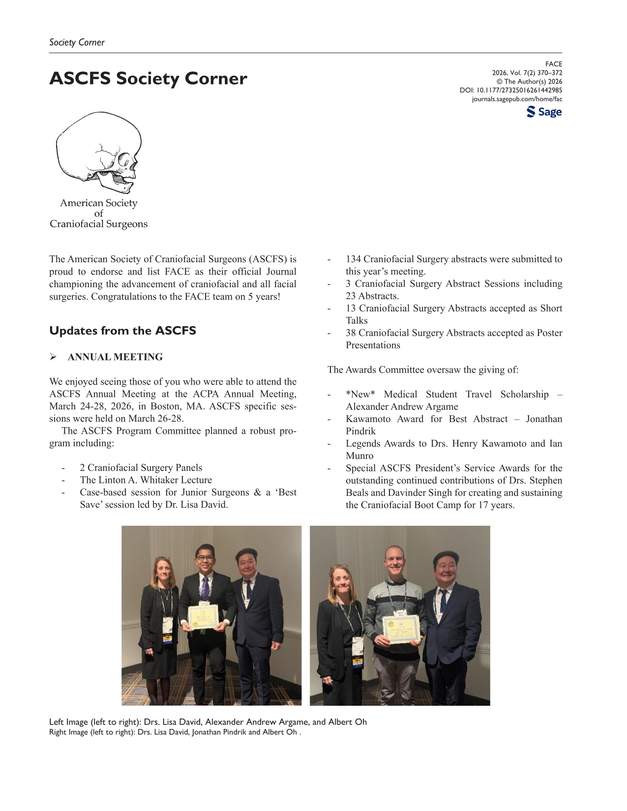
 Restricted accessOtherFirst published June, 2026pp. 370-372
 Restricted accessOtherFirst published June, 2026pp. 373-374