
Abstract
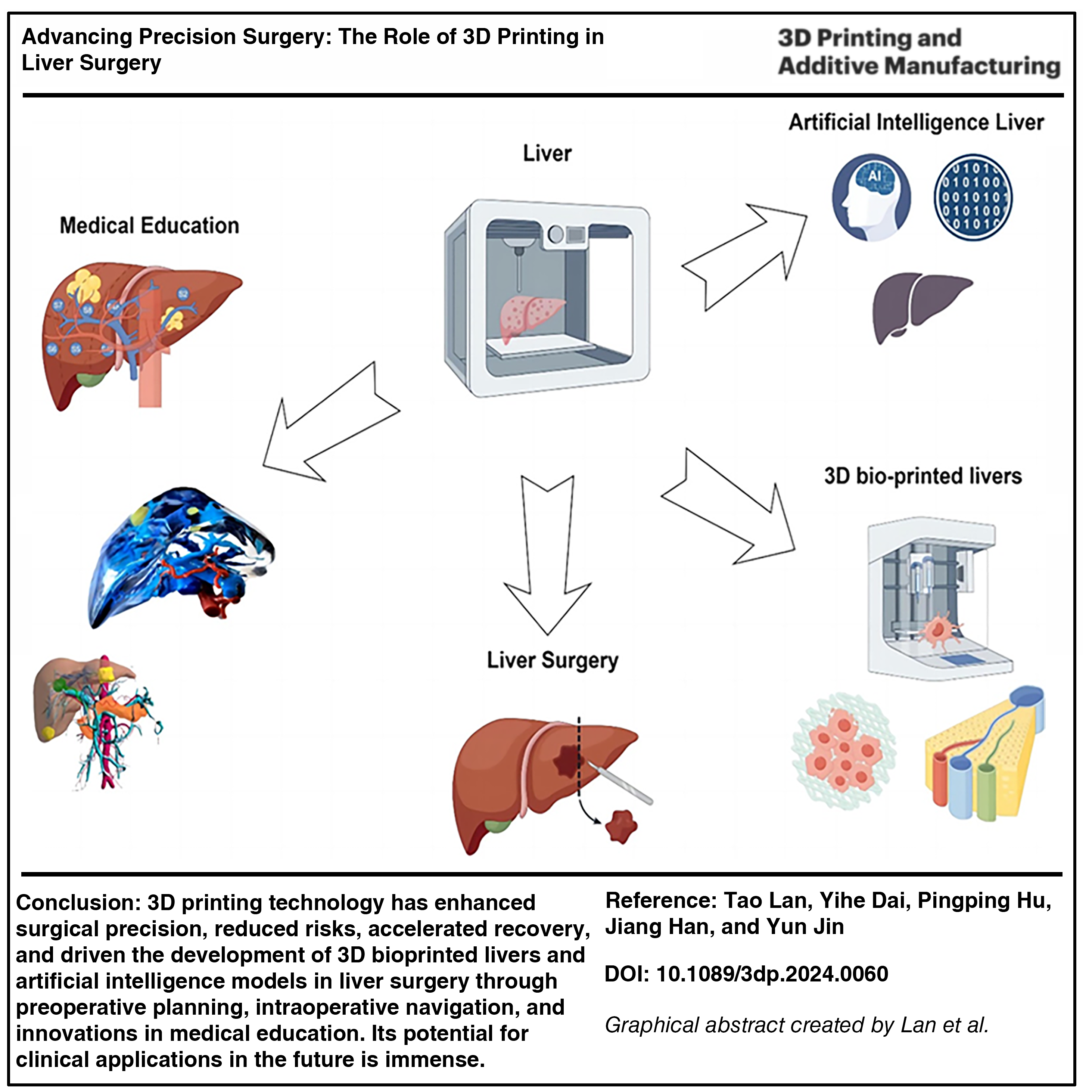
The development of precision surgical procedures is rapidly transforming the treatment methods in complex surgical fields such as liver surgery. 3D printing technology, as a crucial support, has not only redefined surgical planning and execution but also improved medical education and patient communication. This article discusses the application of 3D printing in liver surgery, including surgical planning, intraoperative navigation, education and training, and 3D bioprinting technologies. 3D-printed models, with their ability to accurately display the spatial relationship between tumors and surrounding structures, provide a foundation for surgeons to devise precise surgical plans. This allows surgeons to develop more accurate surgical strategies preoperatively, reducing surgical risks and preserving more healthy liver tissue. Furthermore, this article discusses the challenges faced by 3D printing technology, such as cost and technical limitations, as well as the difficulties in clinical application, and provides a future outlook. Through a review of literature and case studies, this article highlights the significant role of 3D printing technology in enhancing surgical precision, reducing risks, and promoting patient recovery. With technological advancements, the prospects for the application of 3D printing in precision surgical procedures are broad, especially in the field of liver surgery, offering patients safer and more effective diagnostic and therapeutic options.
Introduction
According to the latest global cancer burden data released by the International Agency for Research on Cancer of the World Health Organization in 2022, malignant liver tumor lesions rank sixth in the incidence of malignant tumors worldwide. In China, malignant liver tumor lesions rank fourth in the incidence of malignant tumors, making it the second leading cause of death among patients with cancer. 1 Surgical resection remains the preferred treatment for malignant liver tumors. 2 Due to the liver’s complex anatomy and the diversity of tumors, surgical treatment of liver tumors faces many challenges. First, ensuring the surgery can precisely remove the tumor while preserving sufficient healthy liver tissue to maintain liver function is key to the success of the surgery. 3 Second, avoiding major bleeding during surgery and protecting important vascular and bile duct structures are also major challenges in surgery. Moreover, devising effective surgical strategies for complex or atypical tumors is particularly difficult. With the development of medical technology, the introduction of precision surgery and 3D printing technology offers new possibilities for the surgical treatment of liver tumors. Precision surgery focuses on using medical imaging technologies and computer-aided design to create personalized surgical plans, improving the accuracy and safety of surgery. 4 As an important tool of precision surgery, 3D printing technology transforms patients’ medical imaging data into tangible 3D models, allowing surgeons to understand the tumor’s location, size, and relationship with surrounding structures in detail before surgery, thereby precisely planning the surgical path and reducing uncertainty during surgery.5–7
This review delves into the applications of 3D printing technology in liver surgery, encompassing surgical planning, intraoperative navigation, educational training, and 3D bioprinting technology. The printed models enable accurate visualization of the spatial relationships between tumors and surrounding structures, laying the groundwork for surgeons to devise precise surgical plans. Furthermore, the article addresses the challenges encountered by 3D printing technology, including cost and technical constraints, as well as the hurdles in clinical applications, while also offering future prospects. Through a review of literature and case studies, it underscores the crucial role of 3D printing technology in improving surgical precision, mitigating risks, and facilitating patient recovery. With the continuous advancement of technology, 3D printing technology has broad prospects for application in precision surgery, especially in the field of liver surgery, providing patients with safer and more effective diagnostic and treatment options.
The Evolution of 3D Printing in Medicine
3D printing technology, also known as additive manufacturing, originated in the 1980s. 8 It allows for the direct construction of complex-shaped solid objects from digital 3D models by layering materials until the object is complete. Initially utilized for rapid prototyping, it aided designers and engineers in quickly validating and iterating designs during the early stages of product development. Over time, 3D printing has made significant advancements in accuracy, speed, and the variety of materials available, driving its application across multiple fields, including medicine. At the turn of the century, 3D printing entered the medical field, initially used to manufacture customized prosthetics and medical devices, such as dental implants and orthotic braces. As the technology matured and became more precise, 3D printing began to be used for more complex medical applications, including the manufacturing of human tissue and organ models, as well as serving as a tool for surgical planning and simulation.9–11 These applications not only improved the success rate of surgeries but also offered patients more personalized treatment options.12,13 The breakthrough in surgical applications of 3D printing is partly due to advancements in medical imaging technologies. With improvements in computed tomography (CT) and magnetic resonance imaging (MRI) scanning techniques, doctors can obtain higher-resolution images of patients’ internal structures. These high-definition images can be converted into 3D-printed models, offering new possibilities for surgical planning and simulation.12,14 In addition, developments in material science have supported the application of 3D printing in medicine, enabling the use of various materials (including biocompatible materials) to manufacture models and implants for clinical use. 15 Common biocompatible materials include polylactic acid, poly(lactic-co-glycolic acid) (PLGA), polycaprolactone, gelatin, chitosan, etc. Importantly, advancements in 3D printing technology have significantly reduced the cost and time to manufacture personalized medical devices, making the use of customized tools and implants in emergency surgeries possible. Furthermore, the application of 3D printing technology has also fostered the development of medical education and patient communication by providing physical models to help explain complex medical conditions and surgical procedures.16,17
The progress of 3D bioprinting technology has brought about significant innovation in the medical field. This technology can accurately fabricate biomimetic tissues and organs, such as skin, bones, and hearts, for transplantation surgeries or repairing damaged tissues. Moreover, it plays a vital role in biomedical research and drug development by accelerating the process of developing new drugs through the fabrication of miniature models of human organs. In addition, 3D bioprinting technology can also personalize medical devices, such as prosthetics and scaffolds, thereby enhancing treatment efficacy and patients’ quality of life. The continuous development of this technology has opened up new prospects and opportunities for the medical field.
3D Printing for Preoperative Planning in Liver Surgery
Liver surgery is a highly complex surgical procedure that faces numerous preoperative challenges. The complexity of liver surgery stems from the intricate anatomy of the liver and the high risks involved in the surgical process. The complex network of blood vessels and bile ducts interwoven within the liver necessitates extreme care by surgeons during the operation, aiming to precisely remove tumors while minimizing damage to vital blood vessels or ducts.18–20 This requirement underscores the importance of personalized surgical planning, tailored to devise the optimal surgical strategy for each patient. Furthermore, preserving sufficient healthy liver tissue to support postoperative liver function is crucial for the success of the surgery.21–26 In traditional liver surgery planning, dealing with large tumors poses particular challenges. In many cases, surgeons tend to lean toward conservative treatments such as chemotherapy due to uncertainty about the exact remaining liver volume. However, these tumors can sometimes be curatively removed through surgery. The introduction of 3D printing technology provides a visual representation through 3D reconstruction of the liver and tumors, enabling surgeons to clearly assess the remaining liver volume after tumor removal when planning surgery. 24 This approach not only aids in selecting surgical plans based on real data but also helps in reducing the proportion of large tumors where curative surgery was not chosen. 27
With the application of 3D printing technology, surgeons can now utilize patients’ CT or MRI scan data to print precise 3D models, offering a fresh perspective for surgical planning (FIG. 1). 28 Doctors can thoroughly understand the tumor’s location, size, and its relationship with surrounding tissue structures before surgery, allowing for precise surgical pathway planning, predicting, and avoiding potential risks. 29 Moreover, 3D models can be used for surgical simulation, enabling doctors to practice before the actual surgery, thereby enhancing the safety and success rate of the procedure. 30 3D printing technology not only optimizes the surgical planning process but also provides an effective means to handle unforeseen circumstances during surgery, bolstering doctors’ confidence and consistency in the decision-making process. Through this technology, surgical teams can execute complex surgeries with higher precision, greatly improving patients’ treatment outcomes and safety.31–34
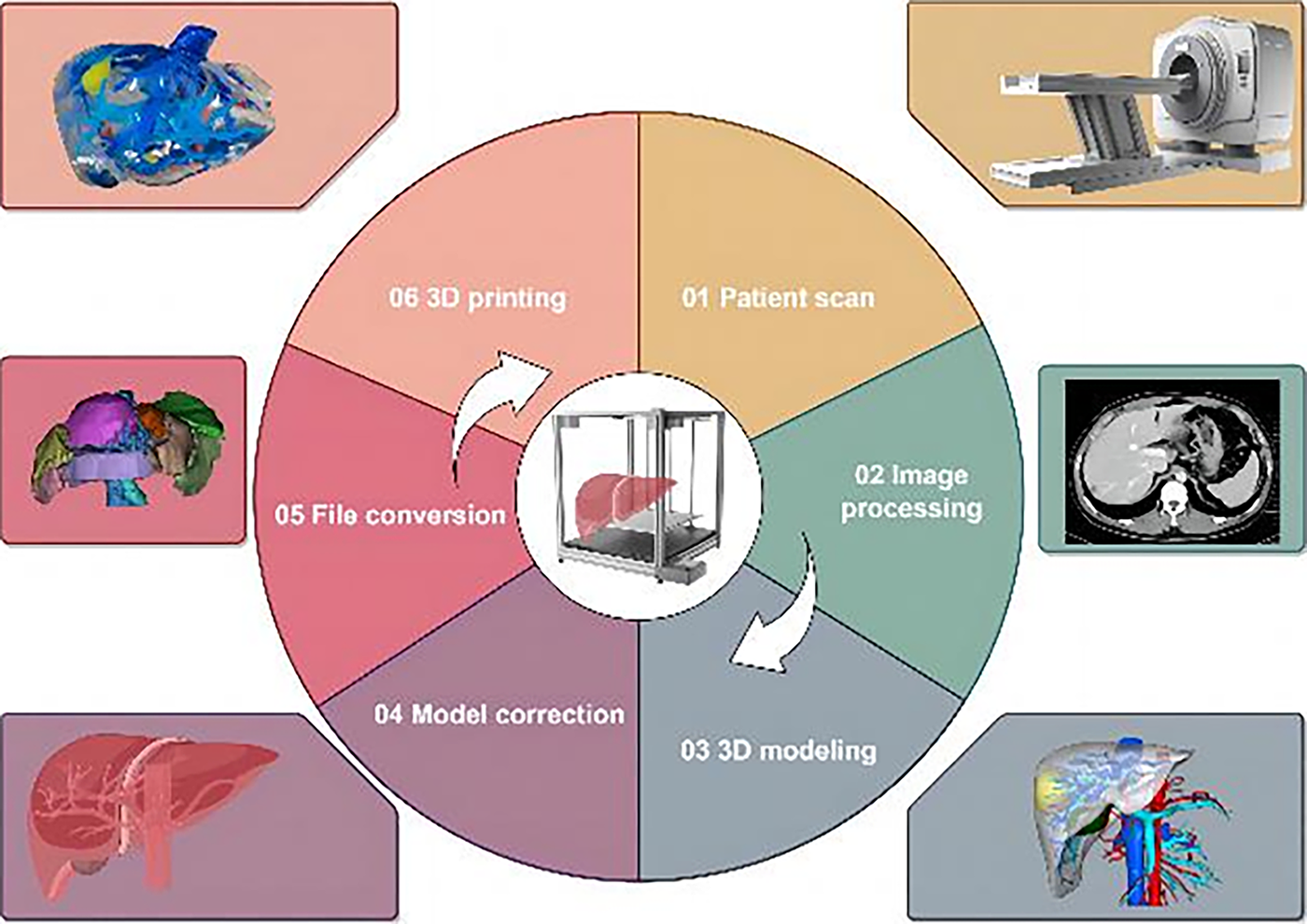
The process of creating a 3D-printed liver model includes patient scanning, image processing, 3D modeling, model adjustments, file conversion, and 3D printing By Figdraw.
Application in Polycystic Liver
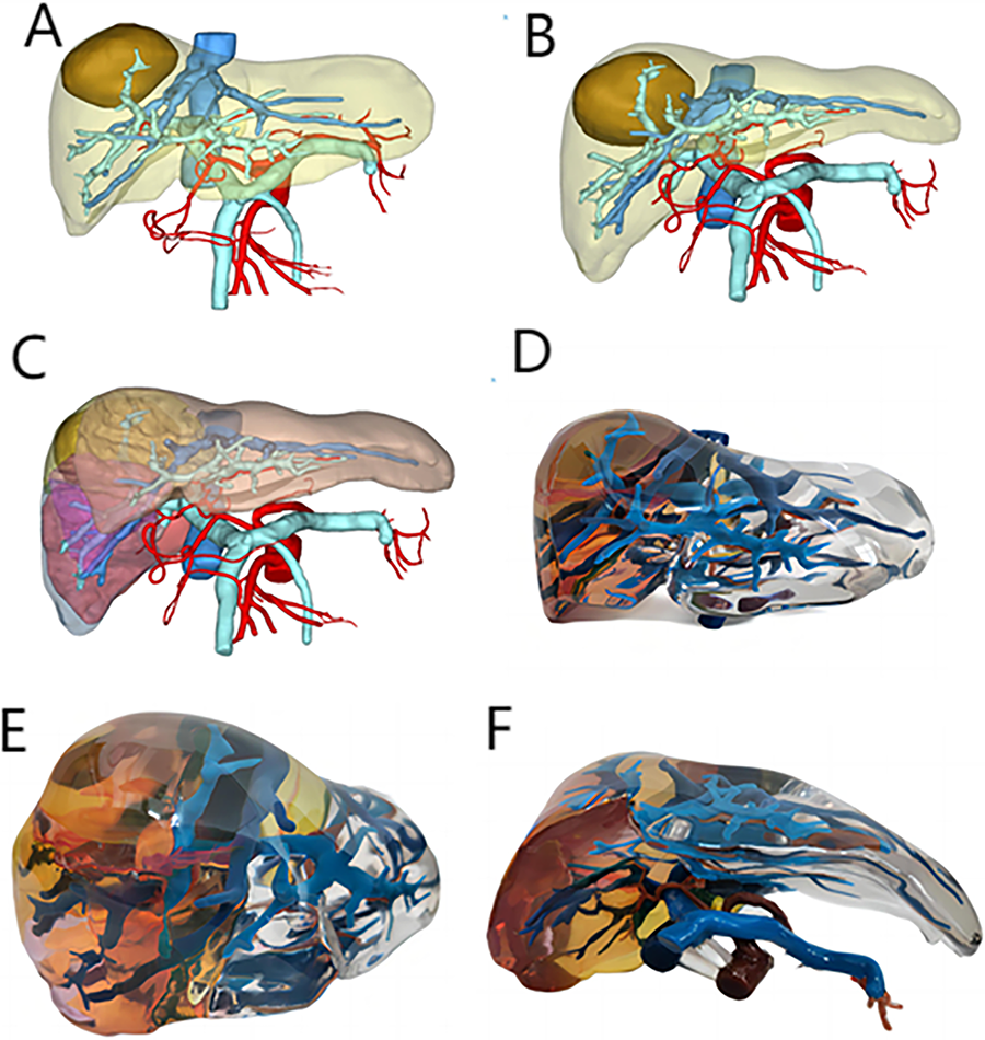
By preoperatively 3D printing a patient’s liver, surgeons can gain a detailed understanding of the exact location of liver cyst lesions and their relationships with blood vessels and bile ducts, providing precise path planning and decision support for the surgery. This meticulous preparation ensures the accurate identification of the liver sections that need to be removed, laying a solid foundation for the successful execution of the surgery. During the operation, surgeons strictly follow the detailed preoperative planning, ensuring the precise removal of the cyst, significantly reducing surgical risks, and enhancing treatment efficacy. 32 The practical application of this technology was further validated by the case of a 41-year-old female patient diagnosed with congenital polycystic liver in our hospital in October 2016. The preoperative 3D printing technology not only provided doctors with detailed surgical planning but also accurately calculated that the residual liver volume accounted for 59.2% of the total liver volume. This preparation work provided important evidence for the patient’s surgery. Based on this plan, the patient successfully underwent partial resection of the left and right liver lobes and cyst fenestration surgery on October 24, 2016. The close integration of preoperative preparation and intraoperative execution demonstrates the tremendous potential of 3D printing technology in improving surgical precision and safety (Fig. 2).
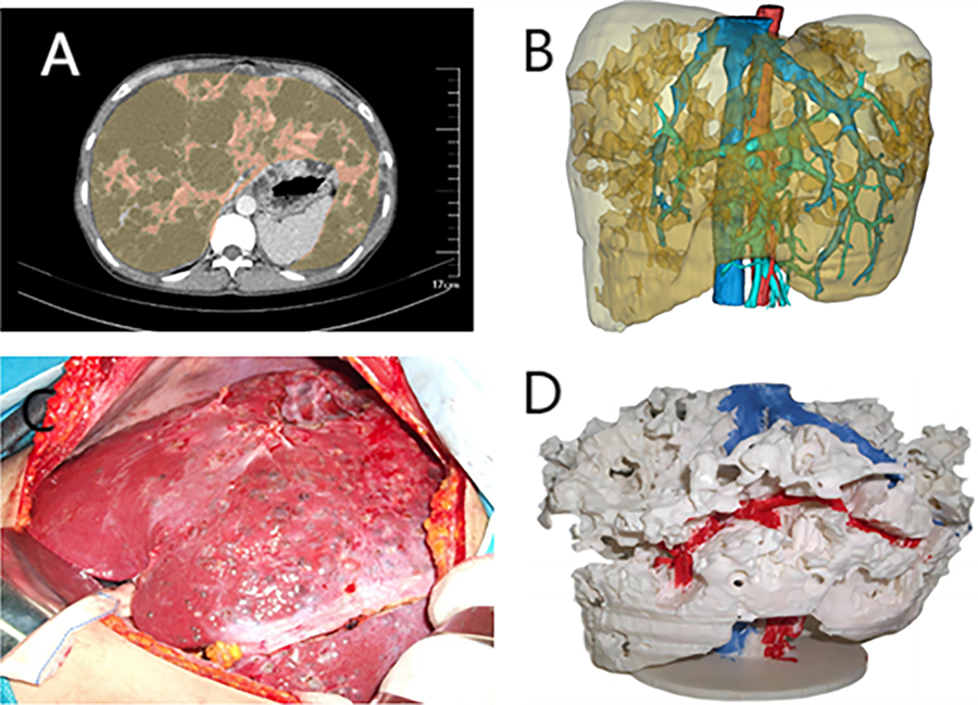
Contains four images
Application in Liver Tumor Resection
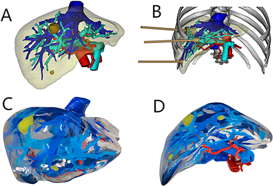
By utilizing preoperative 3D-printed models, surgeons can precisely locate the main blood vessels supplying the tumor, providing accurate guidance for the surgery. This approach allows for the prioritization of these critical vessels during surgery, effectively reducing intraoperative bleeding and ensuring the precise removal of the tumor.35–38 The effectiveness of this method was proven in the treatment of a 60-year-old male patient diagnosed with malignant hepatic melanoma in our hospital in December 2016. Preoperative planning indicated that resecting the right posterior lobe of the liver would preserve 51.8% of the liver volume. Based on this plan, the patient underwent a laparoscopic right hepatectomy guided by 3D printing on December 22, 2016 (Fig. 3).
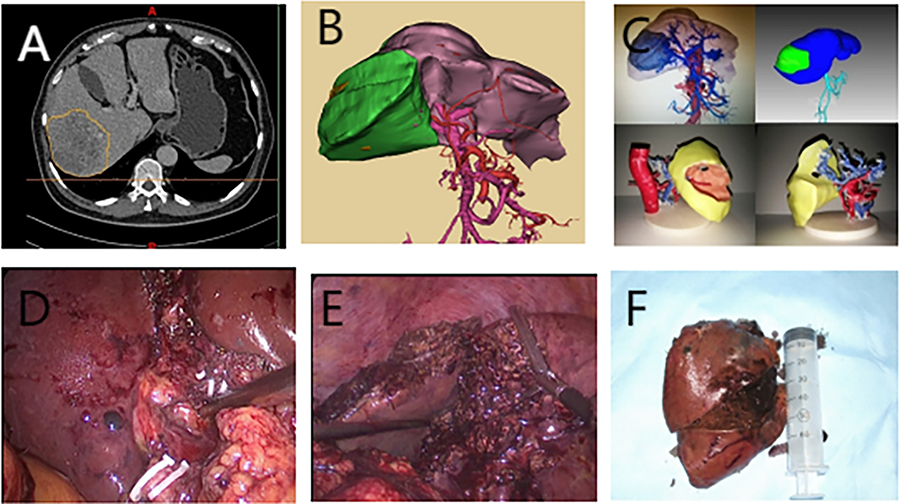
Illustrates the precise preoperative planning using 3D reconstruction and 3D printing models and the entire process of laparoscopic surgery to remove liver tumors. Image
Application of Liver Tumor Ablation
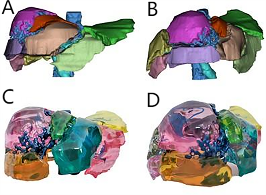
A second patient, a 56-year-old male, was diagnosed with stage CNLCIb hepatic malignancy at our hospital in March 2023. After a thorough preoperative evaluation, it was decided to resect segment VIII of the liver. Preoperative planning showed that this surgical intervention would allow the patient to retain 81.5% of the liver volume. Based on this plan, the patient successfully underwent a segment VIII hepatectomy guided by 3D printing technology on April 3, 2023, demonstrating the significant potential of 3D printing in practical applications (Fig. 4).
Illustrates the application of finely crafted liver models produced using 3D printing technology in liver tumor resection surgery, particularly in planning the resection of segment VIII of the liver. Images
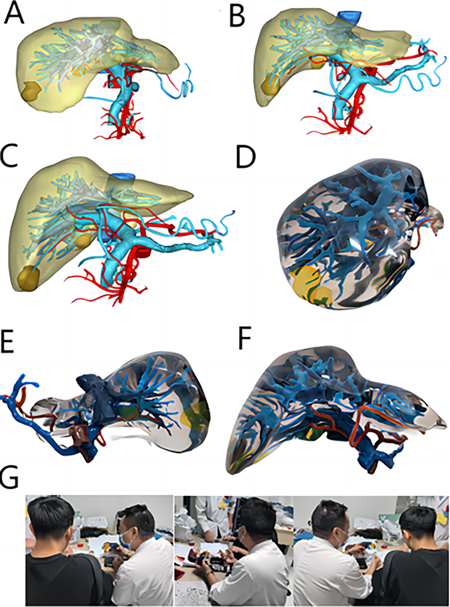
By using preoperative 3D-printed models, we can not only precisely identify the location of tumors within the liver but also design the optimal path and scope for microwave ablation based on this information. This step is crucial as it allows us to formulate a detailed ablation plan, specifying the required duration and frequency. Subsequently, during the surgery, this preparation enables us to accurately perform ablation based on the model, aiming to maximally deactivate the tumor while preserving as much liver functional volume as possible. To illustrate the effectiveness of this method, consider the case of a 52-year-old male patient diagnosed with secondary hepatic malignancy in our hospital in June 2020. Preoperatively, we utilized 3D printing technology and a comprehensive ablation plan for surgical planning, identifying the 7th-8th and 10th-11th intercostal spaces as the surgical entry points. Based on this plan, the patient successfully underwent liver radiofrequency microwave ablation surgery (Fig. 5).
Illustrates the precise planning of liver microwave ablation surgery through detailed 3D- printed models. Image
The Application of 3D Printing in Liver Surgery Medical Education
The liver encompasses four distinct types of vascular systems: the arterial system, portal venous system, hepatic venous system, and biliary system, intricately interwoven within the liver. 39 The foundation of educational use. Through two rounds of randomized tests precise liver surgery lies in the accurate identification of these hepatic structures. Through high-resolution CT scanning and 3D reconstruction, detailed information about the liver’s vasculature and segmental areas can be obtained, facilitating the creation of 3D-printed models. These models are extremely beneficial for medical education as they vividly demonstrate liver segmentation based on the portal vein territory, as well as the arrangement of intrahepatic veins, arteries, and portal venous systems. This aids in a deeper understanding of the anatomical structure and segmentation of liver vasculature, playing a key role in enhancing disease comprehension and formulating treatment plans.40,41 Kong et al. acquired CT images from a 50-year-old female volunteer and printed three different types of 3D liver segment models based on these images: Type I models without parenchyma, Type II models with transparent parenchyma, and Type III models with segmented bile ducts. Subsequently, they compared the learning efficiency of these 3D models with traditional anatomical images, and six experts evaluated the learning efficiency. The results indicated that, particularly, the Type II and Type III 3D liver models significantly surpassed traditional anatomical picture-based learning methods in enhancing learning efficiency and reducing the rate of memory decay. 42
Recently, the development of transparent photosensitive resins has focused on enhancing the material’s transparency and mechanical strength. The new generation of transparent resins not only offers higher transparency but also boasts improved chemical resistance. Such improvements make the visualization of internal liver structures, such as vascular and duct systems, much clearer. This is extremely important for complex surgeries that require meticulous planning and execution. It allows surgeons to perform more precise surgical planning and provides a more effective learning experience for medical education. With clearer visual presentation, surgical rehearsals and teaching processes become more intuitive and efficient. Significant advancements have been made in the development of flexible and elastic materials in recent years. These advancements provide physical properties closer to those of human visceral tissues, especially in simulating the elasticity, tensile strength, and compressibility of the human liver. Innovative materials such as highly pliable thermoplastic elastomers and optimally formulated thermoplastic polyurethanes have been able to deliver outstanding performance in these aspects. The development and application of these materials allow for liver models that not only simulate real surgical operation scenarios, such as vascular dissection, anastomosis, and tumor resection, but also offer a tactile experience very close to real-life handling.33,43
Multimaterial printing technologies, such as MultiJet Fusion and PolyJet, have now made it possible to fuse multiple materials in the same model, paving the way for creating complex and multifunctional models. This technology allows for the simultaneous use of materials with different hardness and transparency in a single model, making it possible to produce complex liver models that display vascular networks (through transparent resins), simulate liver tissue (using flexible materials), and tumors (using rigid materials). Such technological advancements not only bring more flexibility to surgical planning but also greatly enrich the content of medical education and training, making the learning and practice process closer to real situations and more efficient.
With these material and technological updates, the quality of liver surgery education and practice has significantly improved. 3D-printed models can now more realistically simulate surgical scenarios, offer more precise pre-surgical planning, and deepen the understanding of surgical techniques through simulation exercises. These advancements not only help improve the surgical skills of young physicians but also promote patient safety and improve surgical outcomes.32,43–45 As 3D printing technology continues to advance and its applications expand, its role in liver surgery education, and practice will be further strengthened.
Application of Doctor–Patient Communication
Liver disease models for patient education have proven to be particularly effective. These models accurately depict the lesions within the liver and their spatial relationships with surrounding vessels and tissues, providing patients with an intuitive learning tool that significantly enhances their understanding of the diagnosis’s complexity. 3D-printed models enable surgeons to clearly discuss the surgical plan, detailing the steps involved, the liver sections that will be affected, and how the surgery aims to remove or treat the lesion while preserving healthy tissue as much as possible. By offering a physical demonstration of the surgical approach, patients can better visualize what will occur during the operation. Moreover, 3D-printed models play a crucial role in explaining the postoperative recovery process. Demonstrating how the liver will heal and regenerate and the possible changes in liver structure after surgery, gives patients a clearer understanding of their recovery journey. This includes discussing potential postoperative care needs, monitoring for complications, and the expected timeline for returning to normal activities.
In summary, the use of 3D-printed models during the perioperative period not only provides patients with a clear visualization of the surgical process but also deepens their understanding of the treatment plan and recovery process. This fosters effective communication between doctors and patients, laying the foundation for achieving the best possible treatment outcomes.29,46,47 Research has demonstrated that in preoperative consultations, the use of 3D-printed liver models (intervention group), as opposed to relying solely on conventional imaging techniques such as CT scans (control group), significantly improved patient satisfaction (90% vs. 65%). Furthermore, 3D models aided patients in gaining a more comprehensive understanding of their condition, including the number of liver lesions (100% vs. 70%, p = 0.020) and their precise locations (95% vs. 65%, p = 0.044). Simultaneously, 3D models enhanced patients’ grasp of the surgical procedure (80% vs. 55%) and bolstered their awareness of potential postoperative complications (88.9% vs. 68.4%, p = 0.052). 46
The 3D-printed liver models displayed in Figures 6 and 7 provide a comprehensive demonstration of the various vascular systems and liver segments, including the arterial system, portal venous system, hepatic venous system, and bile duct system, and how they intricately intertwine within the liver. These models, fabricated with high-resolution CT scanning and 3D reconstruction techniques, not only vividly showcase the anatomical structure of the liver but also significantly enhance the efficiency of medical education and precise surgical planning. The models in Figure 6 are clearly marked with multiple colors to indicate liver segments and vascular layouts, while Figure 7 demonstrates the practical application of these models in medical practice, showing how they aid surgeons in effectively communicating with patients, discussing lesion locations, adjacent vascular and organ relationships, and surgical strategies in detail. This intuitive display and educational approach not only deepen patients’ understanding of the surgical process but also significantly boost their confidence in treatment plans. With these technological advancements, 3D-printed models can now simulate surgical scenarios more realistically, offering more precise preoperative planning, and deepening understanding of surgical techniques through simulated practice, all contributing to higher surgical success rates and patient satisfaction.
Images
The presentation includes 3D reconstruction images and 3D-printed models of the liver, as well as the practical application scenario where doctors use these models to communicate with patients. Images
3D Bio-Printed Livers
3D bioprinting technology has shown significant potential in the field of biomedical engineering, particularly in liver tissue engineering and regenerative medicine. By precisely controlling the spatial distribution of cells, bioactive molecules, and scaffold materials, 3D bioprinting can construct complex 3D structures that mimic real liver tissue.48–53 This technology not only provides a new experimental platform for studying liver diseases but also holds the promise of addressing the shortage of organ donations, offering customized tissue and organ substitutes for clinical use. In 2013, researchers led by Wang developed a 3D bioprinter equipped with four nozzles aimed at creating complex organs. Utilizing two extrusion nozzles and two injection nozzles, they precisely positioned hepatocytes, Schwann cells, and adipose-derived stem cells (ASCs), with PLGA material serving as the outer covering. Through 3D printing technology, the ASCs were successfully induced to differentiate into endothelial cells within the construct. This marked the birth of the world’s first 3D-printed bioartificial liver with branching blood vessels and a neural network. 54
3D organ bioprinting is an innovative and interdisciplinary field that achieves precise construction of biological structures by adjusting and altering material properties at various levels from molecules and cells to tissues and organs. This field is an interdisciplinary amalgamation, integrating knowledge and skills from multiple scientific and technological domains such as biomaterials science, biology, physics, chemistry, and medicine, relying on close collaboration among these areas. 55 One of the key challenges in 3D bioprinting technology is ensuring the viability and functionality of printed liver tissues in ex vivo cultures. To this end, researchers need to carefully select suitable cell types, such as liver-specific hepatocytes and nonhepatocytes (e.g., endothelial cells and stromal cells), and combine them with specific growth factors and scaffold materials to promote cell proliferation, differentiation, and maturation. 56 In addition, optimizing printing parameters, such as printing speed, temperature, and pressure control, is crucial for maintaining cell viability and the stability of tissue structures.57,58
In the practice of 3D bioprinting the liver, technological challenges primarily focus on several key aspects: First, precise control over cell positioning and distribution during the printing process is essential to ensure cell survival and functional expression within the scaffold material, requiring a deep understanding of the biocompatibility of printing materials; second, constructing a complex vascular network to maintain the long-term survival of printed tissues necessitates the integration of microfluidics and angiogenesis techniques during the printing process; lastly, achieving the liver’s unique multicellular structure, including the orderly assembly of hepatocytes, endothelial cells, and stellate cells, is crucial for mimicking the multifunctionality of the liver.59–62 Although research on 3D bioprinting of the liver is still in its preliminary stages, it has shown unprecedented potential in disease modeling, drug testing, and future clinical applications, such as customized liver transplants.54,63 By simulating liver tissues in specific disease states, researchers can gain a deeper understanding of disease mechanisms, accelerating the development and testing of new drugs.64–66 At the same time, personalized 3D-printed liver tissues hold the promise of significantly reducing rejection reactions in organ transplants, thereby increasing the success rate of surgeries.57,67–69
Looking forward, with further refinement of printing technologies and deeper research into cellular biology, 3D bio-printed livers are expected to achieve more complex tissue structures and advanced functions, paving new paths for the treatment of liver diseases. Moreover, this technology will also advance personalized medicine by offering customized treatment plans, improving treatment outcomes, and enhancing patients’ quality of life.70,71
Conclusion and Future Directions
With the rapid development of 3D printing technology in the medical field, its application in precision surgical operations, especially in liver surgery, has shown tremendous potential. This review article delves into the significant contributions of 3D printing technology in liver surgery planning, simulation, education, and providing customized treatment plans for patients. An analysis of current research and application cases shows that 3D printing technology not only enhances the safety and success rate of surgeries and reduces operation time but also increases patient satisfaction. However, despite significant progress, the application of liver models still faces certain limitations, such as issues with imaging data that may result in the absence of fine blood vessels, the hardness of printing materials that cannot simulate the real touch of the liver, and models being integrated structures after forming, making it difficult to disassemble and distinguish different anatomical parts. The high cost of printing full-size 3D liver models poses a significant challenge. According to related reviews, while some studies have managed to keep printing costs below $100 by using lower-cost materials such as nylon plastic or PLA, these costs can soar to $2,000 when printing full-size liver models with high-quality photopolymer resins.72,73 If the model size is reduced to 50–70%, the cost may drop to between $400 and $980. Therefore, finding ways to reduce the printing costs of these models becomes particularly crucial to enhance the practicality and clinical value of 3D-printed liver models. The preparatory process for printing realistic anatomical models, especially the segmentation and editing of medical imaging data, significantly prolongs production time, with the entire process typically requiring 4 to 5 days. These time constraints challenge the practicality and feasibility of clinical applications of 3D printing technology.74–76
Future research is expected to broaden and deepen the application of 3D printing technology in precision surgical operations. Higher resolution and more refined printing techniques are anticipated to provide unprecedented tools for surgical planning and simulation. Furthermore, coupled with the development of bioprinting technology, especially in the research of biocompatible materials and live cell printing, the possibility of directly printing transplantable liver tissues has been opened, which could fundamentally transform the field of organ transplantation.77,78 However, to realize these potentials, we face multiple technical challenges, including how to enhance the biocompatibility and mechanical properties of printing materials and how to precisely control cell growth and tissue maturation. In addition, ethical and legal norms for bioprinting human organs need to be clarified to ensure the healthy development and application of the technology.79–83 In summary, 3D printing technology is expected to provide safer and more effective treatment options for patients, driving innovation and development in precision surgical operations and bioprinting technology in the future medical field.
Authors’ Contributions
Conceptualization, T.L. and Y.D.; writing—original draft, T.L.; writing—review and editing, all authors; funding acquisition, Y.J.; resources, Y.J. and J.H.; supervision, Y.J. and J.H.
Footnotes
Author Disclosure Statement
The authors declare that they have no conflict of interest.
Funding Information
This work was supported by Famous doctor of Yunnan “Xingdian Talent Support Program” (XDYC-MY-2022-0032); Yunnan Fundamental Research Project (202201AS070002); Reserve Talent Program of Yunnan Province Young and Middle-aged Academic and Technical Leaders (202005AC160017); Key S&T Special Projects of Yunnan (NO. 202402AA310056); Innovation Team Special Program of Yunnan (NO. 202505AS350004).
Ethical Approval
This study does not contain any studies with human or animal subjects performed by any of the authors.