
Abstract
This study aims to systematically review the integration and clinical applications of 3D printing and extended reality technologies in medicine. This offers an in-depth review of technological advances and their application in different areas of medicine such as surgical planning, medical staff training, new ways of visualization, as well as the integration of tailor-made physical and digital elements. Factors that benefit patient safety and promote innovation through technological intervention. The study conducted a systematic review of the literature using the PRISMA methodology, with a selection of studies published between 2005 and 2023. The process included a comprehensive search in academic databases from which articles were extracted that included topics on 3D printing technologies, extended realities, and applied cases in which efficacy and application in clinical settings were proven. As a result, specific inclusion and exclusion filters were established to ensure relevance to the study’s objective. The selected items were subjected to a detailed analysis, based on which the methodologies used, types of studies, areas of impact, population, and clinical impacts were evaluated. The synthesis of the results was carried out using the quantitative and qualitative approaches. This enabled a comprehensive understanding of current trends and potential future applications of these technologies in the field of medicine. The results indicate that the adoption of 3D printing and technologies such as augmented reality (AR) and mixed reality (MR) facilitates more precise and less invasive interventions. It also improves understanding of complex conditions and pathologies through enhanced and enriched interactions and visualizations. In addition, technologies are consolidated as tools for medical strengthening and training through more realistic simulations. Studies show that by having better understanding, visualization, and interaction with components and technologies, there is a reduction in surgical errors and optimized duration times in the surgeon and patient recovery; finally, the use of these technologies also improves the retention of information in training and education contexts. Furthermore, the study reveals that, although the associated costs may be considerable, the gains in accuracy and operational efficiency justify the investment. Among the challenges posed by the use and integration of 3D printing and augmented or MR is the need to further improve the accurate calibration of the 3D images with physical models, which turns out to be a key aspect for the success of these applications in the clinical context. Implementation of technologies such as 3D printing, AR, MR, and virtual reality in clinical practice has been shown to have great benefits in efficiency and safety in surgical procedures. These technologies profile themselves as solutions that promise to revolutionize approaches to learning, surgical planning, and surgery. This offers customization and accuracy features, significantly reducing error margins. Furthermore, medical training is enriched by more realistic simulations and detailed visualizations. However, the construction of innovative tools that consolidate the characteristics of these technologies requires continuous improvement and investment in training, development, and appropriate infrastructure to overcome the challenges associated with the integration of such technologies into clinical practice. This review provides a comprehensive synthesis of current applications and the transformative impacts of 3D printing technologies and extended realities in medicine. Unlike previous revisions, this study carries out a quantitative analysis of its benefits, supported by recent case studies and statistical data. This leads to valuable insights on future directions of medical technology integration.
Keywords
Introduction
Medicine is an area that is constantly evolving, largely due to technological advances and the integration of various technologies. Since the appearance of X-rays in 1896, a key year for the development of radiological diagnostics, 1 research has begun on their specific applications in medical use. Initially, X-rays were used for bone structure analysis. However, the integration and relationship between basic sciences, clinical medicine, and industry has enabled a constant evolution. Thanks to this, we now have tools and procedures such as interventionist radiology, ultrasounds, computed tomography (CT), magnetic resonance imaging (MRI), and positron emission imaging. In this way, the digital image has replaced the traditional medium of contrast with modern agents and the X-ray film, which became the basis and standard for making highly complex diagnostic and surgical planning.
Before carrying out a highly complex surgical procedure, medical specialists perform a series of medical and diagnostic examinations that allow them to understand the patient’s pathology and discern what is the best way to do the different procedures to ensure the success of the surgery and people’s well-being. This is a complex process because it requires the interpretation of a large amount of information, clinical data, diagnostic images, medical history, and a high understanding of anatomy and pathology. 2 Today the evolution of diagnostic imaging allows to analyze the structures of the body in a noninvasive way. Technologies such as MRI, CT, and ultrasound are crucial in the planning process. 3 In addition to these technologies, in the clinical context, 3D printing, augmented reality (AR), and mixed reality (MR) are beginning to be integrated into the planning process as tools that generate value and enrich medical planning, education, and training processes. 4
These technologies, which are not specific to the clinical sector, have been shown to generate great benefits and contributions. In the case of 3D printing, this has gained great relevance in the medical industry, thanks to its ability to materialize and build different pieces using techniques and materials according to the needs of patients. 5 Among its features is the ability to reproduce precise and detailed models, used with different intentions such as planning, simulation, and device development to complement clinical practice. 3 However, MR and AR are becoming important technologies in the clinical context, thanks to the integration of low-cost ease and less time-consuming digital components into their manufacture or process, which changes the ways medical planning has to integrate patient information into projected or holographic environments. 6
Such a situation highlights the importance of understanding the complexity of diagnostic imaging interpretation and surgical planning, as the efficiency and accuracy required in these processes are crucial to the success of medical procedures.
In this context, the present study seeks to answer the following research question: Which are the current main applications of 3D printing and extended realities, and how do they contribute to improving education and clinical practice?
This research aims to document through a systematic search how the incorporation of physical 3D models and data layers in mixed realities can improve or make more efficient the diagnosis of medical specialists. Also, this enables the possibility to explore the potential of these emerging technologies to enrich surgical planning, education, medical training, and the reduction of costs and time required in these processes. In this way, it is intended to contribute to modern medicine, opening new paths for the implementation of advanced technologies in the clinical field, and improving patient care quality.
Materials and Methods
This review uses the PRISMA methodology (Preferred Reporting Items for Systematic reviews and Meta-Analyses) to systematically explore the integration and impact of AR and 3D printing technologies in the medical field. A comprehensive search was conducted in academic databases such as Scopus and ScienceDirect, following a structured four-stage protocol.
At the same time, to map the many indicators and uses in order to create a foundation for comprehending the influence in the different fields and medical specializations, a review was made using the PRISMA methodology (see Fig. 1), which outlines the methodological structure and development in each of the phases. According to the established methodology, the literature search was carried out in databases such as Scopus and ScienceDirect. It is also important to emphasize that a four-stage protocol was designed, which will be described below (see Fig. 1).
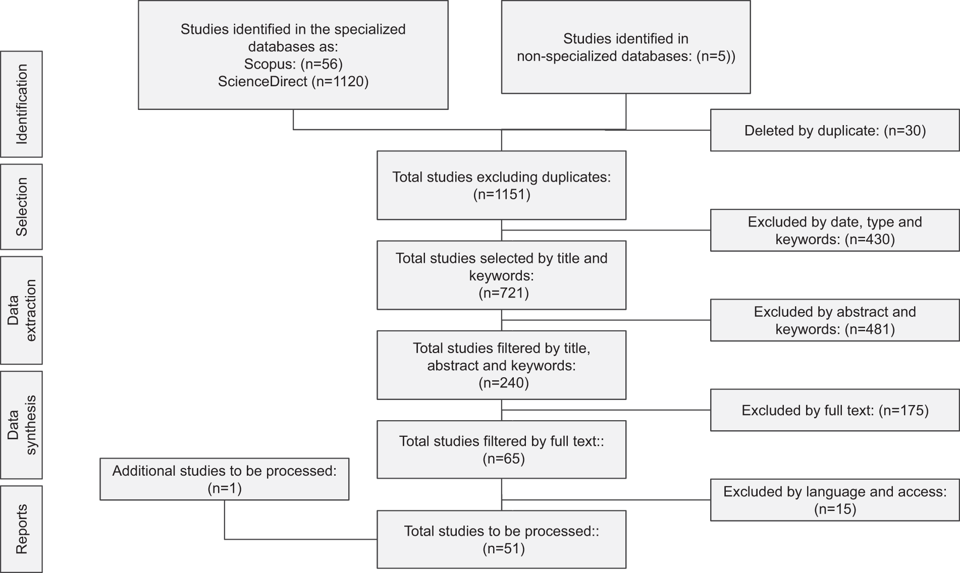
PRISMA flowchart. Source: Self-made.
Step 1. Identification
A literature search strategy was implemented with preestablished parameters and guidelines to delineate and obtain the best-expected results in the databases. To summarize the search and obtain texts aligned with the objective of the research, the following keywords were defined: 3D printing, AR, virtual reality (VR), MR, medicine, surgery, and training. It should be noted that the search was limited to articles published in a temporary frame from 2005 to 2023, to ensure that the information is relevant to the latest medical practice.
Step 2. Selection
Subsequently, a first filter was established in which the inclusion and exclusion factors were defined. This filter excludes duplicate items, by year, document type, and keywords mentioned earlier. Items that met at least one of the following inclusion criteria were selected: surgical study cases, use of 3D-printed models, MR, VR, AR, organs representation, anatomical parts, training, and digital device development. At the same time, the following exclusion criteria were applied: duplicate studies, nonmedical studies, related to education but in the medical field.
Justification of selected studies
Although the total number of selected articles may seem relatively low compared with the total number of studies available, the selection focused on studies with high methodological rigor and proven clinical applications. This approach allowed for a deeper and more relevant analysis, ensuring that the presented results are significant and reliable within the medical context. The priority was placed on the quality of the selected studies to ensure that the findings accurately reflect the current state of 3D printing and extended reality applications in medicine.
Step 3. Data extraction
After the selection and filtering the documents, the basic information from each article was extracted to identify in depth the type of study, the area of specialization, and the kind of process that was included in the documents. It is important to mention that qualitative and quantitative data were extracted. These data were decisive, as they had to be included or excluded from the study according to their categorization and information.
Step 4. Data synthesis
Finally, after having the articles selected for analysis, the relevant information and data were extracted to make it possible to summarize trends, findings, and conclusions. With the information synthesized, the diagrams and tables presented in the study were constructed.
Results
This section analyzes the key findings obtained from the literature review on the use of AR technologies, consisting of VR, AR, and MR, as well as 3D printing. In addition, it examines how they have impacted the medical field by providing a characterization and the analyzed documents' statistical data. The results are analyzed to determine how these emerging technologies are redefining clinical practice, from surgical planning to medical diagnosis and medical training. The above elements are categorized and presented as follows.
Main information about data provides a detailed timeline search overview, the media in which it was published, the type of documents, the average annual growth of the research topic, the authors, etc. This information is relevant because it allows to build the demographic, geographical characterization, applicability in the context, timeline, and the analyzed topic evolution (see Table 1).
Data Description
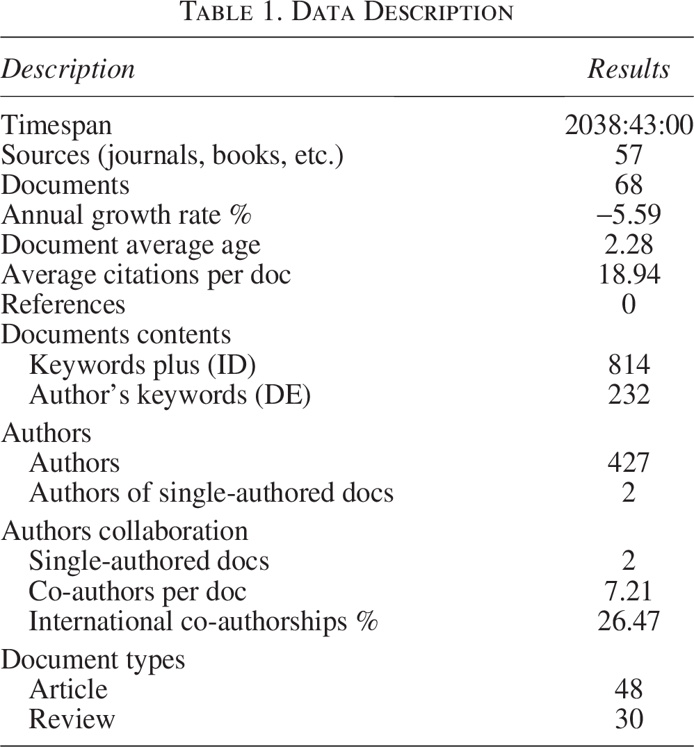
Source: self-made.
However, to understand the evolution, according to the data analysis, it can be evidenced that there is an interest in researching topics related to the technologies of 3D printing, AR, VR, MR applied to the context of health. Graph 2: between 2005 and 2023, there is a pattern of rapid development in annual scientific production (see Fig. 2).
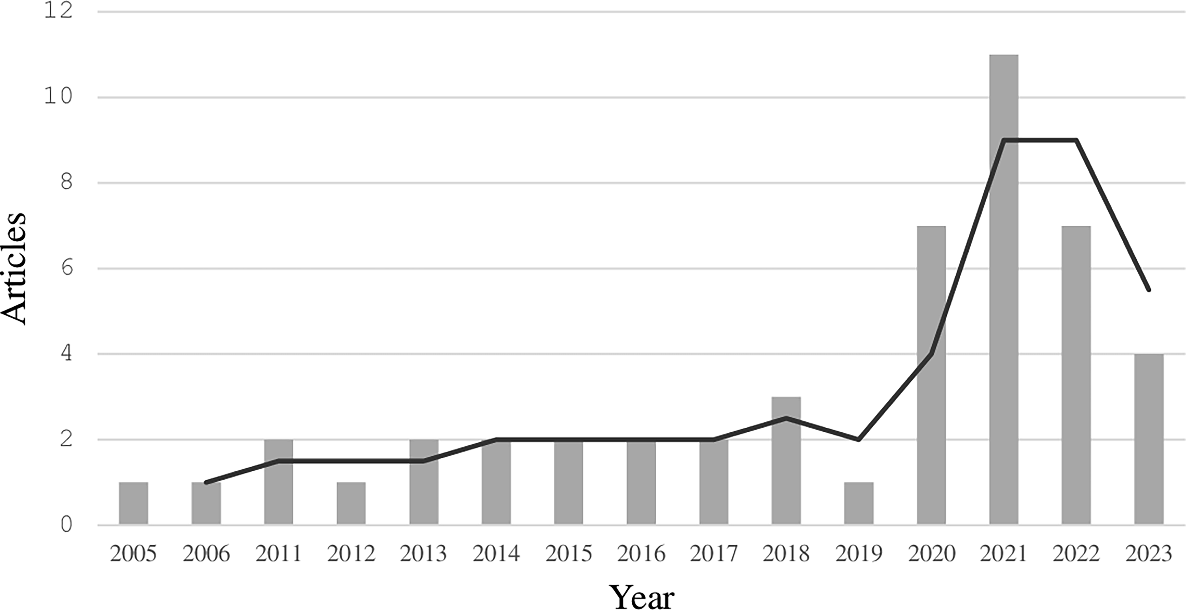
Annual scientific production. Source: Self-made.
Technologies
In this sense, the interaction between science and technology has undergone a series of revolutionary transformations in medicine. This has been a benchmark of innovation throughout history. Indeed, since the discovery of X-rays (1895), medicine has evolved exponentially through the integration of new technologies and developments associated with the time. This study mainly addressed four technologies, which are AR, VR, MR, and 3D printing. From these categories, the most relevant or highly impactful aspects in the medical field were identified.
It is necessary to highlight the development and relevance of the advances in the acquisition and evolution in visualization that have been generated around diagnostic imaging. This is because they are the basis that allows to detect, diagnose, identify, and visualize the anatomy of the human body, conditions, and diseases. 7
Diagnostic images
In 1895, German physicist Wilhelm Conrad Roentgen discovered X-rays from cathode-ray tubes and the application of 2D radiographical plates. Later, this technology was affected by the computers’ arrival, screens, and machine-mediated interaction systems. The technological innovation marks a milestone in the introduction of the digital interface concept, human–machine interface, and user experience. This event significance lies in the fact that it made the specialist physician a user for the first time, whereas his work previously relied on the technical reading and diagnostic images interpretation. 8
Various advances have consolidated diagnostic imaging development, from its use in surgical planning and medical diagnosis. At first, they were visualized on printed plates, then they were transformed into digital systems with the emergence of computing systems. Initially, this interaction was given by specialized commands through a line interface. The graphical user interface systems emergence, the mouse, and icons deployment marked a milestone in user experience, which has prevailed relatively for about 30 years in the radiological context. Indeed, studies state that the mouse and keyboard are still essential for detailed visualization tasks. 9 However, touch technology and tracking sensors have begun to impact the interaction and views with diagnostic images. This provides portability and accessibility with cloud computing systems. 10 Made of the utmost relevance in the surgical context, as it has allowed to find contactless control systems for image visualization and, consequently, reduce the risk of contamination.11,12 This includes technologies such as AR and MR, which, by their characteristics, become valuable tools because they meet the needs of the user and the environment and improve usability in medical diagnosis and treatment. On this subject, Table 2 summarizes the technological developments associated with the evolution of diagnostic imaging, details, and impact in the health sector (see Table 2).
Technological Development and Impact
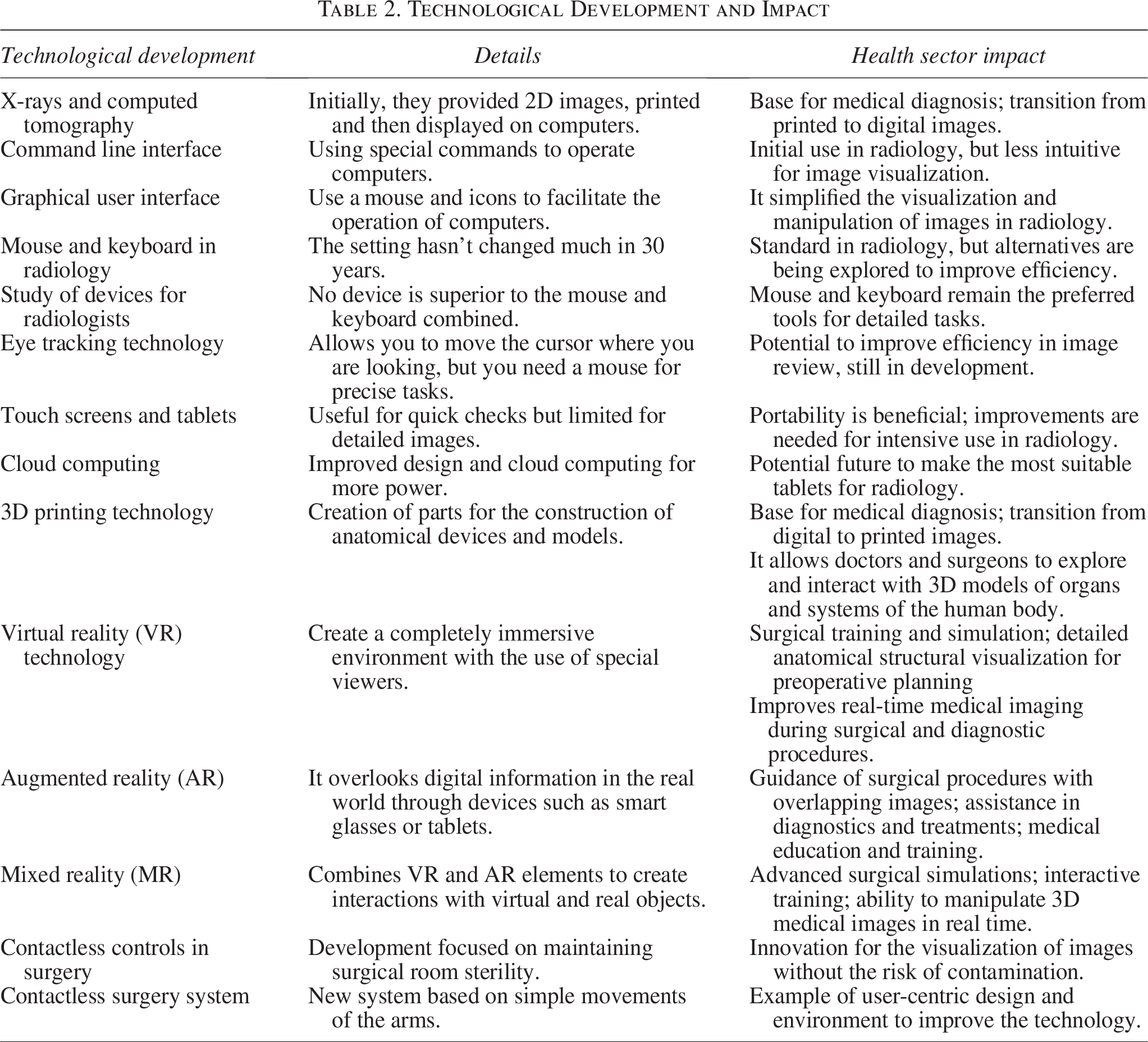
Source: Self-made.
However, it is essential to understand the importance and evolution of diagnostic images, as they become the basis for creation with 3D printing, AR, MR, VR. The process begins with the acquisition of patient images, the postprocessing, and the procurement of 3D digital models. Subsequently, the specialists analyze the images and 3D models for different clinical approaches. This is the standard procedure, without the technology’s integration mentioned in the study (see Fig. 3).
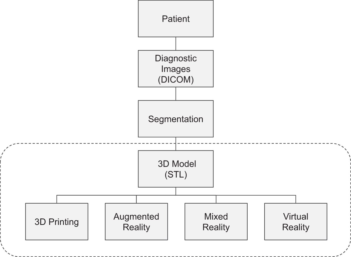
Route integration path for creation with 3D printing and extended realities. Source: Self-made.
3D printing
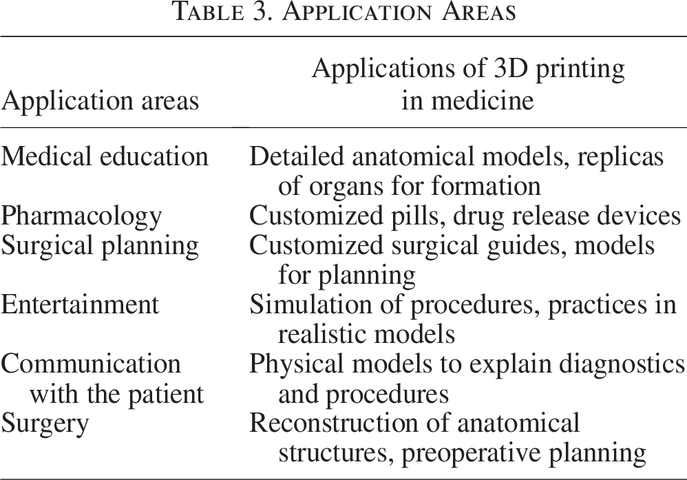
Additive manufacturing technology has seen significant growth in the field of medicine, thanks to its features, it is used to generate value in personalized treatments in patients, impacting clinical processes such as advanced preoperative planning. The creation of surgical guides, implants, prostheses, medical devices, and clinical support elements has proven to have applications in the pharmaceutical sector.13,14 In other words, the process begins with the acquisition of diagnostic images, segmentation, processing, and manufacturing with 3D printing. 15 Due to the ability to build digital models, adding layer-by-layer material, complex structures that are difficult to create with other technologies can be obtained. In this sense, the concept of personalized and patient-centered medicine has managed to consolidate 3D printing with several applications that can be appreciated in Table 3. 16
Application Areas
Source: Self-made.
Table 3 presents the diversity and scope of 3D printing in the field of medicine, highlighting its versatility and potential in several key areas. In medical education and simulation, 3D printing plays a crucial role in providing detailed anatomical models and organ replicas, improving student learning and understanding. 17 In the field of surgery, this technology enables the creation of customized surgical guides and detailed models for accurate and effective preoperative planning. Implementation of these models allows for reduced costs, time in the surgery, and blood loss in the patient.13,14,18 In terms of training, 3D printing is used, and its ability to build models with different materials that provide a wide variety of mechanical properties allows the simulators to create procedures and practices in realistic models. All of these are essential for practical training and for the skill acquisition and development of residents and medical students.19,20
In the field of pharmacology, 3D printing is presented as a tool for the personalization, dosing, and development of devices for the release of drugs. The above is a great tool to customize treatments according to the specific needs of each patient. 21 The use of these technologies in surgical planning has shown great benefits and has enhanced preoperative planning and its accuracy. Additionally, it has reduced planning time and even managed to change the surgical approach by using 3D-printed models. 22 When used in treatments of complex pathologies, 3D-printed models facilitate interdisciplinary communication between the team of surgeons and specialists involved in the operational treatment. 18
Finally, in the field of doctor and patient communication, 3D-printed anatomical models of bodies with complex pathologies become a valuable tool for explaining diagnoses and procedures and improving patient understanding. 3 Each of these points out the contribution of 3D printing technology to medicine from two angles. First, it improves clinical processes and outcomes. While, on the other hand, it enriches education and communication in the field of health.
Augmented reality
AR is a technology that allows the user to overlay 3D virtual elements over objects or the real environment. It is a factor that provides an immersive visual experience 23 and is the result of the advances in traditional computer visualization in recent years. 24 Additionally, the overlapping of information and 3D digital models makes user interaction with these elements more fluid, easy, and natural. The ability to visualize and interact in the surgical field and clinical environments makes it the biggest advantage that technology has over other navigation and planning technologies. Therefore, it is used in specialties such as neurosurgery, 25 cardiology, orthopedics, laparoscopic surgery, 26 oral, maxillofacial surgery, 27 and spinal surgery. 28
However, technology offers great advantages in training and clinical education. The surgical training consists of an approach in which the student, after acquiring the theoretical knowledge of his specialty or area of training, goes through a process of learning by observing and then doing. This means that the student carries out the procedures under the guidance and supervision of the instructor. 29 The suggested model and in situ training are expensive, time consuming, and carry a significant risk of process failures for both the institutions and the patients.29,30 Notwithstanding, AR poses a scenario in which two-way and even remote training can be used, in which the teacher and student can interact in real time. 31 They can even be adapted to different clinical procedures, which reduces time and risk and has a high potential for reuse. 32
Mixed reality
MR is one of the latest technologies that encompasses extended reality. Like AR, it has the capacity to overlap digital elements in the real environment with the ability to interact with them.33,34 The features of the technology have allowed it to be adopted in the health sector quickly. This idea and concept were initially introduced by Paul Milgram and Fumio Kishino in 1994. 35 The authors raised the concept of the virtuality continuum. This refers to the mixture of the different objects presented in the real environment and emphasizes the type of device used to recreate the different scenes and the degree of immersion.
It should be emphasized that every technology needs a means of being recreated. In the case of AR, devices with a screen and a video camera are used to capture the environment and visualize it on the screen. In the case of MRI, different devices have been presented. However, it was only until early 2016, when Microsoft Corp. released the first commercial MR device known as HoloLens. A wearable device type headset that has several integrated sensors, high-definition cameras, accelerometers, and microphones integrated into a computer. 2 The device projects a holographic image into the environment through two semitransparent combined lenses, which are placed in front of the user’s eyes to combine the 3D elements with the actual environment. 36 Interaction and control of data with gestures and voice make it a great tool for surgeons and students in processes ranging from preoperative planning to clinical surgery 37 and professional medical training.38,39
Virtual reality
VR is a technology that allows the visualization of computer-generated 3D images through wearable devices such as headsets. Users can interact with these elements in an immersive experience in a real-time virtual world. Its image visualization features and its ability to create and recreate virtual worlds make it an innovative tool in advanced imaging. 40 Ideal for diagnostic and training processes, it allows students to have active participation, better guidance, and support. 41 In different clinical areas, the implementation of this technology has shown progress. In radiology, it is being used for planning guidance procedures, which improves the processes of doctor–patient communication to communicate information and medical knowledge associated with pathology. This in turn leads to the development of a process of literacy in health anatomical visualization and complex pathologies.42,43 In cardiology, technology is used in processes of surgical simulation, preoperative management, surgery planning, and immersive tele-virtuality. 44
3D printing and MR
In recent years, technological advances have been significant, driving innovative solutions ranging from surgical planning to real-time surgery and medical education. However, it is clear that the synergy between 3D printing and MR represents an innovative fusion where precision and interactivity enhance each other. 45 This combination of digital elements and physical models creates a new visualization mode that is oriented and customized. This allows doctors to observe and understand the details of the injury, navigate the information of digital and physical modeling, improve the accuracy of localizing the lesion, and potentially reduce the risk in surgery. 46 Having said that, if the characteristics of MRI and 3D printing technologies are compared with 2D images, it provides proof that the accurate understanding and evaluation of anatomies have a positive impact on surgical planning, a factor that has its main evidence in the well-being of the patient. 47
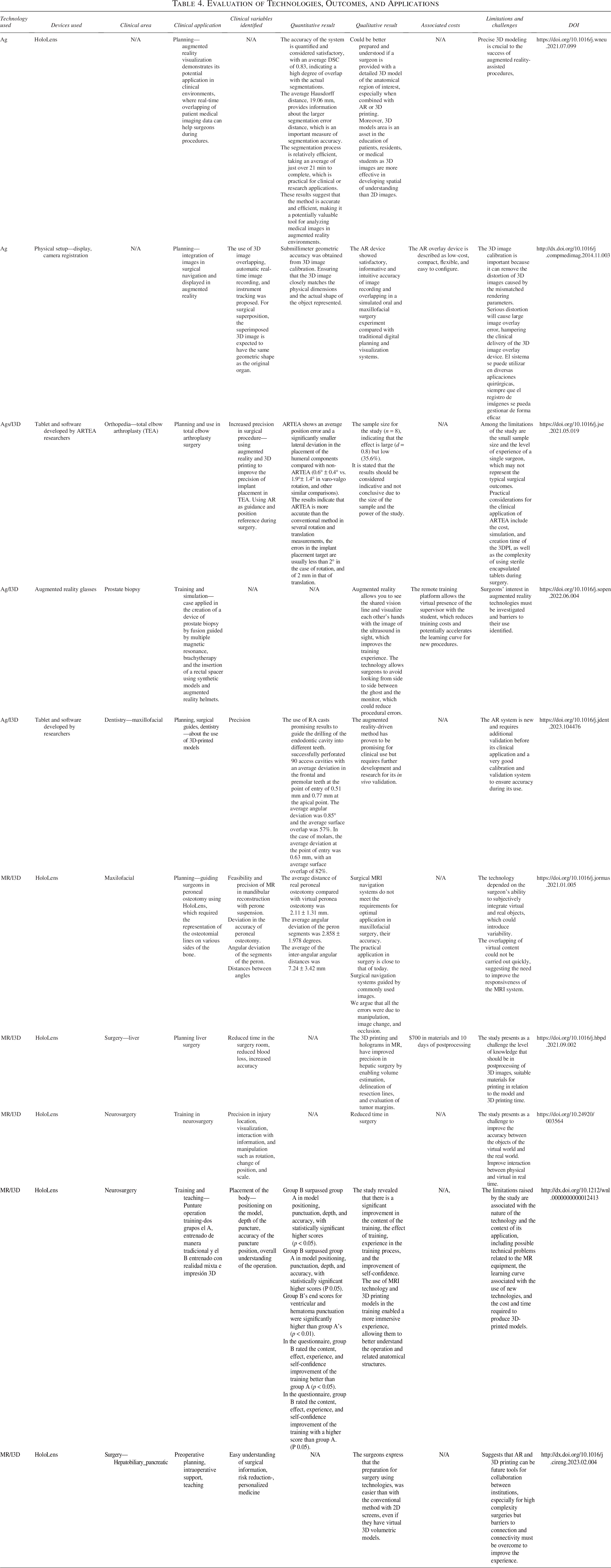
The multi-level combination of these technologies incorporates an additional feature to the processes of simulation, surgical planning, and education. It is only through this approach that the idea of accurate real-time haptic feedback that duplicates the pathology’s anatomy in 3D printing and adds data layers to MR to provide users realistic situations can be conceived. 48 The training takes advantage of these characteristics. In this way, a new concept of modern medical training begins to be consolidated, in which technology reshuffles the way residents learn. 49 At the same time, the mix provides intraoperative advantages with real-time information and interaction, including experts and surgeons connected through devices that can view procedures in virtual scenarios and support surgical procedures48 (see Table 4).
Evaluation of Technologies, Outcomes, and Applications
Source: Self-made.
Discussion
After conducting a systematic analysis of the literature examined, it becomes clear that emerging technologies such as 3D printing, VR, AR, and MR are generating a transformative impact in the various areas of medicine from clinical practice to medical training processes at different levels, optimizing the characteristics and potential of technologies. The rapid development of hardware and software in these technologies enables new applications and solutions to be provided, a factor that expands the range of actions of these technologies in the clinical context. At the same time, the user experience is enriched with new possibilities of interactions and medical applications. 44
Research shows that there is a growing trend toward the integration of these technologies into the health sector and each has a large number of applications. It is evident that the combination of 3D printing technology with some AR platforms, including MR or AR, has the potential to further revolutionize several areas of medicine and lead to significant and innovative developments. In fact, clinical applications with high levels of personalization and precision are currently being created in surgical planning, medical education, and surgery procedures with solutions that integrate physical elements manufactured with 3D printing enriched with digital components.
Such integration can be applied in any of the medical contexts, for example, in the field of practical processes such as surgeries, planning, training, education, or simulation. The aim, in this sense, is that these technologies can become standard tools that accompany the specialists. This would be done through headset-type visualization devices, a real-time visual guide during procedures accompanied by 3D-printed support tools tailored or with a high degree of personalization.
Table 5 details the specific influence and contribution of each of the technologies on the different clinical variables found throughout the research. This provides a clear overview of the benefits that technology brings, from risk reduction to improved accuracy, patient safety, and information visualization. Similarly, it describes how it impacts medical education and simulation, as well as the way in which complete medical procedures are impacted. Understanding these relationships is crucial to appreciating the potential and impact in medical practice (see Table 5).
Variables and Impact of Technology

Source: Self-made.
The integration of technologies itself presents significant improvements around the precision of surgical procedures and a more interactive approach that has demonstrated greater efficiency in medical education processes. However, there are limitations that need to be examined, some aspects revolving around costs. These can be high due to VR, AR, and MR applications development times, long 3D printing times, required infrastructure investment, specialized training, and learning curve on the part of medical personnel.
Although this research does not focus on privacy-related issues, safe handling of patient data, from an ethical and regulatory point of view, is an opportunity for future research to establish clear guidelines on how to handle and protect patient data in these new, advanced clinical and technological contexts.
The inclusion of case studies and applied examples showing the impact of technologies has enabled us to establish a valuable perspective on the potential and practical challenges. In the future, the direct impact of technologies on the patient, and not just on clinical outcomes, must be reviewed in depth. Aspects such as evidence and patient improvement processes or reduced complication rates would be significant indicators of the practical value of technologies in the clinical environment.
Emerging trends in these technologies suggest a landscape in constant evolution. This opens up the possibility of new uses in medicine in the formation of new promising areas in which the needs, experiences, practical challenges, the human dimension understood from patients and medical staff are considered, to integrate them in discussion with 3D printing, VR, MR, and AR technologies.
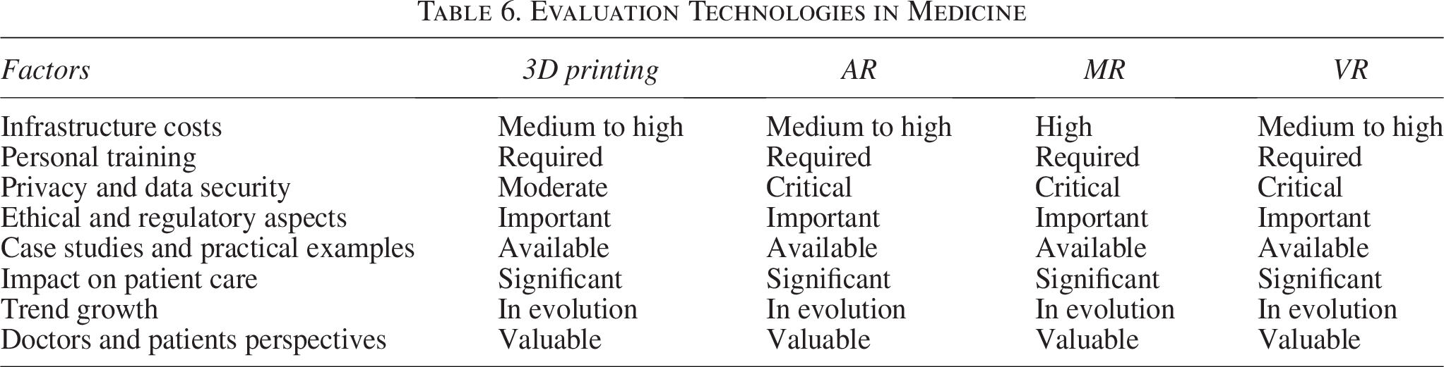
Table 6 shows the relationship of the technologies to the factors mentioned earlier. The information presented in this table enables a deeper understanding of the practical and strategic implications of its use in clinical settings. Opportunities, crucial challenges, and critical considerations are also presented (see Table 6).
Evaluation Technologies in Medicine
Source: Self-made.
Conclusions
Finally, the study has shown that emerging technologies and advances in materials for 3D printing and the constant development in software and hardware for VR, AR, and MR technologies are exerting a valuable transformative impact in the different areas of medicine. Trends are seen as innovations that come together to form useful instruments for improving surgical procedure accuracy, the effectiveness of medical training, and the planning and implementation of specific treatments. In addition, the study also identified challenges, some limitations, and the need for specialized training for the effective use of technologies and the creation of solutions to them.
Optimizing the characteristics of the technologies, being able to extract, generate, and design 3D anatomical models, materialize them with additive manufacturing, and complement them with guided instructions and patient information with AR technologies increase the possibility of clinical applications and allows the experience of the health professional or patient to be enriched and differentiated. Society is moving toward a future in which medicine and digital technologies are increasingly intertwined, as evidenced by the results of this systematic review, which highlights the rapid adoption of 3D, AR, MR, and VR printing technologies. These, although not inherent in the health sector, put at their disposal a potential fully focused efforts on improving the processes of medical practice through continuous adaptation, testing, and transformation toward a more accurate, advanced, and safe medicine for the patient.
Considerations and Limitations
Throughout this study, several key methodological considerations were addressed to ensure the validity and relevance of the results. However, it is important to highlight some inherent limitations of the approach adopted. First, the selection of studies was limited to a relatively small number due to the application of strict inclusion criteria, focusing on studies with proven clinical applications and high methodological quality. While this approach allowed for a deep and relevant analysis, it is possible that some emerging applications in less documented areas were not included. Additionally, the review focused mainly on articles published between 2005 and 2023, which may have excluded recent or ongoing research that could provide new perspectives on the applications of 3D printing and extended realities in the clinical field.
Last, although the study provides a comprehensive overview of the technologies evaluated, longitudinal studies that analyze the long-term impact of these technologies in medical practice were not included. Future studies could focus on evaluating these technologies in a wider range of medical specialties and their large-scale implementation in different clinical contexts.
Footnotes
Author Disclosure Statement
No competing financial interests exist.
Funding Information
No funding was received for this article.