
Abstract
Intact immunoglobulin G antibody has a relatively large molecule size of approximately 150 kDa that remains in the bloodstream for many weeks, which is a considerable disadvantage when it is used to carry radioactive materials for imaging. To lower background activity and increase the contrast of images, we investigated antivascular endothelial growth factor (VEGF) receptor 2 antibody (DC101) conjugated dextran for VEGF receptor 2 imaging in tumor xenografted mice. DTPA-conjugated aminodextran was synthesized, reacted with sulfo-LC-SPDP, and then reacted with DC101. The binding affinity of DTPA-dextran-DC101 to Flk-1 was measured. The gamma imaging and biodistributions of 99mTc-DTPA-dextran-DC101, 99mTc-DTPA-DC101, and 125I-DC101 were studied in B16F10 melanoma xenografted mice. The dissociation values for DC101, DTPA-DC101, and DTPA-dextran-DC101 were 22.48, 3.05, and 14.74 pM, respectively. In gamma images, 99mTc-DTPA-dextran-DC101 showed weak liver uptake and rapid kidney elimination. In biodistribution results, the liver uptake of 99mTc-DTPA-dextran-DC101 was similar with that of 99mTc-DTPA-DC101 at each time point. However, the blood activity of 99mTc-DTPA-dextran-DC101 has shown significant differences, compared with 99mTc-DTPA-DC101 at all time points. The tumor accumulation of dextran-conjugated antibody was increased with time, whereas that of dextran nonconjugated antibody decreased. In particular, the pattern of tumor uptake of 99mTc-DTPA-dextran-DC101 was similar to that of 125I-DC101, so this was thought to reflect the kinetics of DC101, unlike the nonconjugated form. The results of this study suggested that introduction of dextran moiety to make 99mTc-radiolabeled DC101 imaging agent could provide better images with the impaired background and the steady increasing binding to the receptor. However, further studies are necessary to improve clinical pharmacokinetics, such as enhancement of tumor uptake and impaired renal uptake.
Introduction
Antibodies have been used frequently to target neoplasia and other diseases, because of their ability to bind selectively to tumor or disease-associated antigens. Intact antibody of approximately 150 kDa is a relatively large molecule that may remain in the bloodstream for many weeks before being metabolized in the hepatocytes; this is a considerable advantage when acting in the natural setting of removing a foreign invader. 1 However, the direct attachment of a radionuclide to the large immunoglobulin G (IgG) molecule can limit both imaging and therapeutic applications.
For that reason, a variety of techniques, including using smaller antibody fragments or clearing methods, such as a second antibody or extracorporeal immuneadsorption, have been applied to reduce the amount of radiolabeled antibody from the blood and its uptake in the liver or kidney, thereby permitting higher administered doses to visualize metastases in cancer imaging; however, each of these methods has certain limitations. For example, by accelerating the blood clearance, the proportion of radioactivity delivered to the tumor is lowered. 2 –4
For in vivo imaging, direct radiolabeling of these antibodies with radioisotopes, such as 111In, 99mTc, or 123I, has been reported. 1,5–6 However, the accumulation of radiolabeled antibodies in a healthy liver and kidney could limit their clinical usefulness for the detection of primary tumors, as well as metastases, in the chest and abdominal areas. Several researchers binded polymer molecules to the antibody, to reduce nonspecific interaction and to overcome this drawback and optimize the imaging properties of antibodies. 7 –9
Dextran is very hydrophilic, and is metabolized by the liver and excreted through the kidneys. These characteristics of dextran can reduce the background activity of radiolabeled dextran-conjugated antibody. Macromolecules like dextran enable the antibody to accumulate; thus, it can be retained for longer periods in the perivascular regions of solid tumors to a greater extent than in normal tissue, which is known as the enhanced permeability and retention (EPR) effect. 10,11 This effect can provide the chance of binding between receptor and ligand, like antibody. Therefore, the dextran-conjugated antibody has the possibility of enhanced accumulation around and within the tumor. In fact, the dextran-attached antifibrin antibody (99mTc-dextran-MH1 Fab’) has been shown to reduce the background activity, thereby improving pulmonary emboli detection in a rabbit model. 12
Vascular endothelial growth factor (VEGF) and its receptor kinase (fetal liver kinase 1 [Flk-1]) insert domain-containing receptor play an important role in vascular permeability and tumor angiogenesis and occur in many human solid tumors, including bladder, breast, colon, and gastrointestinal tumors, as well as glioma, renal tumor, melanoma, and neuroblastoma. 13
Anti-Flk-1 mAb treatment inhibited tumor growth by suppression of tumor-induced neovascularization and showed the potential for therapeutic application of anti-VEGF receptor antibody in the treatment of angiogenesis-dependent tumors. 13,14 Therefore, in this study, we investigated the tumor targeting efficacy and in vivo characteristics of radiolabeled dextran-conjugated anti-Flk-1 (DC101) antibody in the B16F10 melanoma model.
Materials and Methods
Materials
99mTc pertechnetate was eluted from a technetium generator in our hospital (Samyoung Unitech). ITLC-SG chromatographic strips were purchased from Pall Co. (New York). Dextran (molecular weight, 9000–11,000) and other reagents were purchased from Sigma-Aldrich.
Monoclonal antibody and tumor
The monoclonal rat antimouse VEGF-R2 (Flk-1/KDR) antibody for hybridoma cell line was purchased from ATCC (HB-11534). Hybridoma cells were grown via continuous feed fermentation in 3% serum medium. The monoclonal antibody DC101 was purified from conditioned media by multistep chromatography (rProtein G agarose; Invitrogen). Its purity was assessed by sodium dodecyl sulfate–polyacrylamide gel electrophoresis and the molecular weight was determined to be 147,650 Da by maldi-tof-ms (voyager-DE STR; Applied Biosystems, KBSI). Mouse melanoma (B16F10) tumor was established by injecting athymic nude mice s.c. in the right flank with 2.5×10⁶ tumor cells mixed in matrigel. Tumors were allowed to grow up to 200–250 mm3, before further investigation.
Preparation of aminodextran (2)
Dextran (1 g) was dissolved in 10 mL of 4 N NaOH solution, and then 6-bromohexanoic acid (4 g) was added (i). The mixture was kept at 80°C for 3 hours with occasional stirring. The product was dialyzed for 4 days using a Spectra/Por7 membrane (molecular weight cutoff=1000; Spectrum) against distilled water and was then lyophilized. Dextran carboxylation (
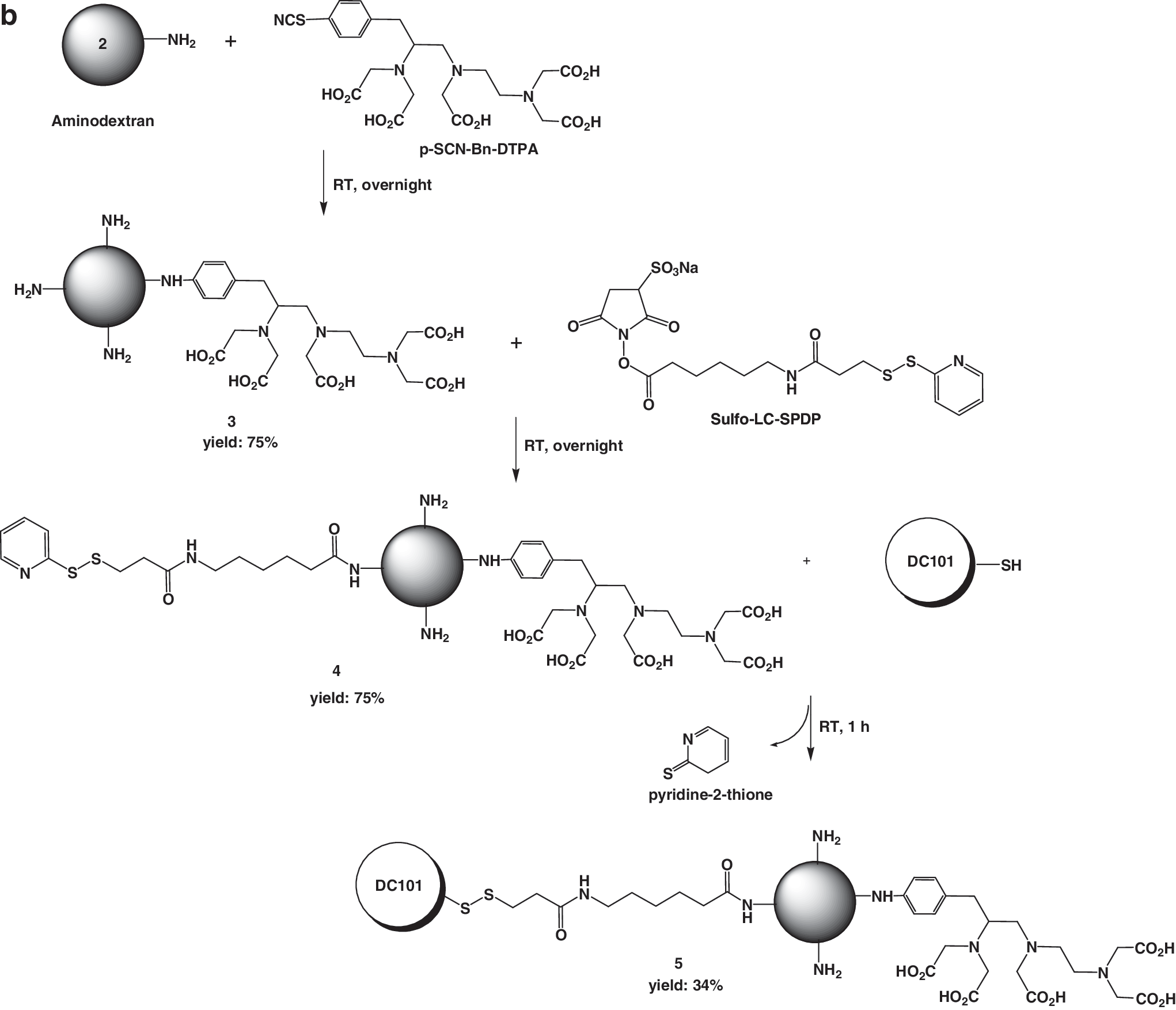
Synthesis of DTPA-dextran-DC101 (5) and DTPA-DC101
Aminodextran (
Radiolabeling
Iodination was performed by the chloramines-T method, previously described. 6 99mTc pertechnetate (37–48 MBq) in normal saline was mixed with 3–5 μg of SnCl2 (in 0.01 N HCl) and 200 μL of DTPA-dextran-DC101 (DC101 concentration=50 μg/mL). The mixture was allowed to react for 15 minutes with shaking. Radiolabeled DC101- conjugated compounds were purified by PD-10 desalting column. 99mTcO4 − and free iodine were separated by chromatography and that was checked on ITLC-SG strips eluted with 0.9% saline and acetone. After development, the chromatographic strips were scanned on an automatic TLC scanner (Bioscan). TLC was performed at various time points.
Binding and blocking assays
Flk-1 protein was purchased from R&D Systems and dissolved in coating buffer (carbonate buffer, pH 9.75). The Flk-1 protein (100 ng/well) was coated onto 96-well plates (16 hours, 4°C) and plates were blocked with blocking buffer (Pierce). After washing with washing buffer (PBS, 0.1% Tween-20), various amounts of DC101, DTPA-DC101, and DTPA-dextran-DC101 in PBS were added to the 96-well plates (Nunc). Then plates were washed three times with PBS containing 0.1% Tween-20. The plates were then incubated with rabbit antihuman IgG-HRP (κ specific) at room temperature for 1 hour. The plates were washed and developed with tetramethylbenzidine substrate and absorbance was measured at 405 nm. EC50 values were calculated by nonlinear regression analysis using the GraphPad Prism computer-fitting program (GraphPad Software, Inc.). Cellular uptake and receptor blocking assays were also performed. Briefly, B16F10 cells were seeded in 24-well plates. After overnight incubation, the medium was removed and cells were treated with Tc-99m–labeled conjugates for 30 minutes at 4°C. For the blocking assay, 300 μg of free DC101 was cotreated. The cells were washed two times with Hank's balanced salt solution. Scintigraphy was performed using a gamma camera (E-CAM; SIEMENS). After gamma imaging, cells were stained with crystal violet solution, and then were dried and dissolved in sulfuric acid. Dissolved cells were transferred to 96-well plate, and absorbance was measured at 450 nm. The percentage of cellular binding was calculated as follows:
Percentage of cellular binding=(Count/pixel in cells)/(Count/pixel in total standard)×100
In vivo imaging study
All animal experiments were performed in compliance with the policies and procedures of the Institutional Animal Care and Use Committee for animal treatment of Chonbuk National University. Mice were anesthetized by subcutaneous injection with a mixture of ketamine (50 mg/kg) and xylazine (10 mg/kg). After sedation, the mice were immobilized on a plate with an adhesive bandage and placed in the supine position under a single-headed gamma camera with a 5-mm pinhole collimator, a window setting of 140 keV, and a width of 20% (Vertex; ADAC Laboratories). The mice were injected with 18.5 MBq of the 99mTc-DTPA-DC101, 99mTc-DTPA-dextran-DC101, and 10 MBq of 131I-DC101 via the tail vein. The gamma camera images were acquired at 5, 60, 180 minutes, and 24 hours postinjection. The static images were stored with a 512×512 matrix size and the acquisition time was 5 minutes.
Biodistribution studies
Female athymic nude mice bearing B16F10 melanoma tumors were injected via tail vein with 3.7 MBq of the 99mTc-DTPA-DC101, 99mTc-DTPA-dextran-DC101, and 1.85 MBq of 125I-DC101 in 100 μL PBS. The mice were sacrificed and dissected at 1, 6, and 24 hours after injection. Blood, tumors, major organs, and tissues were collected and weighted. Tissue radioactivity was measured using a gamma counter (Packard). Results are presented as percentage injected dose per gram (%ID/g). Values are expressed as mean±standard deviation (SD) for 4 mice per group.
Statistical analysis
Quantitative data are expressed as mean±SD. Means were compared by use of the independent samples t test. p-Values of less than 0.05 were considered statistically significant.
Results
Preparation and characterization of DTPA-dextran-DC101
The preparation of aminodextran was carried out by a two-step procedure: (1) the synthesis of dextran-COOH and (2) the synthesis of dextran-NH2. 1H-NMR analyses of dextran derivatives showed the typical signal of bromohexanoic acid (Fig. 1a) and ethylenediamine (Fig. 1b). The yield of dextran-COOH (
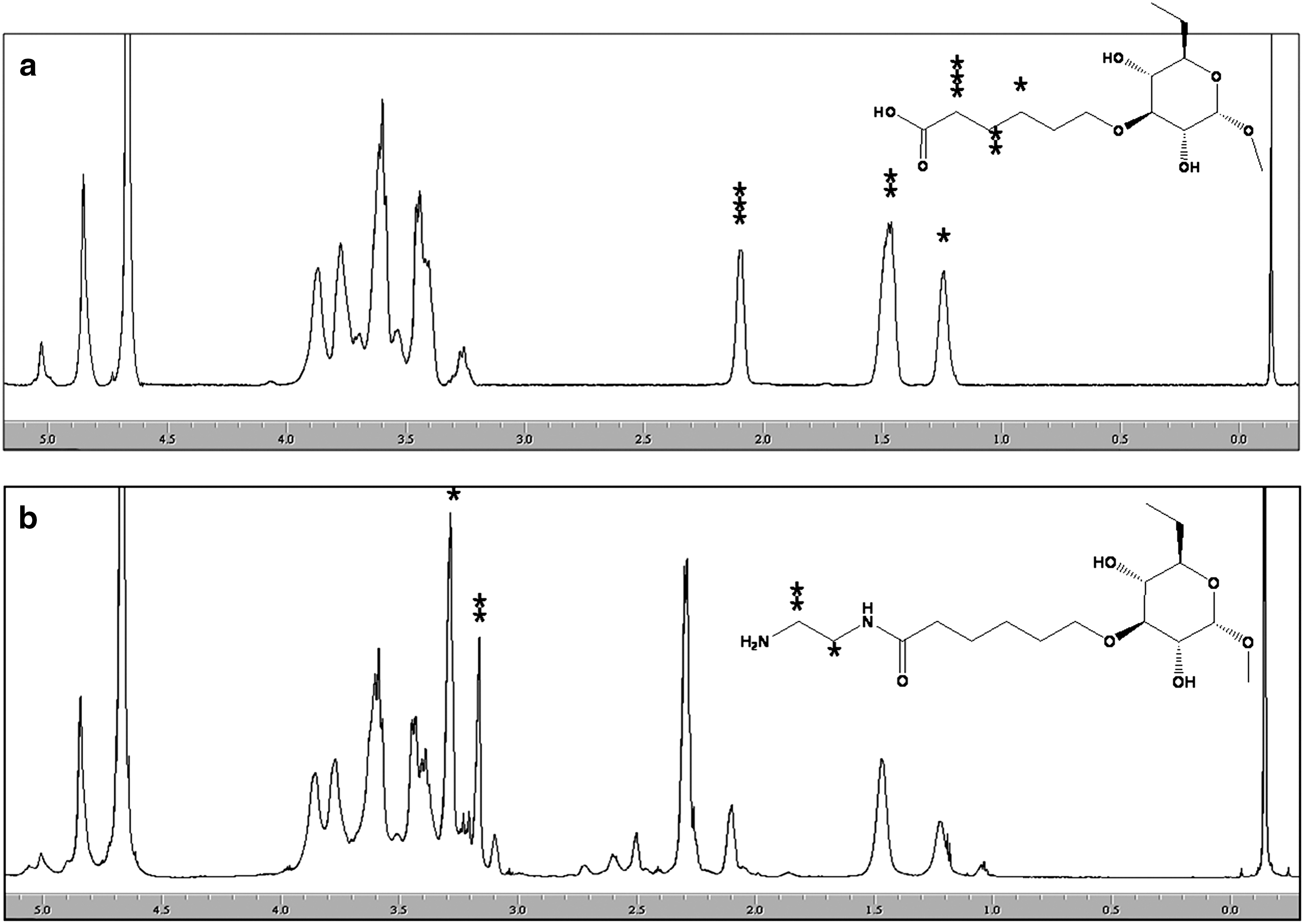
1H-NMR spectra of dextran-COOH
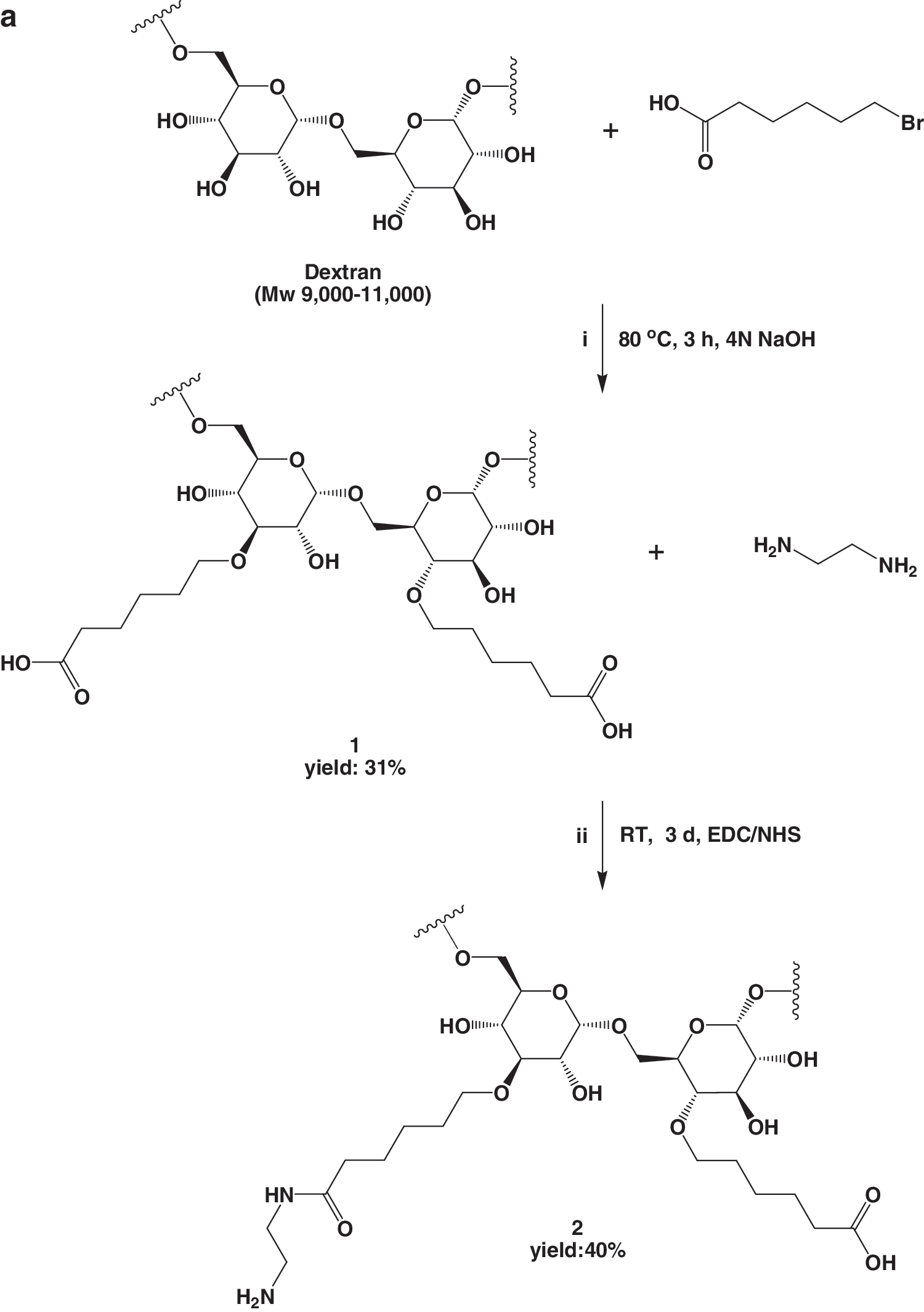
Synthetic schematic of aminodextran
In vitro receptor binding and blocking assays
The antigen-binding efficiencies of the DTPA-DC101 and DTPA-dextran-DC101 on immobilized receptor were determined by enzyme-linked immunosorbent assay. The antibodies bound to KDR in a dose-dependent manner (Fig. 3). The dissociation values (Kd ) for DC101, DTPA-DC101, and DTPA-dextran-DC101 were 22.48, 3.05, and 14.74 pM, respectively. DTPA-dextran-DC101 showed slightly higher affinity than DC101, while DTPA-DC101 showed significantly better affinity. These results might be from the negative charge of DTPA. Cell binding and blocking studies showed that Tc-99m–labeled DTPA-DC101 and DTPA-dextran-DC101 binding were significantly inhibited by free DC101 (*p=0.004, **p=0.001) (Fig. 4a, b). Cell binding and inhibition assays demonstrated that there is no significant difference after dextran conjugation.
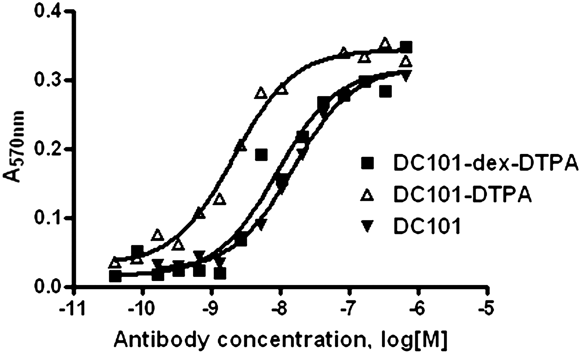
Binding to Flk-1 by DC101, DTPA-DC101, and DTPA-dextran-DC101. Dose-dependant binding of DC101 and DTPA-dextran-DC101 to immobilized Flk-1. All experiments were done in duplicate. The Kd values of DC101, DTPA-DC101, and DTPA-dextran-DC101 were 22.48, 3.05, and 14.74 pM, respectively.
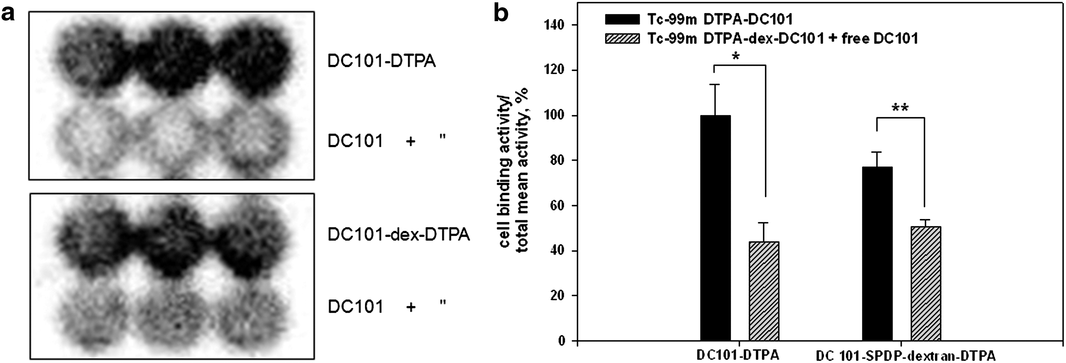
Planar images of 99mTc-DTPA-DC101– and 99mTc-DTPA-dextran-DC101–treated plates
In vivo studies
The labeling yield was >90% and radiochemical purity was 99% and it was stable for 24 hours. Whole-body imaging studies were performed with 99mTc-DTPA-dextran-DC101, 99mTc-DTPA-DC101, and 131I-DC101 in melanoma xenografted mice. Their distributions at 1 hour with whole-body images were shown in Figure 5. As shown in Figure 5, 99mTc-DTPA-DC101 and 131I-DC101 showed strong hepatic uptake, whereas 99mTc-DTPA-dextran-DC101 showed weak hepatic uptake and rapid renal elimination. In the Table 1, 125I-DC101– and 99mTc-DTPA-DC101–injected mice showed high hepatic activity, compared with that of 99mTc-DTPA-dextran-DC101 at early time point. The blood activity of 99mTc-DTPA-dextran-DC101 was lower than those of 99mTc-DTPA-DC101 and 131I-DC101 at all time points. The p-values for blood activities between 99mTc-DTPA-dextran-DC101 and 99mTc-DTPA-DC101 at 1, 6, and 24 hours were 0.05, 0.005, and 0.02, respectively. Significant difference was shown after 1 hour. The renal uptake of 99mTc-DTPA-dextran-DC101 was higher than those of 99mTc-DTPA-DC101 and 125I-DC101 at all time points. Tumor uptake of 99mTc-DTPA-dextran-DC101 was increased with time, whereas 99mTc-DTPA-DC101 was decreased with time until 24 hours. However, there were no significant differences in the tumor uptake by dextran conjugation until 24 hours between 99mTc-DTPA-dextran-DC101 and 99mTc-DTPA-DC101.
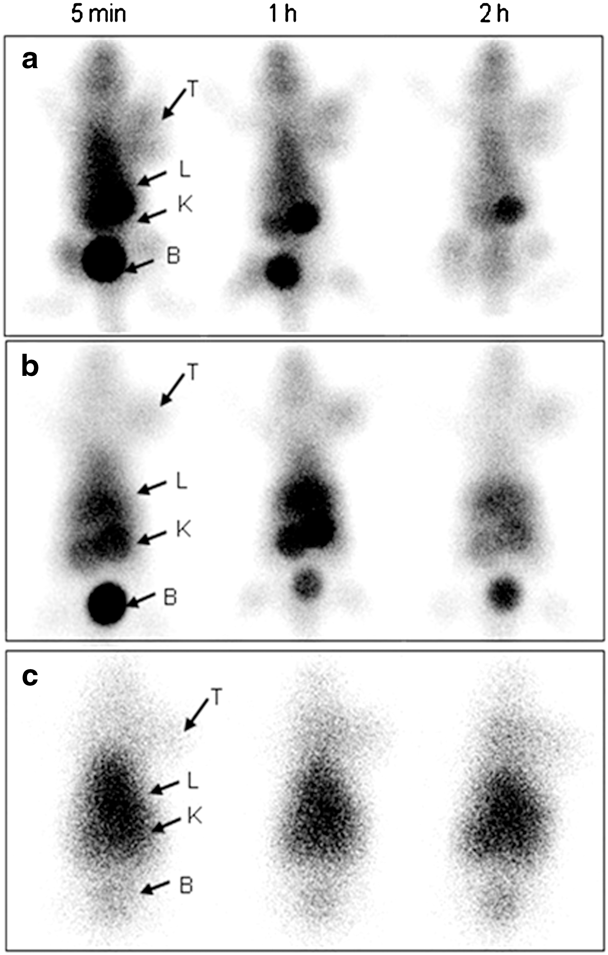
In vivo imaging of athymic nude mice bearing B16F10 melanoma xenografts at 1 hour after i.v. injection of 99mTc-DTPA-dextran-DC101
ID, injected dose; SD, standard deviation.
The P value between 99mTc-DTPA-dextran-DC101 and 99mTc-DTPA-DC101 was 0.005.
The P value between 99mTc-DTPA-dextran-DC101 and 99mTc-DTPA-DC101 was 0.02.
Discussion
Therapeutic antibody-based imaging has become an important component of the therapeutic plan for an increasing number of human malignancies. To be effective tumor imaging agents, tumor-targeting radiolabeled antibodies must provide both high tumor uptake and low background activity. However, monoclonal antibodies (150 kDa) have a relatively slow blood clearance (T 1/2=48–72 hours), which allows ample time to achieve high accumulation in tumor. 1 Because of this, imaging agents using enzymatically derived antibody fragments or pretargeting methods have been investigated. And also, for enhanced target affinity due to prolonged retention and reduced immunogenicity, polysaccharide-masked protein and antibody have been studied. 15 –17 However, these properties have not been examined in a tumor model. In the current study, we investigated the usefulness of a 99mTc-dextran–conjugated antibody to lower the blood concentration and increase the tumor-to-blood ratio during circulation.
In this study, for dextran conjugation, we initially prepared aminodextran and this was conjugated with anti-Flk-1 antibody (DC101). The aminodextran-conjugated DC101 was then labeled with technetium-99m and administered to melanoma xenografted mice.
As shown in Figure 3, receptor binding assays demonstrated that for Flk-1 binding, DTPA-dextran-DC101 is similar to DC101 and low compared with DTPA-DC101. The binding affinity of DTPA-dextran-DC101 for Flk-1 receptor was not improved by dextran conjugation. The reason for increasing the affinity of DTPA-DC101 than DC101 might be increased nonspecific interaction. On the other hand, DTPA-dextran-DC101 might show reduced nonspecific interaction to Flk-1 than DTPA-DC101, because of highly water-soluble dextran. Although the cellular uptake of 99mTc-DTPA-dextran-DC101 and 99mTc-DTPA-DC101 was not perfectly inhibited by free DC101 in our in vitro inhibition study, the values showed significant differences (Fig. 4).
In planar images (Fig. 5), dextran-conjugated antibody showed lower hepatic uptake and faster renal elimination than nonconjugated antibody. However, the biodistribution of hepatic uptake at 1, 6, and 24 hours did not show significant differences. There was discrepancy between whole-body images and %ID/g value in liver uptake. This might be due to high blood activity of 99mTc-DTPA-DC101 than 99mTc-DTPA-dextran-DC101, because the liver is a blood pooling organ. As shown in Table 1, dextran-conjugated antibody shows low blood activity and high renal activity, compared with nonconjugated form. In case of dextran-conjugated antibody, other organ activities were also relatively low, compared with the nonconjugated form. As a result, the background activity was rapidly decreased and this might be caused by the characteristics of dextran. Although the reason of high renal uptake was not clearly understood, it might be due to metabolism of some conjugates by enzymes existing in the blood or renal cells. 18 In our previous study, we investigated whether 99mTc- or Cy5.5-labeled chitosan-DC101 conjugates could identify VEGF-R2 overexpressed lesion of ischemic area. 19 99mTc- or Cy5.5-labeled chitosan-DC101 conjugates also showed high renal uptake. 99mTc-labeled DC101 conjugates might be degraded by lysosomes in the liver and kidney and the radiometabolites were excreted from the kidney. And also in that study, we reported that, in vivo, the chitosan-DC101 specific binding to the VEGF-R2 was not mediated by EPR effect in the ischemic mice.
Moreover, our results in this study revealed that dextran conjugation reduced the background activity, as previously reported. 12 The decreased blood radioactivity of antibodies conjugated with dextran has shown significant differences after 1 hour, compared with nonconjugated form. However, in this study, the tumor uptake did not show significant differences between 99mTc-DTPA-DC101 and 99mTc-DTPA-dextran-DC101 at each time point, in spite of relatively higher uptake. Only the tumor uptake of 99mTc-DTPA-dextran-DC101 was increased with time, similar to 125I-DC101 tumor uptake pattern. Therefore, although tumor uptake did not show significant differences in this study whether dextran was conjugated or not, 99mTc-DTPA-dextran-DC101 had the possibility of much more uptake to the tumor than the unconjugated form and might reflect the kinetics of DC101 similar to iodinated DC101. On the other hand, the tumoral uptake of 99mTc-DTPA-DC101 showed decreased pattern with time. Therefore, the tumor uptake of 99mTc-DTPA-DC101 with 1.63 %ID/g in early phase (1 hour) might not represent the binding capacity of this antibody onto receptors. The key finding of the current study was the increasing tumor accumulation of the dextran-conjugated antibody, whereas that of the dextran nonconjugated form decreased (Table 1). 99mTc-DTPA-DC101 was more taken up into the stomach and intestine until 6 hours. This finding could be explained by the fact that radioactivity was dissociated from the antibody within 1 to 6 hours.
In conclusion, instead of the direct DC101 radiolabeled with 99mTc-DTPA or radioiodine, we designed the DTPA-dextran–conjugated system to obtain better images with enhanced renal excretion of background activity and reduction of blood activity. Dextran conjugation to the antibody could provide more opportunity of binding of anti-VEGF antibody onto its receptor with time, similar to the radioiodinated antibody in the melanoma xenografted mouse model. However, additional experiments are necessary to improve clinical pharmacokinetics, such as decreased kidney uptake and prolonged tumor uptake.
Footnotes
Acknowledgments
This article was supported by research funds of the Chonbuk National University in 2008 and a grant from the National R&D Program for Cancer Control, Ministry for Health, Welfare and Family Affairs (0620220) and the National Research Foundation of Korea (NRF) grant funded by the Korea government (MEST) (No. 2011–0028581).
Disclosure Statement
No competing financial interests exist.