
Abstract
Breast cancer is the most common cancer in women worldwide. Molecular imaging plays an important role in breast cancer diagnosis, staging, and treatment response evaluation. Positron emission tomography (PET) and single-photon emission computed tomography (SPECT) are the main clinical molecular imaging modalities that are based on the detection of radiotracers. This article discusses the typical radiotracers used for breast cancer imaging by PET and SPECT. In addition, radiotracers that are currently applied for human breast cancer imaging or under clinical trials are also reviewed in compliance with the categories of tumor-specific targets to which they are aimed at.
Introduction
Breast cancer is the most common malignancy among women. In 2010, a total of 207,090 new cases of invasive breast cancer, along with 54,010 new cases of noninvasive types, were expected to be diagnosed in women in the United States. Moreover, about 39,840 women were projected to die from this disease that year. According to the National Cancer Institute, the 5-year survival rate of breast cancer ranges from 23.4% in patients with stage IV disease to 98% in patients with stage I disease, highlighting the importance of early detection and diagnosis of breast cancer. 1
Molecular imaging not only serves as an essential tool in breast cancer diagnosis and staging but also provides a significant proportion of information for surgical management, radiation planning, chemotherapeutic assessment, and follow-up evaluation of patients. The major functional molecular imaging modalities used in the clinic are positron emission tomography (PET), single-photon emission computed tomography (SPECT), and their combinational use with CT. Both PET and SPECT are based on the detection of radiolabeled ligands, termed “radiotracers,” which are assumed to target tumor-specific characteristics at the molecular level. The accumulating understanding of the breast cancer molecular biology has highlighted pivotal factors that are critical for breast cancer progression, which has allowed researchers to select suitable targets for developing tumor-specific radiotracers (Fig. 1). For instance, given that sustained tumor growth demands elevated glucose consumption for energy production in the regions with lesions, PET radiotracer 18F-labeled glucose analog 18F-fluoro-deoxy-glucose (18F-FDG) is commonly used to visualize the glucose metabolism of breast cancer cells. 2 Similarly, 18F-fluoroestradiol (18F-FES) is used to image the estrogen receptor (ER), which is highly overexpressed in a large proportion of breast tumor tissues. 3 Many other radiotracers have been designed to image cell proliferation, apoptosis, angiogenesis, hypoxia, and other features of breast tumors (Table 1). Because breast cancer treatment has become more individualized in addressing the distinct biological characteristics of tumors from each patient, more target-specific molecular imaging radiotracers may play a key role in guiding treatment selection and evaluating treatment response in the early stages of the disease.
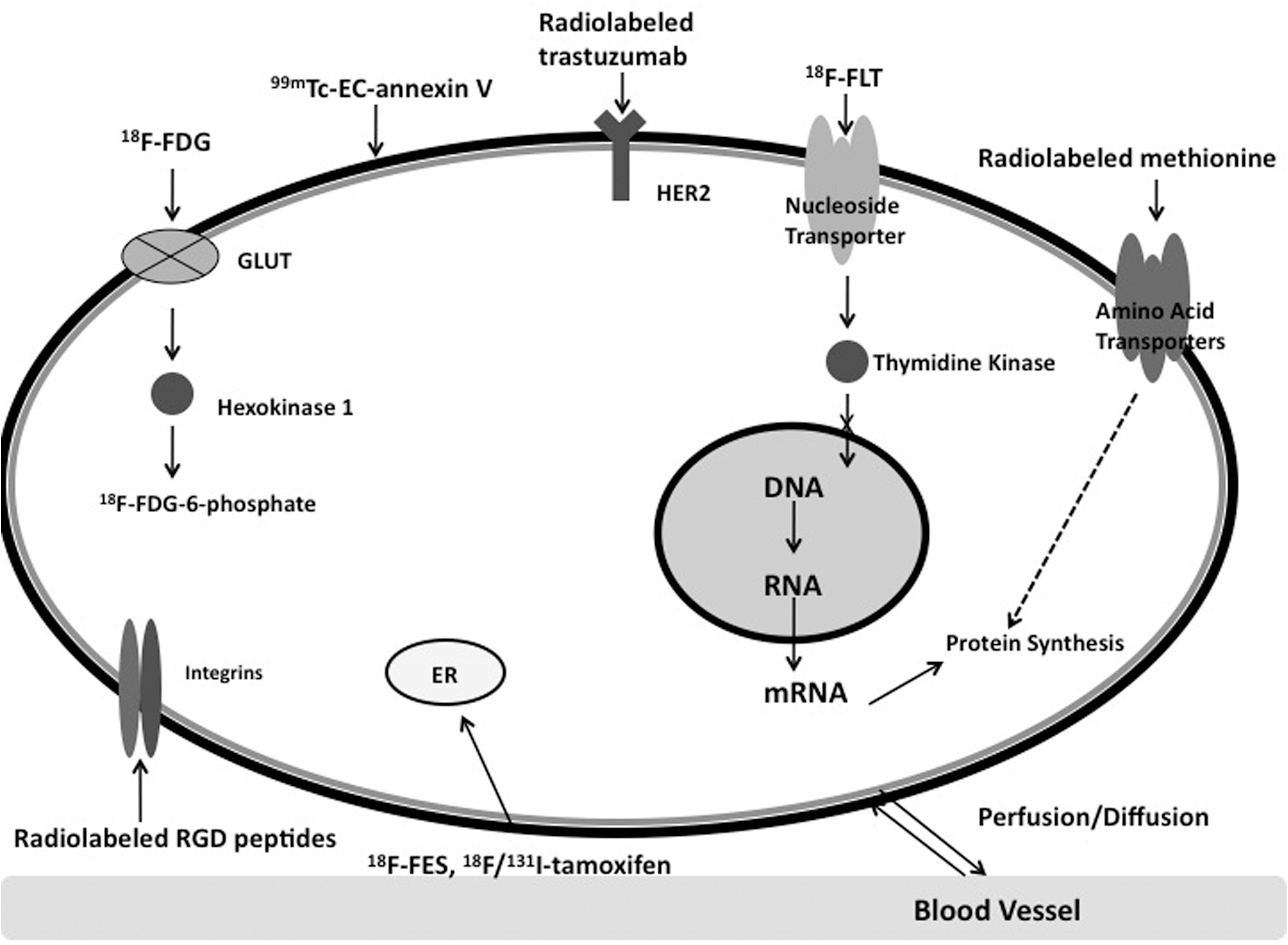
Selected cellular targets and their corresponding nuclear imaging radiotracers. ER, estrogen receptor; 18F-FDG, 18F-fluoro-deoxyglucose; 18F-FLT; 18F-fluorothymidine; 18F-FES, 18F-fluoroestradiol; GLUT, glucose transporters.
The radiotracers selected in the table have been applied to first-in-human studies, which include the commercialized products, the agents under clinical trials, or under Radioactive Drug Research Committee approval.
F-FDG, 18F-fluoro-deoxy-glucose; 18F-FES, 18F-fluoroestradiol; 18F-FLT, 18F-fluorothymidine; 18F-galacto-RGD, 18F-galacto-arginine-glycine-aspartic acid; 99mTc-MDP, 99mTc-methylene diphosphonate; SPECT, single-photon emission computed tomography; PET, positron emission tomography; HER2, human epidermal growth factor receptor 2.
Reviewed in this article were the most commonly used radiotracers for breast cancer imaging in the clinic, 18F-FDG for PET, and 99mTc-sestamibi for γ-imaging. Also discussed are other radiotracers (listed in Table 1) that currently applied for breast cancer imaging, such as radiolabeled trastuzumab targeting human epidermal growth factor receptor 2 (HER2) and 18F-fluorothymidine (18F-FLT) imaging cell proliferation. Those radiotracers have been described according to the categories of the tumor-specific targets.
The Most Common Radiotracers for Breast Cancer Imaging in Clinic
18F-FDG for PET/CT and positron emission mammography
In recent years 18F-FDG, which is an 18F-labeled analog of glucose, has become the most common and attractive radiotracer for PET scanning. Both 18F-FDG and glucose are transported across the cell membrane by glucose transporters. 18F-FDG is phosphorylated by hexokinase to 18F-FDG-6-phosphate, whereas glucose is phosphorylated to glucose-6-phosphate. Unlike glucose-6-phosphate, 18F-FDG-6-phosphate cannot be further metabolized and therefore is trapped and accumulates steadily in tumor cells. 2 Hence, 18F-FDG is able to provide high sensitivity and specificity for detecting, staging, and restaging tumors by imaging high glucose metabolic rates in tumor cells. For breast cancer cells, increased glucose utilization is caused by the overexpression of the glucose transporters Glut-1 and Glut-3 and by increased hexokinase activity. The rate-limiting step in the uptake of 18F-FDG in breast cancer appears to be the phosphorylation process by hexokinase, particularly hexokinase I. 4
Although 18F-FDG-PET generally has high sensitivity and specificity in detecting malignancies, whole-body 18F-FDG-PET is not quite suitable for primary breast cancer diagnosis, especially for low-grade tumors and tumors smaller than 1 cm in diameter. 5 In addition, it is not appropriate for breast cancer screening. Therefore, 18F-FDG with positron emission mammography (PEM) has been introduced as an alternative. Compared with PET, PEM has a much higher spatial resolution because it has two opposite detector heads on each side of the breast, which minimizes the distance between the radiation source and the detectors. Schilling et al. have reported that PEM can detect tumor as small as 1.5 mm in diameter, with much less breast compression compared with traditional mammography. 6 They have also indicated that PEM is not affected by breast density and it has good sensitivity (90%) in detecting ductal carcinoma in situ, which is not readily detected by PET.
For locoregional staging of breast cancer, 18F-FDG-PET has shown high sensitivity in axillary staging of patients with late stage cancer, but it is not sensitive enough in detecting early-stage micrometastases and small tumor-infiltrated axillary lymph nodes. 7 Therefore, in this case 18F-FDG-PET is not appropriate to replace the sentinel lymph node biopsy. However, 18F-FDG-PET appears to be suitable for distant (systemic) staging; it is also well suited staging of locally advanced breast cancer, which usually has a large primary tumor (>5 cm in diameter) or advanced axillary disease without clinically apparent distant metastases. 8 Moreover, several studies have demonstrated that 18F-FDG-PET is superior to traditional bone scintigraphy in the detection of osteolytic and intramedullary metastases but is inferior in the detection of primary osteoblastic lesions. 9
An increasing number of studies have used 18F-FDG-PET to evaluate treatment response. Although 18F-FDG has not yet been recommended as a routine assessment agent, it has been proven to be an accurate early predictor of poor response to therapy. In a study showing that high 18F-FDG uptake was associated with a low blood flow or perfusion rate in tumors, the patients with high uptake were more likely to have poor response and early relapse. 10
99mTc-sestamibi for scintimammography and breast-specific γ-imaging
The use of 99mTc-methoxyisobutylisonitrile, which is also called 99mTc-MIBI or 99mTc-sestamibi, in scintimammography has become more popular for assessing suspicious lesions of patients with negative or indeterminate results from mammography performed in the clinic. 11 This small lipophilic cation was originally developed as a myocardial perfusion agent. The first report of its application in breast cancer detection was published in 1992 by Aktolun et al. 12
The cellular uptake mechanism of 99mTc-sestamibi is unclear, although it is known that its uptake is driven by a negative transmembrane potential and that most of the radioactivity is found in mitochondria. 13 99mTc-sestamibi scintimammography has higher sensitivity (85%) and specificity (87%) than traditional mammography, and its sensitivity is independent of breast density. 14
99mTc-sestamibi is a substrate of the transmembrane P-glycoprotein, which is a member of the MDR/TAP subfamily involved in multidrug resistance. Therefore, the uptake, clearance, and retention of 99mTc-sestamibi have been investigated as predictors of response to chemotherapy in human breast cancer. 15 In a study of 45 patients who had primary breast cancer, Cayre et al. concluded that negative 99mTc-sestamibi scintimammography results predicted chemoresistance with a specificity of 100%. 16 In addition, 99mTc-sestamibi uptake was inversely correlated with the expression of the multidrug resistance protein MDR1 in invasive ductal carcinomas (p<0.05). 16
However, 99mTc-sestamibi scintimammography demonstrates relatively low sensitivity in detecting nonpalpable lesions or tumors smaller than 1 cm. Therefore, high-resolution small field-of-view breast-specific gamma imaging (BSGI) has been developed as an alternative. Similar to PEM, the single-head detector used in BSGI is mounted opposite to a compression plate so that the patient's breast is compressed between the detector and the plate. According to a 6-year BSGI study performed by Hruska et al. at the Mayo Clinic, the sensitivity of 99mTc-sestamibi-BSGI for tumors larger than 1 cm was 97% and that for smaller tumors was 74%. 17 Although the average sensitivity of PEM is slightly higher than that of BSGI (93% vs. 89%), BSGI has a much higher negative predictive value (100% vs. 88%). 18 Thus, both of these imaging modalities have demonstrated a great ability for detection, and there has been no proven clinically significant advantage to one modality over the other.
Other Radiotracers Imaging Breast Cancer Molecular Biomarkers
Here, this study has introduced and discussed the radiotracers currently applied for breast cancer imaging other than 18F-FDG and 99mTc-sestamibi, according to the categories of the tumor-specific targets (Table 1).
Radiotracer targeting hormone receptors
18F-FES targets ER
More than 70% of breast tumors are positive for hormone receptors such as ER. ER-directed therapeutic agents against breast cancer, such as tamoxifen and aromatase inhibitors, are highly effective for patients with ER-positive tumors and have fewer side effects than traditional chemotherapy. 19 To date, 18F-16α-17β-fluoroestradiol (18F-FES) has been proven to be the most successful PET radiotracer in determining ER status and the prognosis of ER-directed hormonal therapy in breast cancer patients. 3
For clinical use, an automated synthesis of 18F-FES can be achieved in a decay-corrected yield of 30% at 60 minutes after the end of bombardment with high radiochemical purity (>99%). 2018F-FES has a high binding affinity to ER, especially the ERα subtype. 21 In both humans and rodents, 18F-FES is rapidly taken up and metabolized by the liver. Twenty minutes after injection, 80% of the 18F-FES is converted to radiolabeled glucuronide and sulfate conjugates in blood. The recommended injection dose and imaging time of 18F-FES is 6 mCi at 30 minutes after injection. 22 Peterson et al. demonstrated excellent agreement (r=0.99) between 18F-FES-PET imaging results and ER expression as assayed by immunohistochemical analysis in 17 patients. 23 In addition, their study indicated that the ER-negative tumors had a partial-volume-corrected standardized uptake value (SUV) of <1.0, whereas that of ER-positive tumors was above 1.1.
Radiolabeled tamoxifen imaging ER
Tamoxifen, a selective ER modulator, is a standard therapy for ER-positive breast cancer. It is an ER antagonist in breast tissue and an agonist in other tissues, such as bone marrow. Yang and colleagues were the first to radiolabel tamoxifen with 18F and 131I for PET and SPECT, respectively, for the imaging of mammary tumors in rat models. The synthetic yields of 18F-fluorotamoxifen (FTX) and 131I-tamoxifen were 30%–40% and 20%–25%, respectively. 24 Two years later, Inoue et al. reported the first-in-human study of FTX in 10 patients with of 23 ER-positive suspected primary or metastatic lesions. 25 In that study, FTX uptake in 19 of the lesions was correlated with the response of tamoxifen therapy. In addition, the average SUV of FTX in the tumors with good response was significantly higher than those with poor response (2.46±0.62 vs. 1.37±0.59, p<0.05).
Radiotracers imaging HER 2
Radiolabeled trastuzumab
HER2 is a transmembrane glycoprotein that forms heterodimers with other epidermal growth factor receptor family members to activate distinct signaling pathways involved in cell growth, survival, differentiation, adhesion, and migration. HER2 is overexpressed in many types of cancers. Because about 25%–30% of human breast tumors have HER2/neu gene amplification and/or HER2 protein overexpression, 26 HER2 has become an attractive target for breast cancer imaging and treatment. The humanized monoclonal antibody trastuzumab has been successfully used for HER2-positive breast cancer treatment. By labeling trastuzumab with different radionuclides, HER2 expression and localization in breast cancer patients can be assessed noninvasively and thus patients with HER2-positive tumors that are qualified for trastuzumab treatment can be identified. 27
Zirconium-89 (89Zr; t 1/2=78.41 hours) is used for trastuzumab labeling because it has the longest half-life among the positron-emitting radionuclides, allowing PET to be conducted for up to 7 days after injection. 28 Antibodies such as trastuzumab often have a high molecular weight and thus a slow metabolism and clearance rate. Hence, radionuclides with long half-lives are suitable for obtaining better tumor-to-blood ratios even several days postinjection. Dijkers et al. recently reported on their first clinical trial of 89Zr-trastuzumab PET in 14 patients with HER2-positive metastatic breast cancer. They determined the optimal imaging time to be 4–5 days postinjection and the optimal dose to be 50 mg for trastuzumab-naïve patients or 10 mg for patients already on trastuzumab treatment. 29
Trastuzumab has also been labeled with 124I, 86Y, and 76Br for PET, 30 –32 and with 111In for SPECT. 33 To date, only 89Zr-trastuzumab and 111In-trastuzumab have been applied to humans in clinical trials. Although the tumor uptake levels are similar, 89Zr-trastuzumab demonstrates better image quality due to its higher spatial resolution and sensitivity in PET.
Radiotracer imaging cell proliferation
18F-fluorothymidine
18F-fluoro-3′-deoxy-3-
Because of the radiotracer's low uptake in the normal brain tissue, 18F-FLT-PET is commonly used in detecting brain tumor in clinical studies. 38 Its high uptake in bone marrow and liver limits its diagnostic application, especially in the assessment of liver and bone metastases. Further, 18F-FLT-PET is not superior to 18F-FDG-PET for cancer staging because it has low tumor-to-background ratios. For these reasons the imaging potential of 18F-FLT in breast cancer has been evaluated more often in the prediction and assessment of tumor response to a certain treatment. For instance, Pio et al. scanned 14 breast cancer patients before and 2 weeks after the first cycle of the treatment and found that 18F-FLT uptake was strongly correlated with the percentage change in CA27.29 tumor marker levels (r=0.79; p=0.001). In addition, the change in 18F-FLT uptake after the chemotherapy course was predictive of late change in tumor size as assessed using CT scans (r=0.74; p=0.01). 39 In a more recent study, Kenny et al. assessed 18F-FLT response to a combined chemotherapy using 5-fluorouracil, epirubicin, and cyclophosphamide in 13 breast cancer patients with 17 discrete lesions. Their observations indicated a significant difference in 18F-FLT uptake change between the responders and nonresponders. The average decrease in responding lesions at 90 minutes was 41.3% for SUV and 52.9% for net irreversible plasma to tumor transfer constant (Ki); the corresponding values for nonresponding lesions were 3.1% and 1.9%. 40
To meet clinical application needs, however, significant improvements are required in18F-FLT radiosynthesis. So far there are two synthesis methods using different precursors, but neither has achieved optimal radiochemical yield within a short preparation time. By using precursor 3′-O-nosyl thymidine with its pyrimidine ring protected with N-BOC, Yun et al. achieved a high radiochemical yield of 42%±5.4% within 60 minutes at a radiochemical purity >97%. 41 Machulla et al. used 5′-O-(4,4′-dimethoxytriphenylmethyl)-2,3′-anhydrothymidine as the precursor to achieve the preparation time within 10 minutes at 160°C, but the yield was only 14.3%±3.3%. 42
Radiotracers imaging amino acid transporters and protein synthesis
Radiolabeled methionine
Amino acid-based radiotracers are being developed for tumor imaging, as tumor cells uptake and consume more amino acids than normal cells do to sustain their uncontrolled growth. Most of these radiotracers are reported to enter the tumor cells via amino acid transporters, such as the Na+-independent L-type amino acid transporter system LAT and the Na+-dependent transport systems A and B0. 43
11C-methionine, which is also called 11C-MET, has been synthesized with an automation module within 20 minutes after the bombardment for a radiochemical yield of >30%. The specific activity was 3.3 Ci/mmol, and the radiochemical purity was >96%. 44 This radiotracer was recently evaluated by Lindholm et al. for its potential in assessing early response to therapy in advanced breast cancer. Twenty-five out of 26 metastatic sites from 13 patients could be detected by 11C-MET-PET. The SUV in all six responding metastatic sites decreased by 30%–54% (p<0.05), whereas that of nonresponding sites did not decrease significantly (11%–13%; p=NS). 45
99mTc-labeled methionine has also been successfully used to detect breast cancer. In a recent clinical trial with 47 patients that was performed by Sharma et al., 99mTc-methionine was proven to have the same biological properties as 11C-methionine, but the longer half-life of the in-house generator-produced radioisotope 99mTc (t 1/2= 6 hours) provides a simpler and more affordable way to image breast tumor using conventional scintimammography. 99mTc-methionine was synthesized by conjugating methionine with diethylene triamine pentaacetic acid and 99mTc, and the radiochemical yield was >95%. The sensitivity, specificity, and positive predictive value of 99mTc-methionine in this clinical trial were 87.8%, 92.8%, and 96.6%, respectively. 46
Radiotracers imaging angiogenesis
Radiolabeled arginine-glycine-aspartic acid peptides
The formation of new blood vessels, angiogenesis, is required for a solid tumor to obtain essential oxygen and nutrients for growth. Integrins, which are located on cell surfaces, are extremely important in angiogenesis because they mediate cell–cell and cell–extracellular matrix interactions. 47 At this time 24 integrins have been reported. Among these, integrin αVβ3 is the best-studied subtype as a molecular marker for targeting the angiogenic cascade. A small peptide sequence consisting of arginine-glycine-aspartic acid (RGD) has been identified as the motif that integrins bind to with their αV subunit, including αVβ3. 48 Several radiolabeled RGD peptides have been developed to image breast tumors in humans. Two (2) of these are 99mTc-NC100692 for SPECT and 18F-galacto-RGD for PET.
NC100692 is a cyclic synthetic ligand containing an RGD binding site with high affinity to integrin subunits αVβ3 and αVβ5, which are upregulated during angiogenesis. 99mTc-labeled NC100692 is being tested in a phase II clinical trial by GE Healthcare, Ltd. In the proof-of-concept study performed by Bach-Gansmo et al., 19 of 22 malignant lesions from 20 breast cancer patients were detected by 99mTc-NC100692 scintigraphy (86%). 49 In the Phase 2a study performed by Axelsson et al., in 10 patients with breast cancer 99mTc-NC100692 scintigraphy detected 1 of 7 metastases in liver, 4 of 5 in lung, 8 of 17 in bone, and 1 of 1 in the brain. 50
The radiotracer 18F-galacto-RGD was first developed by Haubner et al. in 2001. 51 By adding a sugar amino acid to the RGD peptide, the lipophilicity of the tracer could be reduced, resulting in less uptake in liver and increased uptake in tumor. The overall radiochemical yield of 18F-galacto-RGD was 29%±5%, and the purity was >98%. 52 In a study of 18F-galacto-RGD-PET in 16 patients with invasive ductal breast cancer, Beer et al. observed that all of the invasive carcinomas could be identified (SUV=3.6±1.8, tumor-to-muscle ratio=6.2±2.2), but only three of eight lymph node metastases were detected. 53 The 18F-galacto-RGD images were found to represent αVβ3 expressed in both tumor cells and endothelial cells, so this agent was not tumor specific. Results from several preclinical and clinical studies have indicated that 18F-galacto-RGD-PET may not be suitable for differentiating tumor from inflammation because αVβ3 is also highly expressed in macrophages and other inflammatory lesions. 54
Radiotracers imaging apoptosis
99mTc-EC-annexin V
During the early phase of cell apoptosis, phosphatidylserine redistributes from the inner cytosolic leaflet to the outer leaflet of the plasma membrane. This transfer of phosphatidylserine represents a hallmark in the detection of dying cells. Annexin V (molecular weight ∼36 kDa, 319 amino acids) is a natural human phosphatidylserine-binding protein with nanomolar affinity in the presence of physiological concentrations of calcium. By radiolabeling Annexin V, anti-cancer drug-induced apoptosis can be visualized and the efficacy of therapy can be assessed in an early stage. 55 Kurihara et al. conducted a 99mTc-labeled annexin V (99mTc-EC-annexin V) imaging study using SPECT in 10 patients with primary breast cancer (stage II–III) and evaluated its potential in detecting treatment-induced apoptosis. A significant difference was observed in the mean tumor-to-normal tissue ratio between the patients who were receiving chemotherapy (2.6±0.5, n=5) and those who were not (1.5±0.2, n=5). 56
Radiotracers for bone scan
99mTc-methylene diphosphonate
The most common site of breast cancer metastases is bone, mainly spine and pelvis. The distribution of bone metastases is a prognostic factor of breast cancer. 57 Bone scintigraphy using 99mTc-methylene diphosphonate (99mTc-MDP) is the standard initial imaging technique for assessing bone metastases. The uptake mechanism of 99mTc-MDP in bone is its chemical absorption onto the surface of hydroxyapatite and then incorporation into the crystalline structure of hydroxyapatite. 58 99mTc-MDP bone scintigraphy is readily availability and affordable, allows rapid generation of whole-body images, and has a specificity and sensitivity for breast cancer bone metastases of 78%–100% and 62%–100%, respectively. 57 Because its detection rate in patients with early stage breast cancer is very low (0.82% for stage I disease), routine screening with 99mTc-MDP is recommended only for patients with advanced stage disease. 59 In addition, 99mTc-MDP bone scintigraphy may not be suitable for monitoring hormonal therapy response because of the increased 99mTc-MDP uptake caused by new bone formation during the repair process after the therapy (flare phenomenon). 60 In summary, 99mTc-MDP bone scintigraphy is best used for screening patients with stage III or IV breast cancer rather than for diagnosis. In addition, instead of bone scintigraphy, 99mTc-MDP using SPECT with or without CT has higher resolution and greater accuracy, and therefore is more suitable for diagnosing breast cancer bone metastases. The specificity and sensitivity of 99mTc-MDP-SPECT are 91%–93% and 87%–92%, respectively. 61
18F-fluoride
18F-labeled sodium fluoride (18F-fluoride) is a nonspecific PET radiotracer for whole-body bone metastases imaging. Its uptake is via the exchange of hydroxyl ions in the hydroxyapatite crystal, mainly at the surface of the skeleton. 62 18F-fluoride PET has higher resolution, sensitivity (99%), and specificity (97%) than 99mTc-MDP-SPECT. 63 In addition, the absolute uptake of 18F-fluoride in normal bone is twice as high as that of 99mTc-MDP. Although 18F-fluoride is not tumor specific, it can differentiate tumor and benign tissue better than 99mTc-MDP bone scintigraphy can because of the superior spatial resolution of PET scanner. Eighty percent of benign abnormalities detected by 18F-fluoride PET are caused by endplate factures and arthritis of the articular facets, both of which have a typical 18F-fluoride uptake pattern. 64 In 18F-fluoride PET images, lesions that are not located at joint surfaces and do not display the typical pattern of endplate fracture, osteophytes, or serial rib fractures are suspicious for metastases.
Conclusion
Molecular imaging modalities, such as PET and SPECT, play an important role in the diagnosis, staging, and treatment response evaluation of breast cancers. The focus of this study was on the radiotracers commonly used in molecular imaging of breast cancer, according to the classification of the tumor-specific characteristics to which they are targeting. To date, 18F-FDG and 99mTc-sestamibi are still the two most extensively used radiopharmaceuticals in breast cancer for PET and gamma scintigraphy, respectively. Advances in molecular cancer biology have led to increased understanding of the cancer biomarkers that are tightly associated with cancer progression and thus to the rapid development of more personalized and specifically tumor-targeted treatments. The demand for molecular imaging and tracer development to help direct and assess the treatment response in an early stage is increasing.
Footnotes
Acknowledgment
This work was supported in part by sponsored research agreement made by Cell >Point L.L.C (MDA LS01-212) and the John S. Dunn Foundation.
Disclosure Statement
All authors have no commercial associations that might pose a conflict of interests in connection with the submitted article.