
Abstract
Background:
The currently available radiopharmaceuticals are not specific for tumor imaging.
Purpose:
The present study was conducted to radiolabel doxorubicin with Technetium-99m ([99m]Tc) as a scintigraphic marker of high DNA turnover/intercalation in malignant cells.
Methods:
Labeling was done by direct method and the developed radiotracer was subjected to quality control tests. The blood kinetics, scintigraphy of tumor-bearing mice, and biodistribution were studied after intravenous injection of about 7.4 MBq of [99m]Tc-doxorubicin. The isotime (5 minutes) anterior images were acquired at different time intervals of 1.5, 3, and 4 hours.
Results:
The labeling efficiency of [99m]Tc-doxorubicin was estimated to be more than 95%. The protein-binding efficiency was greater than 88% and in vitro stability was up to 24 hours. The biodistribution data support the clearance of the radioligand by dual (renal and hepatic) pathways. A semiquantitative data analysis of the anterior images indicated that a focal concentration of the radiotracer was seen in the tumor at 1.5 hours, which persisted in 3-hour and 4-hour images, respectively.
Conclusions:
This scintigraphic approach, therefore, could be a powerful tool for cancer detection at early stage. The technique, however, needs further validation through animal experimentation and clinical studies.
Introduction
Molecular imaging plays an important role in the detection, diagnosis, and treatment of cancer. 1 The conventional radiopharmaceuticals used for tumor imaging like [99m]Tc-Methoxyisobutyl-isonitrile ([99m]Tc-MIBI), [99m]Tc-Tetrofosmin, and [201]Thallium ([201]Tl) have some limitations. The tumor uptake mechanism of [99m]Tc-MIBI is related to many factors and not yet clearly understood. It has been reported that the tumor uptake of [99m]Tc-MIBI is not a tissue-specific process, but is mainly dependent on cell metabolism and affected by the metabolic processes, mitochondrial and plasma membrane potentials. 2 It had been reported that retention of [99m]Tc-Tetrofosmin in cancer cells depends on the activity of 170-kDa P-glycoprotein coded on the multidrug resistance 1 (MDR1) gene, which functions as an adenosine triphosphate-dependent efflux pump for many chemotherapy drugs. 3 The primary disadvantage of [201]Tl imaging is the background uptake by the salivary and thyroid glands that might interfere with the accurate evaluation of lesions harboring the neck region. 4
[18]F-Fluorodeoxyglucose ([18]F-FDG) positron emitting tomography (PET) imaging has been primarily used for the localization, staging, and the treatment response monitoring of different cancer pathologies. 5 A nonspecific [18]F-FDG uptake in lesions, such as benign tumors or inflammatory processes, leads to false-positive results. Therefore, [18]F-FDG uptake is not particular for tumors only and therefore lacks specificity as reported. 6,7 The growing clinical utility of [18]F-FDG PET imaging is attributed to its remarkable sensitivity and also to the recognition of uptake patterns in different disease conditions. The nonspecificity of the available radiopharmaceuticals, therefore, defines the clinical need to develop more specific radiopharmaceuticals for an early cancer diagnosis and treatment response monitoring. The development of single photon emission computed tomography (SPECT) radiopharmaceuticals is of special importance at least in the developing countries. The current strategies in molecular imaging focus on the development of newer radiopharmaceuticals for targets of nuclear DNA proliferation, mRNA, or protein molecules (receptors) on the cell surface, all of which are hallmark of early cancer development. 8 These scintigraphic probes can be employed as specific molecular targets for the assessment of treatment response. 9 Molecular imaging can detect the cancer process at an early stage as the functional changes precede the anatomical ones. 10 Doxorubicin is a natural glycoside whose antitumor activity against a relatively broad spectrum of human cancers has been demonstrated. 11 –13 Anthracyclines most likely interact with topoisomerase II by stabilization of the ternary DNA–enzyme–drug complex, which hinders the relegation of the DNA strands and ultimately induces irreversible DNA breaks. 14 The donor group (hydroxyl and nitrogen) present as structural units of doxorubicin could be exploited to form a coordinate bond with a radioactive metal like [99m]Tc, which could be used for the scintigraphic localization of tumors. In the present study, radiolabeling of doxorubicin with [99m]Tc was carried out effectively and the formulation was further tested experimentally for blood kinetics, animal biodistribution, and its localization in the tumors by scintigraphic technique.
Materials and Methods
Labeling procedure
A pure salt of doxorubicin as a generous gift was procured from Dabur, India. All other chemicals and reagents were purchased from Sigma-Aldrich. Fresh pertechnetate ([99m]TcO4 -) eluted from ([99]Molybdenum-[99m]Technetium Column Generator (Isorad, Israel) was used for the labeling procedures. The variable concentrations (20 μg, 50 μg, 100 μg, 200 μg, 400 μg, and 1000 μg) of stannous chloride (Sncl(2).2H(2)O), different pH (2–9) conditions, and variable incubation times (5 minutes, 10 minutes, 20 minutes, and 30 minutes) were tested. [99m]Tc-doxorubicin was prepared by dissolving 2 mg of doxorubicin in 1 mL distilled water followed by addition of a standardized concentration of 100 μg stannous chloride dihydrate and pH was marked to 6.0. The contents were filtered through a 0.22 μm membrane filter (Millipore) into a sterile vial. About 40.0 MBq radioactivity of pertechnetate was added to the mixture and incubated for 15 minutes. The resultant radioligand [99m]Tc-doxorubicin was then subjected to various quality control tests.
In vitro quality control procedures
Radiochemical purity of the labeled complex was determined by instant thin layer chromatography (ITLC) using 100% acetone and 0.9% sodium chloride as solvents. Briefly, 20.0 μL of radiocomplex was dropped onto the ITLC strip at the marked origin point and put into the solvent chamber at room temperature. The percent labeling of [99m]Tc-doxorubicin was calculated at 15 minutes, 1 hour, 4 hours, and 24 hours by ITLC method. The percentage of free pertechnetate, hydrolyzed pertechnetate, and bound pertechnetate was calculated. The serum stability assay was evaluated by incubating 1.8 mL of normal human serum mixed with 0.2 mL solution of [99m]Tc-doxorubicin in a vial at 37°C. About 20.0 μL of the solution was taken out from the sample at different time intervals of 15 minutes, 1 hour, 2 hours, 4 hours, and 24 hours and the percentage of the labeled complex and free pertechnetate was determined by ITLC. Any increase in free pertechnetate was considered as the degree of degradation.
Plasma protein binding and lipophilicity
In vitro protein binding of [99m]Tc-doxorubicin was carried out in human plasma by protein precipitation with 10% trichloroacetic acid (TCA). About 0.9 mL of fresh human plasma and 0.1 mL of the labeled complex were mixed and incubated for 1 hour at 37°C followed by addition of 1 mL of 10% TCA and centrifugation at 500 g for 5 minutes. The supernatant was decanted; the pellet was resuspended in 1.0 mL of 5% TCA and centrifuged again at 500 g for 5 minutes. The supernatant was collected in a separate tube during each washing. Radioactivity was measured in both the precipitate and the supernatant fractions by using a well-type gamma spectrometer (GR611M; Nucleonix). Protein binding of the complex was expressed as percent fraction of protein bound radioactivity of the total radioactivity.
The lipophilicity was measured by using organic
Blood kinetics and biodistribution studies
Blood kinetics was studied in an adult rabbit after intravenous (dorsal ear vein) injection of about 20.0 MBq of the radiopharmaceutical. Blood samples were collected from the contra-lateral ear vein at different time intervals (1, 2, 3, 4, and 24 hours). The samples were weighed and radioactivity was expressed in percentage of injected dose per gram (%ID/g) of the sample. About 7.4 MBq of the radiopharmaceutical was injected intravenously (i.v.) into the penile vein of rats. The biodistribution and excretory route of [99m]Tc-doxorubicin was studied in male Wistar rats (6–8 weeks, 100±20 g). The rats (n=4) were sacrificed at each time point of 1, 4, and 24 hours after radiotracer administration. Different organs were removed, washed in saline, made free from adhering tissues, dried on filter paper, and kept in a weighed test tube. The activity (count per second) in different organs was recorded by using a gamma spectrometer and expressed as %ID/g of organ.
Tumor induction
Balb/c mice (6–8 weeks, 25±5 g) were used for the study. Ehrlich ascites tumor (EAT) cell line (NCCS) was maintained in the peritoneum of the mice in the ascites form by serial weekly passage. Exponentially growing cells were harvested, washed, and resuspended in phosphate-buffered saline. A fixed number of EAT cells (about 1.5×10⁷ cells) were injected subcutaneously in the right thigh region of each mouse. The mice were used for study after about 10 days when the tumor had grown to about 1 cm in diameter.
Scintigraphic studies
The tumor-bearing mice were injected i.v. (tail vein) with 7.4 MBq of [99m]Tc-doxorubicin. Static whole-body imaging was performed using a single-head gamma camera (ECAM; Siemens) with a low-energy high-resolution parallel hole collimator. The isotime (5 minutes) anterior images were acquired at different time intervals of 1.5, 3, and 4 hours in 256×256 matrix. The images acquired at different time points were subjected to a semiquantitative analysis to calculate the lesion-to-background ratio (LBR) of the radiotracer. For this purpose, a region of interest (ROI) was drawn around the lesion and the same-sized ROI was drawn around the contra-lateral thigh to calculate the background activity.
Results
Our results indicated that stannous chloride dihydrate showed excellent results with 100 μg of concentration (Fig. 1). Doxorubicin has carboxyl, amine, and amide groups that form coordinate bonds with [99m]Tc and satisfy the octahedral geometry of [99m]Tc (III). The putative structure of [99m]Tc-doxorubicin is presented in Figure 2. Best labeling efficiency was achieved at pH 6.0 and the efficiency deteriorated with pH in the alkaline range (Fig. 3). An incubation time of 10–20 minutes was found to be adequate for complete labeling (Fig. 4). In vitro stability assay showed that [99m]Tc-doxorubicin preparation remained stable (>95% labeling efficiency) for up to 24 hours as only a small fraction (1%–2%) of the preparation degraded over a period of 24 hours (Fig. 5). The radioligand showed more than 95% labeling efficiency up to 24 hours in serum (in vitro). Only 2%–3% degradation was noticed (Fig. 6). The plasma protein binding was found to be 81.17%±1.33%. The lipophilicity was found to be 16.41%±0.33%. Blood clearance studies carried out in rabbits indicated that [99m]Tc-doxorubicin had a biexponential clearance pattern (Fig. 7). Nearly half (50%) of the radiotracer cleared from the circulation at 1.2 hours and only 0.97% of the tracer activity remained in blood at 24 hours. The clearance (t1/2) by fast and slow components was observed to be 2.4±0.2 hours and 13.3±0.3 hours, respectively. The biodistribution studies (Fig. 8) carried out in rats indicated that maximum uptake of the radiotracer was seen in kidneys (3.1%) at 1 hour followed by spleen (1.3%) and liver (0.8%). The biodistribution data support the clearance of the radioligand by dual (renal and hepatic) pathways. The activity in lungs washed out very rapidly. A semiquantitative analysis on the static (anterior images) scintigraphic data was carried out, which indicated that lesion (thigh tumors) to background ratio (LBR) of 2.5 was seen in 1.5-hour images. An increasing LBRs of 3.0 and 3.3 were observed in tumor lesions at 3 hours and 4 hours, respectively. The scintigraphic data are presented in Figure 9.
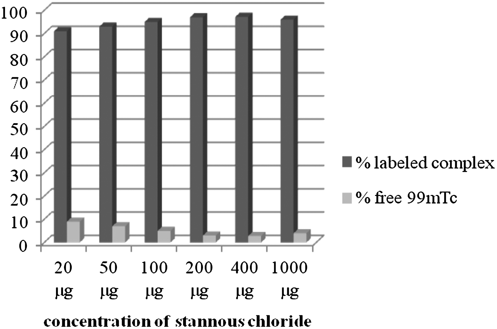
Standardization of concentration of stannous chloride dihydrate for optimum labeling.
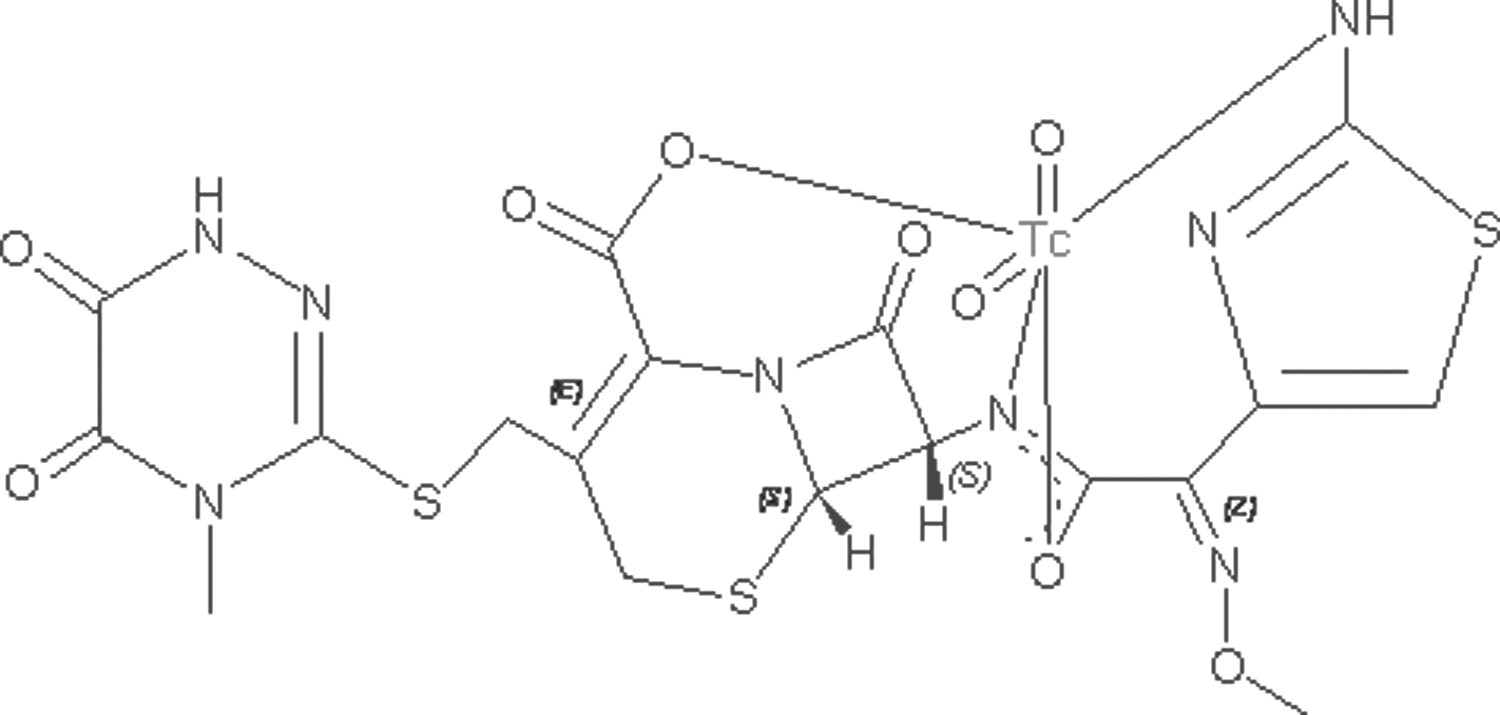
Putative structure of [99m]Tc-doxorubicin with site of [99m]Tc attachment.
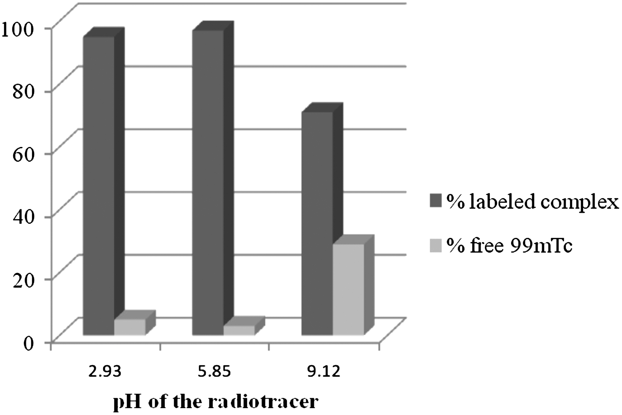
Standardization of pH for optimum labeling.
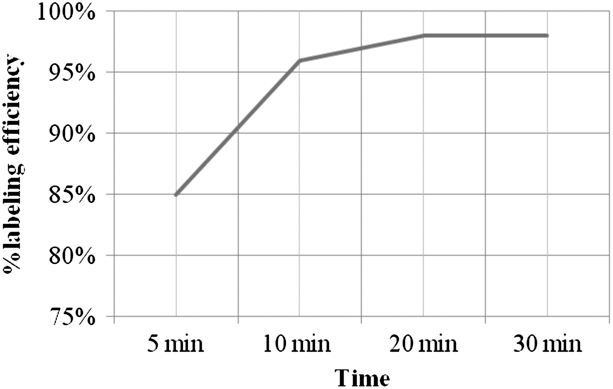
Optimization of incubation time for maximum labeling.
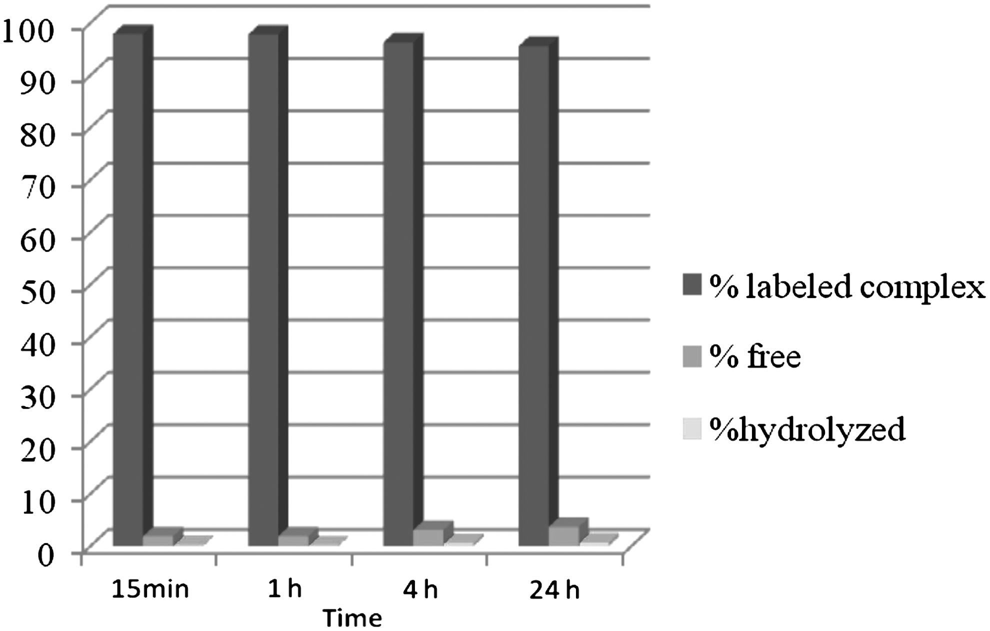
In vitro stability of [99m]Tc-doxorubicin as a function of time.
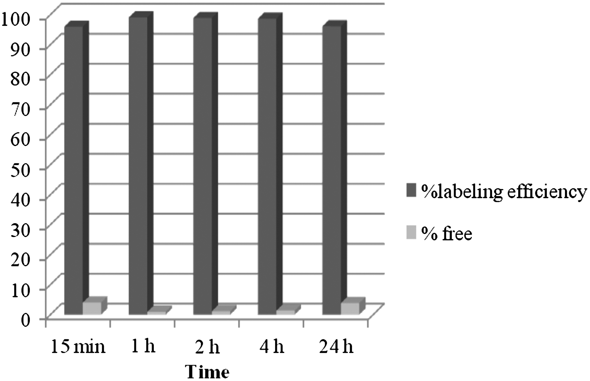
Serum stability of [99m]Tc-doxorubicin as a function of time.
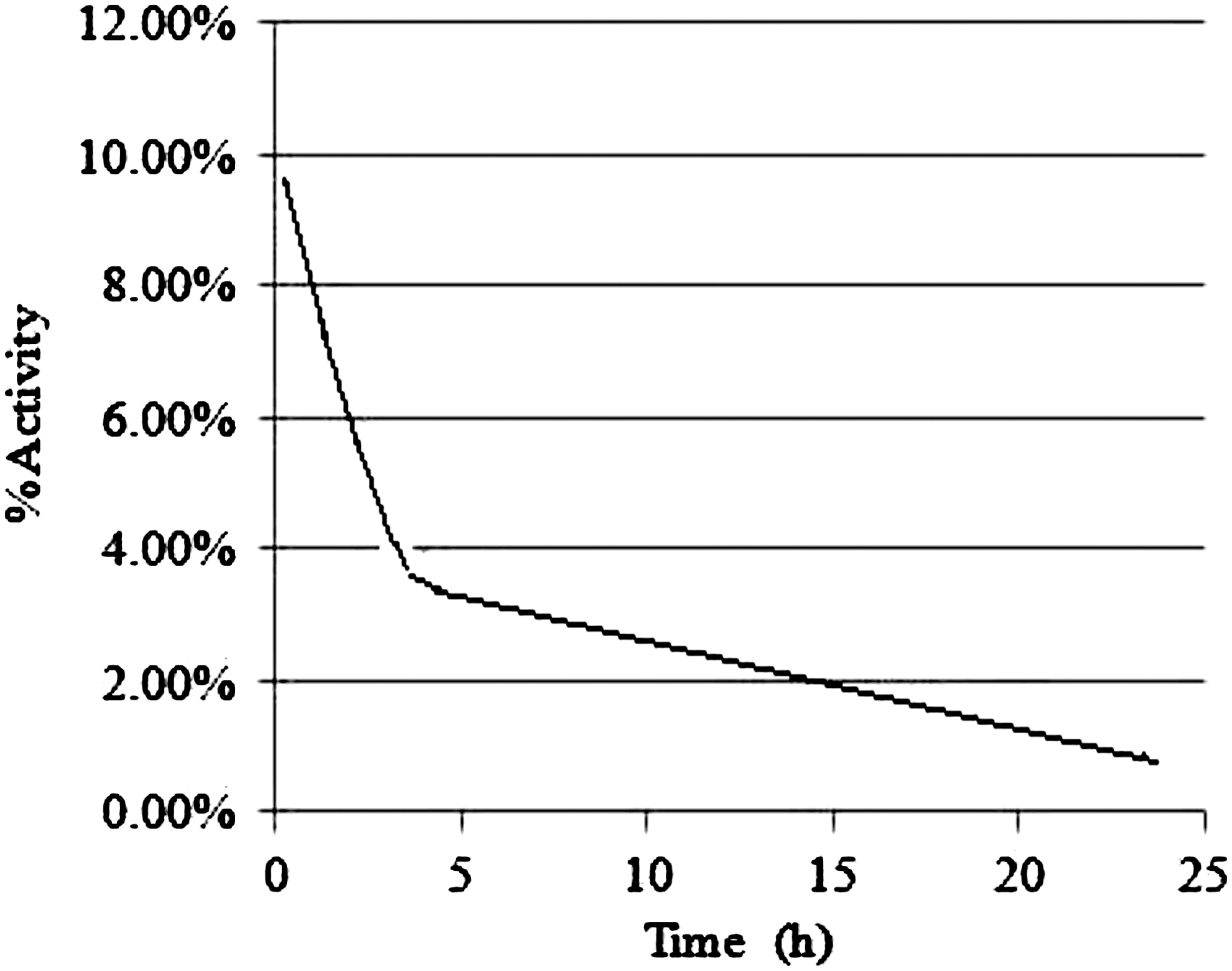
Blood kinetics of [99m]Tc-doxorubicin as a function of time (expressed as percent of ID%/g of blood).
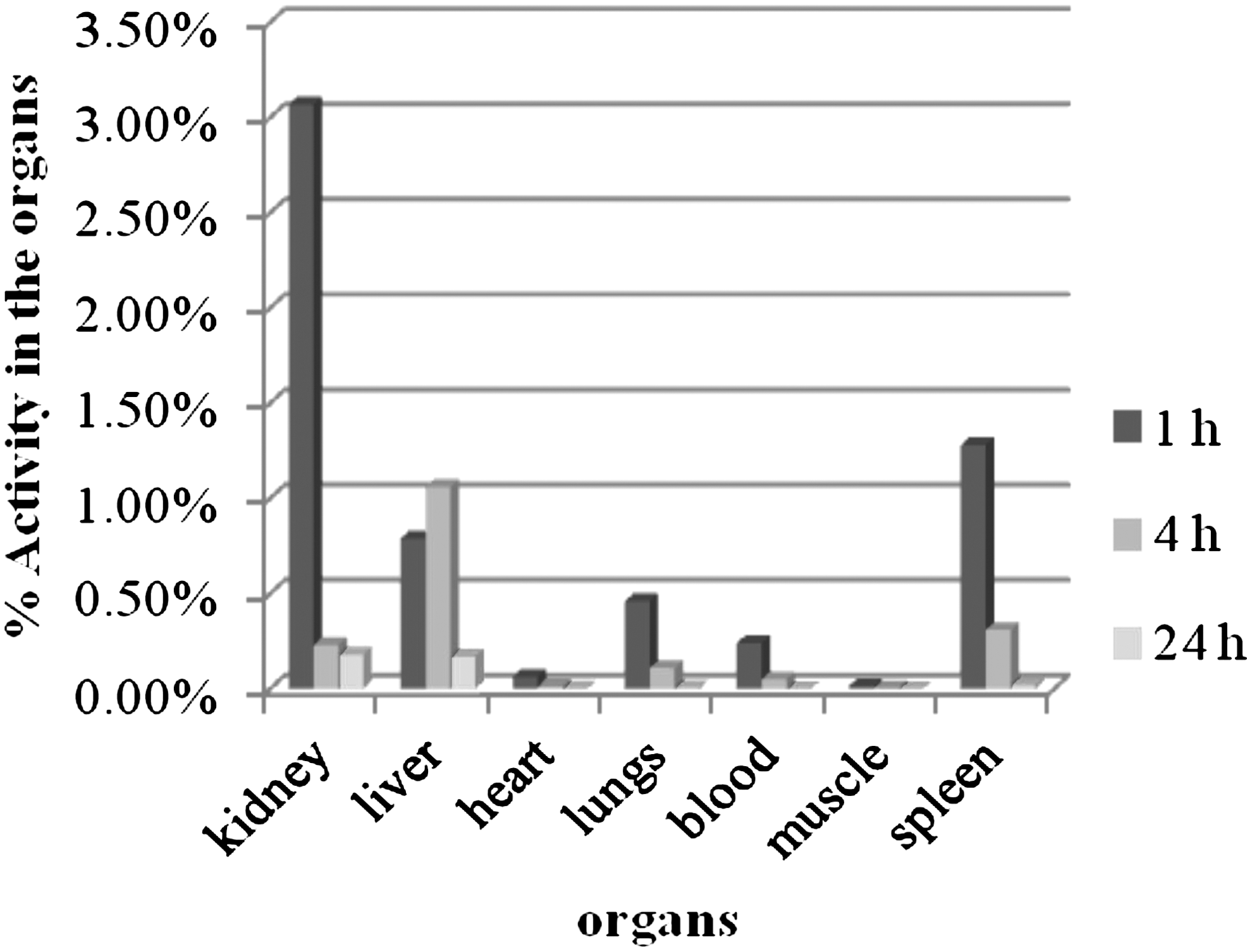
Biodistribution of [99m]Tc-doxorubicin in different organs in rats at 1-, 4-, and 24-hour time intervals (expressed as %ID/g of tissue).
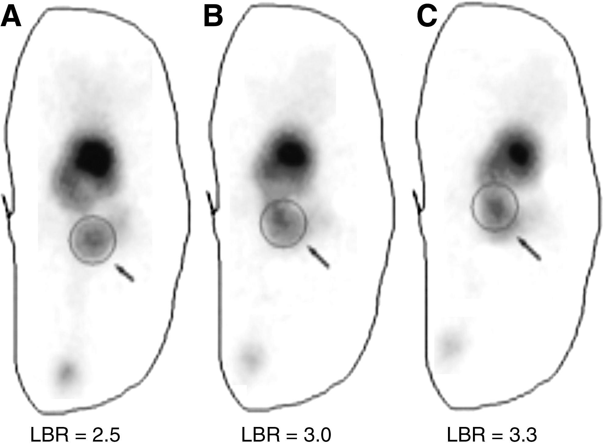
Whole-body static images of tumor-bearing mice at 1.5 hours
Discussion
The rapid multiplication of cancer cells is an exact reflection of the biological behavior of their DNA, whose physical characteristics as well as biological activity (DNA duplication and transcription) are elevated, compared with the DNA of healthy cells. 15 Various tumor-imaging agents have been developed to date using PET- or SPECT-based technology for early detection of both primary and secondary malignancies. However, a deeper probe into the mechanism of uptake reveals that these represent either hypermetabolism ([99m]Tc-MIBI, [201]Tl, Gallium-67 ([67]Ga), etc.) or cellular turnover ([18]F-FDG). 16 –19 This makes them actually cell markers rather than tumor markers and the result is compromised sensitivity and specificity of the available agents for the tumor detection. In the present study, we successfully labeled doxorubicin with [99m]Tc by direct labeling method. The radiolabeled preparation was proved to have an excellent labeling efficiency (>95.0%) and good serum stability, and the preparation remained stable for up to 24 hours. The proposed mechanism of radiolabeling of [99m]Tc is through the coordinate bond formation of amine and hydroxyl groups available on doxorubicin. The preliminary data on the radiolabeling procedure of [99m]Tc-doxorubicin, in vitro quality controls, and its preclinical utility have been reported by us earlier. 20 The complex showed very low lipophilicity that confirmed hydrophilic nature of the complex. However, the compound showed higher protein binding (≈80%) similar to the unlabeled doxorubicin salt. 21 The blood kinetics studies showed biphasic (t1/2 of fast component=2.4 hours, t1/2 of slow component=13.0 hours) blood clearance of the radiotracer. The hydrophilic nature of the radiocomplex may be the possible explanation for the fast component. On the other hand, a slow release of the radiotracer from different body organs into the blood pool may be the possible reason for the slow component. The biodistribution of unlabeled doxorubicin was reported mainly in liver, spleen, kidney, and lungs. 22 The biodistribution studies of [99m]Tc-doxorubicin demonstrated that the radiotracer gets excreted through hepato-renal routes like unlabeled doxorubicin. 23 The accumulation supports that like inert doxorubicin, it gets metabolized in the liver. 24,25 The scintigraphic data analysis indicated that the radiotracer gets concentrated in the tumor lesions and the uptake rises consistently till 4 hours of imaging. The rise in the LBR with time could be due to greater DNA telomerase activity in the tumors. This scintigraphic approach, therefore, could be a powerful tool for cancer detection at early stage especially in developing countries. The technique, however, needs further validation through animal experimentation and clinical studies.
Footnotes
Acknowledgments
The authors are thankful to the Life Science Research Board (LSRB), DRDO, for the funding of the project. Technical help provided by Mr. Komal Saini is greatly acknowledged.
Disclosure Statement
There is no conflict of interest between the authors of this article.