
Abstract
This study aims to develop a new agent, the 32P-chromic phosphate–poly(
Introduction
Prostatic cancer (Pca) is one of the most common diseases in elderly men in western countries and has an increasing incidence in Asian countries. 1 The principal problem arising from Pca is its propensity to metastasize, 2 in which lymph node metastasis is an important factor for the progression of Pca. Harisinghani et al. 3 reported that lymph node metastasis can be found in 41% of diagnosed Pca cases. Among these cases, 70% died of lymph node metastasis. Bader et al. 4 reported that pelvic lymph node metastasis in Pca plays an important role in progress and survival. Furthermore, the biochemical recurrence of Pca, symptomatic progress, and incidence of cancer-related deaths are also related to lymph node metastasis. 4 Small primary tumors usually develop proximal and/or distant lymph node metastasis with a poor prognosis in the early stages. Once pelvic lymph node metastasis occurs, radical therapies fail to improve prognosis and can also lead to side effects. 5 Evidence-based consensus on the treatment of Pca with lymph node metastasis is currently lacking, 6,7 because no clear optimal treatment has been proven safe and effective for treating Pca with lymph node metastasis.
Implantation brachytherapy is designed to implant radioactive seeds directly into tumors and infiltrate nearby tissues (e.g., lymph nodes). Localized ionizing radiation causes tumor vascular networks to shrink with the induction of cellular apoptosis and necrosis of tumor tissues. Treatments of cancers, such as liver, pancreas, 8 head and neck, lung, 9 prostate, 10 breast, 11 and other tumors, are developing rapidly, especially in the approach of biodegradable radioactive implants. Most studies focused on how to effectively improve radioactivity at the target and effective half-life, and how to reduce the distribution to other organs. Some studies explored the degradation or dissolution in seeds. Implantation brachytherapy plays an important role in clinical practice. However, adverse events such as bone marrow suppression, organ perforation, and migration along the blood circulation to the lungs and other organs and tissues may occur. 12 –15
The present study aims to evaluate the therapeutic effect of 32P-chromic phosphate–poly(
Methods
Animals
Six-week-old male athymic BALB/c-nu/nu nude mice (purchased from Experimental Center, Shanghai Institute of Oncology) with a mean body weight of 20 g were raised in a sterile environment at a room temperature of 20°C–21°C and a relative humidity of 30%–60% with 12 hours of sunshine. The mice were fed with food pellets and sterilized water in accordance to the guidelines approved by the China Association of Laboratory Animal Care and the Institutional Animal Care Committee.
Cell culture
The human Pca cell line PC-3M, purchased from the Department of Pathology, Peking University, was cultured in a 1640 medium (GIBCO-Invitrogen) supplemented with 10% fetal calf serum, 100 U/mL penicillin, and 100 mg/mL streptomycin at 37°C in a humidified atmosphere of 5% CO2. The cells were harvested and suspended at an optimal concentration of 2–3×10⁵ cells/mL and allowed to grow until preconfluence before subculture. The cells were subcultured once a week. The cells were detached from culture flasks with 0.25% trypsin, centrifuged at 100 g, and resuspended in a supplemented medium.
Preparation of 32P-CP-PLLA seeds of sustained release
32P-CP-PLLA seeds of sustained release were synthesized using 32P-CP colloid and biodegradable PLLA. PLLA was highly biodegradable and biocompatible in vivo. PLLA manufactured by the controlled-release laboratory of the Hefei University. Different quantities of the PLLA accessories were taken to be mixed with 1 mL 32P-CP colloids (China Hi-Tech isotope Co. Ltd., 1850 MBq/mL) at a specified percentage and dispersed with amount of dehydrated alcohol. Polysorbate as the surfactant was added to modify the compatibility. These ingredients were blended at a constant pace at an ambient temperature (60°C) for 1 hour and dried in a vacuum-drying oven for 9–12 hours. Magnesium stearate of 1 mg was added, served as the surface lubricant before the 32P-CP-PLLA seeds were sealed in lead cans. The diameter and length of the seeds were approximately 0.85–0.9 mm and 2.2–2.5 mm, respectively.
Animal model establishment, grouping, and implantation
The mice were anesthetized with 2% pentobarbital natrium via abdominal injection and immobilized in a supine position under a sterile cover. An incision was made 3-mm above the pubic symphysis, and the bladder and seminal vesicles were carefully lifted to expose the dorsal prostate. Subsequently, 2×10⁶ human Pca cells (PC-3M) were slowly inoculated into the membrane of ventral prostate through the dorsal prostate at an angle of 45° to avoid urethra. If leakage was observed in the peritoneal cavity or urethra/bladder, the mice were excluded in the experiment. The exteriorized organs were then returned into the abdominal cavity. After inoculation, the abdominal muscle layer and skin were closed using a 4-0 absorbable suture and a 4-0 nonabsorbable suture, respectively. After 4 weeks, the tumor mass attained a diameter of 1.0 cm. Thirty tumor nude mice were randomly assigned into five groups (n=6 per group). 32P-CP-PLLA seeds of 3.7, 7.4, 14.8, and 0 MBq (nonradioactive seeds as control) and 32P-CP of 14.8 MBq were implanted to each group through B-type ultrasonic-guided.
Single-photon emission computed tomograph imaging
Seven and 14 days after implantation, the nude mice in each group received a static scan through single-photon emission computed tomography (SPECT) for 20–60 minutes, with a low-energy general-purpose parallel-hole collimator, peak energy of 70 keV, and a window width of 30%. Dynamic distribution of seeds was observed.
Blood toxicity
Blood was collected from the tail vein 0, 7, 14, and 28 days after implantation. White blood cells (WBCs) and platelets were counted for blood toxicity. Changes in the percentage of blood cell count (N%) between 0 and 14 days were calculated as follows: (N 14−N 0)/N 0×100%, where N 14 refers to the blood count of the 14th day, and N 0 is the pretreatment blood count. Simultaneous body weights were also measured for the change in the percentage of weight in the first 14 days as follows: (W%)=(W 0−W 14)/W 0×100%, where W 0 is the pretreatment weight, and W 14 is the weight 14 days after treatment.
Pathology
Three mice in each group were sacrificed 14 days after implantation. Tumor weights were measured, and rates of tumor suppression were calculated as follows: (W 1−W 2)/W 1×100%, where W 1 is the average tumor weight of the control group, and W 2 is the individual tumor weight of the treatment group. Bilateral inguinal lymph nodes and tumor tissues were sampled from several sites. Tissues (1 mm3) without necrosis and normal tissues were immediately fixed in 10% formaldehyde and embedded in paraffin for light microscopy (Olympus) observation. The morphological changes in the tumor were observed, and the rates of tumor cell necrosis in 14 days were calculated (the ratio of death and the total cells).
Electron microscopy
The rest of the samples were prepared routinely in super thin slices of 0.05-μm thick and observed via model H-600 transmission electron microscopy. Ultrastructure changes in the tumor cells and morphology of apoptotic cells were observed.
Statistics
Results were expressed as mean value±standard deviation using a two-tailed Student's t-test; p<0.01 was considered to indicate statistical significance.
Results
32P-CP-PLLA seeds
The radioactive seed was a light green regular cylinder, 0.85–0.9 mm in diameter and 2.2–2.5 mm in length. No significant differences were found between the masses or radioactivity of the seeds among groups.
SPECT imaging
Seven and 14 days after implantation, SPECT static imaging revealed that the seeds mainly gathered in the tumor and abdominal para-aortic lymph nodes of the nude mice. No significant imaging was found in other organs or tissues, indicating local retentions of the agent. Part of the sustained-release 32P-CP could migrate through the lymphatic system into the abdominal aortic lymph nodes, showing a high uptake of radioactivity (Fig. 1).

SPECT static imaging of 7
Blood toxicity
WBCs, PLT, and body weight varied with increasing activities. However, no significant differences were observed in these variables between treated and antithetical groups (p>0.01) (Table 1).
For 14 days, WBCs, PLT, and weight in each treatment group by a single-factor analysis of variance, compared with the control group, were not statistically significant (p>0.01).
Light microscopy
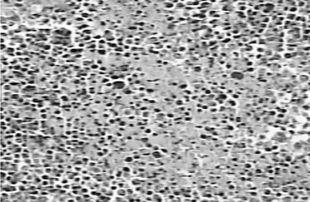
Half of the animals in each group were executed after 14 days. The tumor weight, rates of tumor cell necrosis, and rates of tumor suppression are shown in Table 2. Light microscopy results showed that the nuclei of the nest-shaped tumor cells in the tumor-bearing mice of the control group were pleomorphic and deeply stained. Pathological mitosis and atypia were also observed (Fig. 2). Abdominal aortic lymph nodes were infiltrated, and partial lymphatic structures were damaged (Fig. 3). Medium- and high-radioactivity groups of 32P-CP-PLLA showed necrotic and fragmentary cancer cells, particularly in high-radioactivity groups (Fig. 4). Retroperitoneal lymph nodes were seriously damaged, assuming red-staining fine granules instead of fine granular tissue (Fig. 5). Microscopy results showed that vital organs such as the heart, liver, lungs, and spleen were essentially normal in each group.
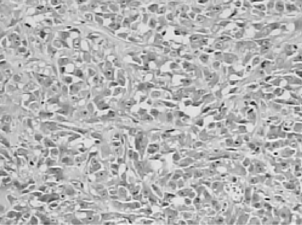
Pathological changes of tumor cells in the control group. (HE×100) Nest-shaped tumor cells and the nucleus were deeply stained.
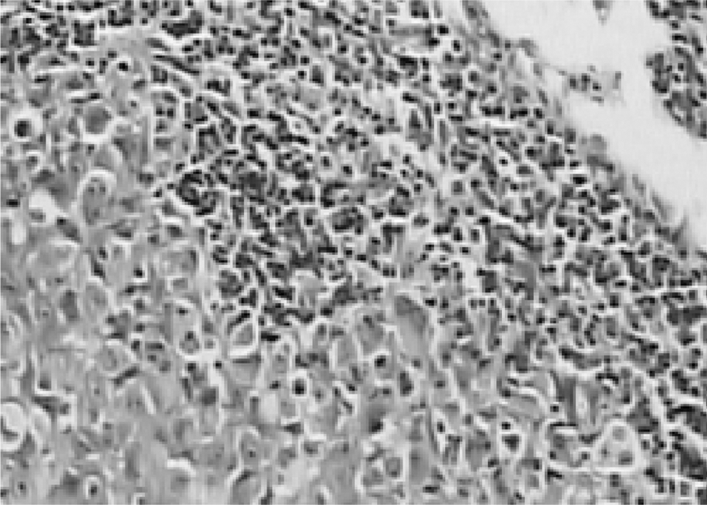
Pathological changes of retroperitoneal lymph nodes in the control group. (HE×100) The lymph nodes were infiltrated by tumor cells.

Pathological changes of transplanted neoplasm in the high-activity group. (HE×200) A great quantity of fragmentary cancer cells and some fine unstructured tissues.
Pathological changes of retroperitoneal lymph node in the high-dose group. (HE×100) Retroperitoneal lymph nodes are heavily damaged, instead of fine granular tissue.
Tumor cell necrosis rate in each treatment group by analysis of variance after square-root transformation, compared with the control group, were statistically significant (p<0.01).
Transmission electron microscopy
In the control group, cancer cells were rich in the rough endoplasmic reticulum. Mitochondrial morphology and structure containing abundant glycogen were normal. The nucleus was round, with exuberant chromosomes and prominent nucleoli. By contrast, no cell structure, but plenty of cell debris, was observed in the group with high dosage of 32P-CP-PLLA. The small number of residual cells includes incomplete karyothecas and disappeared intracytoplasmic cellular organelles (Fig. 6).
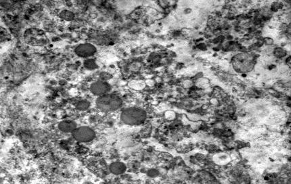
Ultrastructural changes of transplanted neoplasm in the high-dose group (×5000). There was no cell structure, but plenty of cell debris instead. Those small numbers of residual cells were of incomplete karyothecas and had disappeared intracytoplasmic cellular organelles.
Discussion
Pca is diagnosed in more than 500,000 men worldwide each year. 16 Metastasis occurs in 30%–50% of these diagnosed patients, which results in a high mortality. In advanced Pca, 25%–42% patients have metastases, 3,17 which are incurable until now. For the past 10 years, great progress has been made in brachytherapy for solid tumor with 32P-CP colloid and 32P glass microspheres. Main uptake and longer retention of the radionuclides in the tumor tissue were reported in animal studies. The path of radionuclide migration consists of metastatic tumor cells along the lymph circulation. Therefore, the radionuclide can exert a killing effect on lymphatic metastatic foci, with a slight toxic side effect on nearby normal tissues. However, their distributions outside the tumor and reduction of toxic side effects are yet to be solved. Shorter effective half-life of 32P-CP colloids at the target and increase in side effects due to local dosage increase are both important deficiencies. These side effects include higher toxicity to the liver and bone marrow, weight loss of experimental animals, and subcutaneous-bleeding complications of 32P-CP colloidal, and so on. 18 –21
Accordingly, we developed homemade 32P-CP-PLLA seeds of sustained release using 32P-CP colloid and biodegradable PLLA. In contrast to the permanent implantation of 125I titanium metal seeds, PLLA is highly biodegradable and biocompatible in vivo. 22 In addition, PLLA has several advantages. First, its dosage is easy to control. Second, the seeds are confined to the target, and radioactivity is unlikely to overflow. The released 32P-CP is mainly distributed in the seeds and the target organ, which effectively reduces the presence of blind spot. Third, its slow release not only significantly increases the radiation distribution ratios of the implanted target sites to the surrounding normal tissues and effective half-life but also significantly decreases distribution in important organs involved. As a result, their toxic side effects are drastically reduced. Fourth, the seeds would fragment and disappear soon after degradation, without permanent retention in the body. Hence, severe complications could be avoided. Finally, radiation protection procedures are easy to perform. 23 Studies have found that the 32P-CP-PLLA particle could significantly suppress tumor growth in nude mice bearing pancreatic carcinoma. 24 Bioevaluation study of 32P-CP-PLLA particle brachytherapy in a rabbit VX2 lung tumor model has confirmed that the 32P-CP-PLLA particle can efficiently suppress the growth of tumors, which was in agreement with the previous study. 25 In addition, a study about therapeutic potentials of lymph metastasis after 32P-CP-PLLA seeds transplanted into hepatoma H22 xenograft model found that the degradation of the 32P-CP-PLLA seeds in vivo was slow. They could continuously release low-dose 32P-CP particles. A small amount of those could continue to migrate along the lymphatic fluid. Therefore, the treatment of lymphatic metastasis of tumor with 32P-CP-PLLA seeds was secure. 26
The present study aims to examine the therapeutic effect of 32P-CP-PLLA seeds of sustained release for the growth, invasion, and lymph node metastasis of prostate tumor. For this purpose, we established a model of orthotopic PC-3M prostate tumors in nude mice. 27 The findings of the present study showed that the inhibition rates and necrosis rates of tumor cells were positively correlated with radioactivity. Necrosis in solid tumors and lymph node metastases in groups with medium and high activity of 32P-CP-PLLA 14 days after implantation suggested that 32P-CP-PLLA could kill cancer cells not only locally but also nearby lymph nodes. The heart, liver, lung, spleen, and other important normal tissues showed no adverse effects. A certain degree of bone marrow suppression was observed after the seed implantations in the first phase, which were positively correlated with radioactivity. However, bone marrow suppression could be restored to a level close to that of pretreatment after 28 days. That is, radiation injury is reversible in short term. In conclusion, 32P-CP-PLLA seeds are effective for the treatment of Pca with lymphatic metastasis and might be used for Pca patients in a clinic in the near future.
Footnotes
Acknowledgments
Our study was supported by the National Basic Research Program of China (863 program) (2007AA02Z471), National Natural Science Foundation of China (No. 30670588), the key project of Science and Technology Research Support of Nanjing Medical University (2010NJMUZ36), and Science and Technology Research Support of Nanjing Public Health Bureau, Science and Technology Bureau (201108029).
Disclosure Statement
There are no existing financial conflicts.