
Abstract
44Sc is a promising positron emission tomography (PET) radionuclide (T1/2 = 4.04 hours, Eβ+average = 632 keV) and can be made available, using a cyclotron production route, in substantial quantities as a highly pure product. Herein, the authors report on a first-in-human PET/CT study using 44Sc-DOTATOC prepared with cyclotron-produced 44Sc. The production of 44Sc was carried out through the 44Ca(p,n)44Sc nuclear reaction at Paul Scherrer Institut, Switzerland. After separation, 44Sc was shipped to Zentralklinik Bad Berka, Germany, where radiolabeling was performed, yielding radiochemically pure 44Sc-DOTATOC. Two patients, currently followed up after peptide receptor radionuclide therapy of metastatic neuroendocrine neoplasms, participated in this proof-of-concept study. Blood sampling was performed before and after application of 44Sc-DOTATOC. PET/CT acquisitions, performed at different time points after injection of 44Sc-DOTATOC, allowed detection of even very small lesions on delayed scans. No clinical adverse effects were observed and the laboratory hematological, renal, and hepatic profiles remained unchanged. In this study, cyclotron-produced 44Sc was used in the clinic for the first time. It is attractive for theranostic application with 177Lu, 90Y, or 47Sc as therapeutic counterparts. 44Sc-based radiopharmaceuticals will be of particular value for PET facilities without radiopharmacy, to which they can be shipped from a centralized production site.
Introduction
In recent years, 44Sc has been proposed as a novel radionuclide for positron emission tomography (PET).
1,2
The physical properties of 44Sc (T1/2 = 3.97 hours—recently determined as T1/2 = 4.04 hours
3
; Eβ+average = 632 keV; I = 94.3%) are attractive to use this radionuclide for diagnostic imaging purposes, dosimetry, and monitoring therapy response.
2
As a trivalent metal, with similarities to lanthanides and rare earth elements, the chemical properties of 44Sc enable stable coordination with a DOTA chelator.
4
This allows the use of targeting ligands, such as somatostatin analogs (e.g., DOTATOC), which are subsequently employed for therapeutic purposes using 177Lu or 90Y. Moreover, in combination with the therapeutic match, 47Sc (T1/2 = 3.35 days, Eβ-average = 162 keV, Eγ = 159 keV, I = 68%), one of the first truly theranostic radionuclide pairs could be clinically established in the near future.
5
47Sc can be efficiently produced at high-power electron linear accelerators, generating Bremsstrahlung as proposed previously.
6,7
It is proposed that clinically significant activities can be produced in the 48Ti(γ,p)47Sc or 48Ca(γ,n)47Ca
These characteristics give 44Sc a unique potential for clinical PET application and have encouraged several research groups to perform proof-of-concept studies in preclinical settings with a number of tumor-targeted biomolecules. There is, however, only a single report of a clinical application of 44Sc in a patient to date, in which 44Sc was obtained from a 44Ti/44Sc generator. 1
In preclinical studies, there are a number of peptide-based ligands (e.g., somatostatin, 8 –10 bombesin, 11 and arginine-glycine-aspartic acid (RGD) analogs 12 ) and other small-molecular-weight targeting agents (e.g., puromycin, 13 folate, 14 and prostate specific membrane antigen (PSMA) ligands 15 ), as well as antibodies (cetuximab 16 ), which were used for labeling with 44Sc and subsequently investigated with regard to complex stability, determination of tissue distribution profiles, and tumor targeting in mice, including preclinical PET imaging. The most important findings underlined the superiority of DOTA over NOTA, NODAGA, and DTPA for stable coordination of 44Sc and the similar in vitro/in vivo behavior of 44Sc- and 177Lu-labeled compounds in mice. 4,10,14,15 On the other hand, 44Sc- and 68Ga-labeled DOTA conjugates showed slightly different in vitro and in vivo characteristics in some cases, which can be attributed to the different coordination chemistry of the two metals. 8,10,11,15
The preclinical PET image quality was convincing and allowed excellent visualization of tumors, as well as healthy organs and tissues in which the 44Sc-labeled conjugates accumulated. 2,10,12 –16 To compare the image resolution that can be obtained with 44Sc and other PET nuclides, phantom studies were recently performed with a preclinical PET scanner. 17 It was revealed that the resolution of 44Sc was superior to that of 68Ga, but inferior when compared with 64Cu and 89Zr, an assessment in good agreement with the β+-energy of these PET nuclides. For clinical purposes, it is thus expected—and confirmed by the first clinical PET scan with 44Sc 1 —that the image resolution will be similar to what can be achieved with 68Ga-based PET.
In view of a routine 44Sc clinical application, the question of availability of this novel radionuclide has to be addressed. In this regard, production of 44Sc at a cyclotron, by irradiating natural or enriched Ca targets, was previously proposed with the nat/44Ca(p,n)44Sc nuclear reaction. 18,19 Over the last few years, substantial quantities of activity were produced through this method at Paul Scherrer Institut (PSI), Switzerland. 2 A method for the separation of 44Sc from 44Ca target material was developed at PSI and recently optimized. The excellent quality of the resulting 44Sc was shown by high radionuclidic (<1% 44mSc) and chemical purity, enabling the performance of several preclinical studies, in which tumor-targeting ligands were labeled with 44Sc for subsequent in vitro and in vivo application. 10,14 This production method has, therefore, been considered as superior in terms of yield, cost, and feasibility over the use of a 44Ti/44Sc generator, developed by Rösch et al. 1,20
Given the promising decay properties and resolution capability using PET, 44Sc has the potential to be used as an alternative to the short-lived 68Ga (T1/2 = 68 minutes) for PET imaging of neuroendocrine neoplasms. 44Sc may be advantageous with regard to the half-life, as it opens the possibility of a centralized production of 44Sc-labeled peptides and their shipment over several hundred kilometers to hospitals with PET centers that do not have a radiopharmacy on site. This may be of particular interest in countries where cyclotrons are not densely distributed, as is the case in the United States and several countries in Asia and Africa.
In this proof-of-concept study, the authors report on the first-in-human cases of PET/CT imaging, using DOTATOC labeled with cyclotron-produced 44Sc, performed at the Theranostics Center for Molecular Radiotherapy and Molecular Imaging, Zentralklinik Bad Berka (ZBB), Germany. The aim of this study was to demonstrate the proposed concept of using cyclotron-produced 44Sc in a clinical setting. In this regard, the authors set out to investigate the potential of 44Sc-DOTATOC for the management of neuroendocrine neoplasms by demonstrating the physiological and pathological tissue uptake in these patients. Two male patients, previously receiving peptide receptor radionuclide therapy (PRRT) for the treatment of metastatic, pancreatic neuroendocrine neoplasms, underwent post-therapeutic restaging of the disease using PET/CT performed at several time points after administration of 44Sc-DOTATOC.
Experimental
Production and separation of 44Sc
44Sc was produced by the 44Ca(p,n)44Sc nuclear reaction at Injector 2 cyclotron facility at PSI in Switzerland, as previously reported. 2,14 The 72 MeV proton beam was degraded (with niobium) to ∼11 MeV for the irradiation of targets at a beam current of 50 μA over a period of 90 minutes. The separation of the produced 44Sc from the target material was carried out by chromatographic methods using non-branched N,N,N′,N′-tetra-n-octyldiglycolamide (DGA) extraction resin and strong cation exchange (SCX) resin to provide 44Sc at a high quality and quantity, as previously reported. 2 The separated 44Sc was shipped over 600 km to ZBB in Germany.
Radiolabeling of DOTATOC
Radiolabeling of the somatostatin analog DOTA0,Tyr3-octreotide (DOTATOC), was performed in the radiopharmacy at ZBB. Ammonium acetate buffer (0.8 M, pH 5.0) was freshly prepared using ammonium acetate dissolved in ultrapure water (Merck Darmstadt) and concentrated HCl. Radiolabeling was performed by mixing the 44ScCl3 solution (361 MBq, 700 μL) with ammonium acetate buffer (700 μL) and a stock solution of DOTATOC dissolved in ultrapure water (500 μL, corresponding to 250 μg DOTATOC). The reaction mixture was incubated at 91°C for 30 minutes, followed by cooling down to room temperature and neutralization of the solution by addition of sodium phosphate buffer (3 mL, 1 M, pH 7.2). After sterile filtration of the resulting solution, quality control of the radiolabeled product was performed according to the guidelines applied for 68Ga-labeled somatostatin analogs. 21 Radiochemical purity was determined using thin layered chromatography (TLC) and high performance liquid chromatography (HPLC). The final solution was checked with regard to the pH value, sterility, and endotoxins.
Ethical and regulatory issues
44Sc-DOTATOC was administered in compliance with the German Medicinal Products Act (section 13, subsection 2b) and the 1964 Declaration of Helsinki. An institutional review board (European Neuroendocrine Tumor Society-certified tumor board) approved the study and both patients signed a substantial written informed consent before the investigation, which was performed in accordance with the regulations of the German Federal Agency for Radiation Protection. 22 Additionally, before commencement of PRRT, written informed consent forms were signed by the patients for the purpose of collection and storage of their data in the institutional databank and national registry, the anonymized evaluation, and publication of these data.
Patient selection and characteristics
Two male patients were selected for PET/CT investigations using 44Sc-DOTATOC for restaging after five and three cycles of 177Lu- or 90Y-based PRRT at ZBB, respectively. The patient characteristics are presented in Table 1. Patient 1 was 70 years old and suffering from well-differentiated, nonfunctional neuroendocrine neoplasm of the pancreatic tail with hepatic and lymph node metastases. At initial diagnosis, the disease was at stage IIIb. The PET/CT study reported herein was performed for restaging of the disease 6.5 years after his fifth PRRT. Patient 2 was 63 years old and suffered from a well-differentiated, functional neuroendocrine neoplasm of the pancreas head, initial WHO stage IIIb, with bilobar liver and paraaortic lymph node metastases. The PET/CT study reported herein was performed for restaging 27 months after his third PRRT.
PET, positron emission tomography; PRRT, peptide receptor radionuclide therapy.
PET/CT imaging and reconstruction parameters
PET/CT imaging was performed using a Biograph mCT Flow 64 scanner from Siemens Medical Solutions AG. Whole-body PET/CT scans were acquired in the supine position from vertex to feet at various time intervals. The PET scans were acquired using Speed 10 in flow motion. The reconstruction was performed using recon TrueX+ToF software with 3 iterations and 21 subsets decayed, applying decay correction for 44Sc. The images were prepared using a Gaussian filter (4 mm at full width half maximum (FWHM)) and a matrix size of 200. The computed tomography (CT) scans were acquired at 100 kV and at pitch of 1.5. CareDose4D was applied using 0.5-second gantry rotation time, 5 mm slice thickness, and a matrix size of 512 × 512.
PET/CT imaging using 44Sc-DOTATOC
The preparation of the patients included blood sampling for laboratory analysis, the morning before PET imaging, as part of the routine restaging procedure. To accelerate renal excretion of the radiotracer, furosemide (20 mg) was administered intravenously 30 minutes before the administration of 44Sc-DOTATOC, followed by adequate oral hydration (0.75–1.0 L) with bottled drinking water. The patients received an intravenous bolus injection of 78 MBq (Patient 1) and 96 MBq (Patient 2) 44Sc-DOTATOC, respectively. Eight sets of PET/CT scans were acquired with each patient. The time points of image acquisition for Patient 1 and Patient 2 are given in Table 2. Low-dose CT was performed for attenuation correction after oral administration of Mucofalk™ (1 L), a negative intestinal contrast agent.
The indicated times refer to the time after injection of 44Sc-DOTATOC.
Data (image) analysis
The PET/CT images obtained with 44Sc-DOTATOC were interpreted by two physicians (one board-certified nuclear medicine physician and one board-certified radiologist, each with over 10 years of experience in reporting PET/CT studies with somatostatin analogues) independently. A qualitative and semiquantitative evaluation of the scans was performed by analyzing the somatostatin-avid lesions on the transverse, coronal, and sagittal sections. Standardized uptake values (SUVmax) were obtained by drawing circular regions of interest around each somatostatin-avid lesion, which were automatically adapted (40% isocontour) to a three-dimensional volume of interest (VOI), with the software provided by the vendor. The quantitative assessment of somatostatin-avid lesions was obtained exclusively from the transverse sections. Tumor-to-background ratios were calculated using SUVmax for the target lesions and SUVmean for the normal background organs and tissues (right gluteus muscle, liver, right kidney, and spleen) based on a reasonably large VOI drawn over a homogeneous region. Patient 1 had a previous splenectomy, thus, tumor-to-spleen ratios were not determined.
As part of the routine clinical practice of restaging patients with neuroendocrine neoplasms being treated or followed up at ZBB, the PET/CT images obtained with 44Sc-DOTATOC were compared with those of the previous PET/CT performed with 68Ga-DOTATOC. The final written reports of the concurrent ultrasound (US), magnetic resonance imaging (MRI), and CT (bone window) studies documented by the radiologist were also taken into consideration for disease restaging.
Results
Production of 44Sc and synthesis of 44Sc-DOTATOC
Due to logistical issues, the target irradiation and chemical separation took place in the early morning hours, with 2 GBq of radionuclidically pure 44Sc being made available for shipment to ZBB. The transport left PSI ∼1 hour later to arrive in Bad Berka a further 8 hours later, which corresponded to a total decay of two and a half half-lives. The labeling of DOTATOC was performed immediately upon arrival of the 44Sc in the radiopharmacy at ZBB to obtain a specific activity of ∼1.4 MBq/μg peptide. The quality control of the 44Sc-labeled product revealed a radiochemical purity of >99% based on TLC and HPLC methods. After addition of sodium phosphate buffer, the final pH of the injection solution was 6.2. Sterility and absence of endotoxins were confirmed by sterility test and limulus amebocyte lysate (LAL) test.
Pathological uptake
The PET/CT images obtained after administration of 44Sc-DOTATOC demonstrated excellent uptake of the radiopeptide in lesions of both patients (Fig. 1). The best image quality was achieved at 4 hours after administration of 44Sc-DOTATOC (Fig. 2).
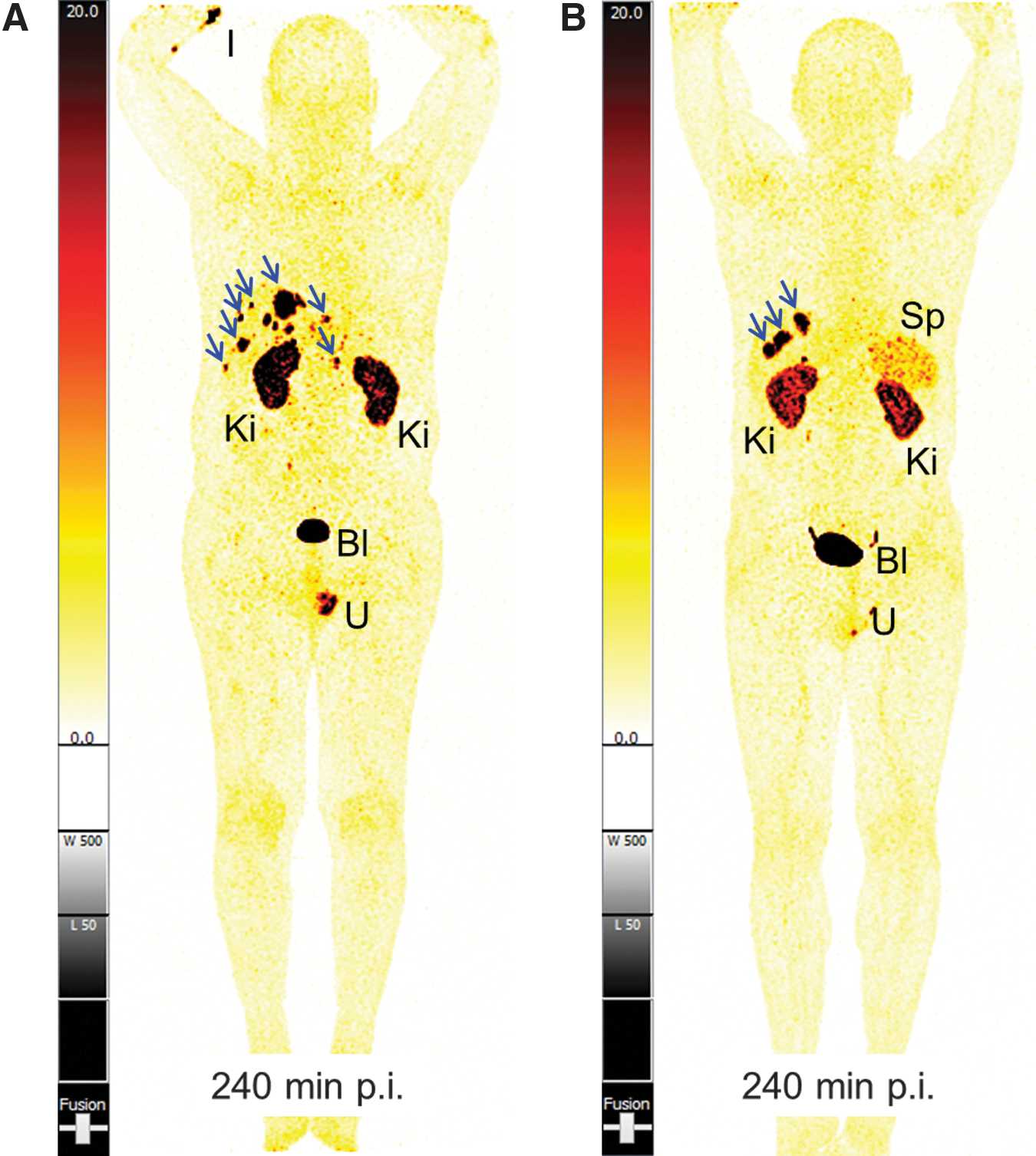
Whole-body PET scans presented as MIP of
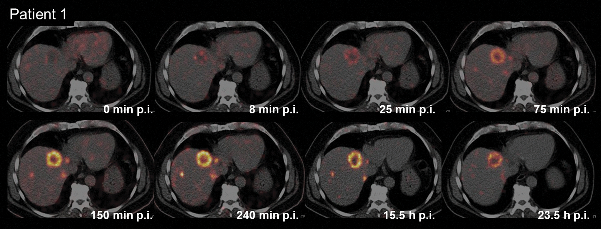
PET/CT images (transverse sections) obtained with Patient 1 at different time points after injection of 44Sc-DOTATOC.
In Patient 1, the scans performed with 44Sc-DOTATOC allowed identification of a new target lesion in the dorsal aspect of segment VII of the liver in comparison with the previous PET/CT images obtained with 68Ga-DOTATOC (Fig. 3A, B). It was coregistered as a lesion with 12 mm diameter on the concurrent MRI (Fig. 3C). As a consequence, the patient was restaged as having progressive disease. Another cycle of PRRT, however, was not indicated due to an insignificant increase in overall tumor burden. The PET/CT scan, performed with 68Ga-DOTATOC for restaging 6 months later, demonstrated stable disease (Fig. 3D). The images allowed detection of the lesion in the dorsal aspect of segment VII of the liver, which was previously seen on the PET/CT scan performed with 44Sc-DOTATOC (Fig. 3C).
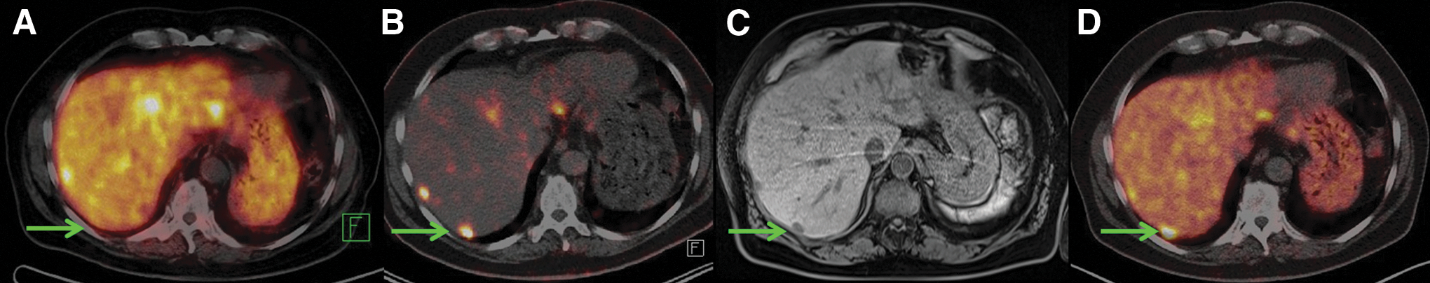
Comparison of serial images of the transverse section of liver, representing the lesion in segment VII (green arrow), obtained by PET/CT imaging of Patient 1 using somatostatin analogs for restaging after the third cycle of PRRT (PRRT-3).
In Patient 2, the PET/CT images obtained with 44Sc-DOTATOC demonstrated progressive disease, with best image quality seen at 4 hours after injection (Fig. 4).
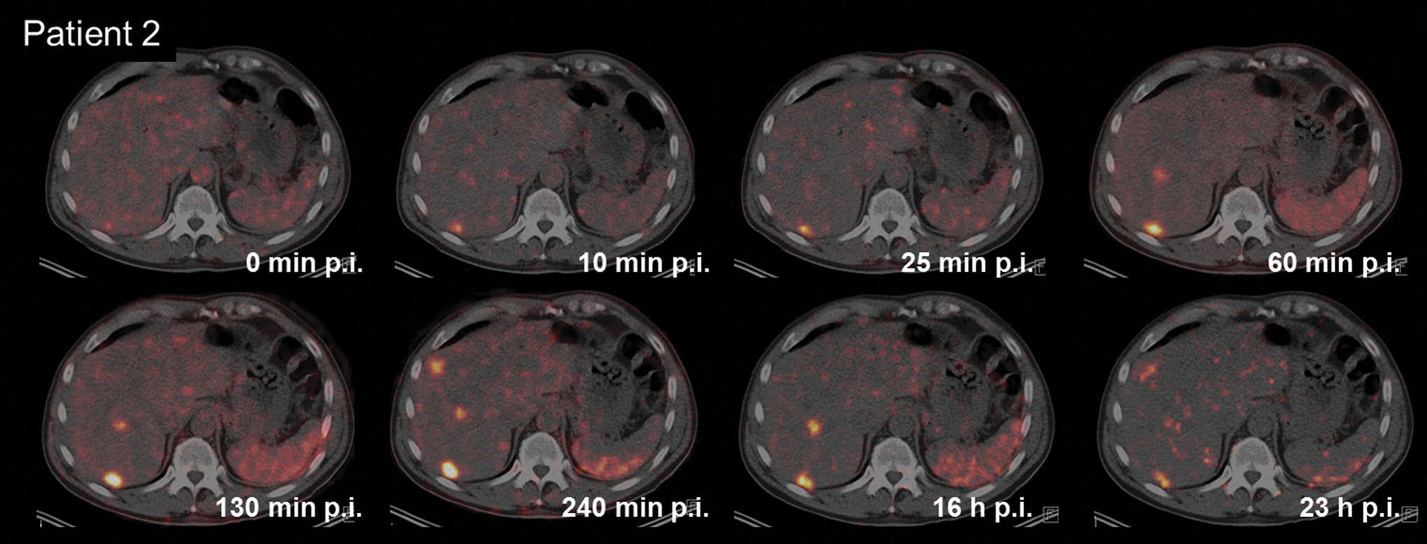
PET/CT images (transverse sections) obtained with Patient 2 at different time points after injection of 44Sc-DOTATOC.
In comparison with the PET/CT scan performed 9 months earlier with 68Ga-DOTATOC, a small new lesion in the apicolateral aspect of segment VIII of the liver was detected with 44Sc-DOTATOC PET/CT (Fig. 5A, B). This lesion was not seen on the concurrent MR and US images obtained within 24 hours after the PET scan (Fig. 5C). In this patient, PRRT was not performed due to the clinically insignificant increase in tumor burden. A follow-up PET/CT performed with 68Ga-DOTATOC for restaging 9 months later, however, enabled detection of this lesion in apicolateral segment VIII of the liver, which was previously seen on PET images obtained with 44Sc-DOTATOC (Fig. 5D).

Comparison of serial images of the transverse section of liver, representing the lesion in segment VIII (green arrow), obtained by PET/CT imaging of Patient 2 using somatostatin analogs.
Physiological uptake of 44Sc-DOTATOC
Early PET/CT images demonstrated uptake of 44Sc-DOTATOC in the normal blood pool, including the heart and blood vessels. Mild, but decreasing, uptake of the radiopeptide was observed as background activity in the normal tissue throughout the image series. Moderate accumulation of radioactivity was visualized in the liver of both patients as well as in the spleen (Patient 2). Uptake and retention of radioactivity in the kidneys, ureters, and the urinary bladder were a consequence of renal excretion of 44Sc-DOTATOC. It is interesting to note that there was no significant uptake of 44Sc-DOTATOC in the pituitary, parotid, and salivary glands, as well as the thyroid and the intestines.
Tumor-to-background ratios
Tumor-to-background ratios were determined for both patients based on SUVmax of the target lesion compared with SUVmean of physiological tissue, including gluteus muscle, liver, and kidney; as well as the spleen for Patient 2 only, measured over a reasonably large area. Increasing tumor-to-background values were observed over the first 4 hours after injection of 44Sc-DOTATOC (Fig. 6A, C). At later time points of image acquisition (15 to 24 hours p.i.), the tumor-to-background ratios decreased (Fig. 6B, D). These findings correlated with the visual assessment of acquired images, with the best image quality seen at about 4 hours after injection of 44Sc-DOTATOC. The relatively high tumor-to-gluteus muscle ratio indicated lower accumulation of radioactivity in the soft tissue compared with the uptake in tumor tissue. The tumor-to-liver ratios were high between 3 and 4 hours after injection, representing the most adequate time period for detection of liver metastases. The tumor-to-kidney ratios remained relatively low, however, without significant variations over time, suggesting constant accumulation of radiopeptide in the kidneys, followed by persistent elimination through urine for the entire duration of investigation in this study. In Patient 2, the tumor-to-spleen ratios remained relatively low with only marginal increase during a 1–4-hour period after injection, which suggests a higher residence time of 44Sc-DOTATOC within the spleen.

Clinical safety of 44Sc-DOTATOC
The imaging procedure was well tolerated by both patients. No clinical adverse effects manifested by symptoms such as nausea, emesis, rash, erythema, pruritus, fever, etc. were observed or reported by either patient during, immediately after, or at follow-up checks of the patients after administration of 44Sc-DOTATOC. Laboratory analyses, indicative for hematological, renal, and hepatic functions, remained unchanged relative to the administration of 44Sc-DOTATOC (Table 3). According to the Common Terminology Criteria for Adverse Events (CTCAE v4.03), there were no significant changes in the relevant blood test values representing the hematological, renal, and hepatic functions at 6 months (Patient 1) and at 9 months (Patient 2), respectively. None of the laboratory values investigated during follow-up blood tests, performed after the PET/CT study with 44Sc-DOTATOC, were significantly altered.
Before = 2 hours before the PET/CT scan performed with 44Sc-DOTATOC; After = at the time of the next study for restaging.
ALT, alanine transaminase; AST, aspartate transaminase; eGFR, estimated glomerular filtration rate; GGT, gamma-glutamyl transpeptidase; MDRD, modification of diet in renal disease.
Discussion
In this proof-of-concept study, the authors were able to demonstrate, for the first time, the application of cyclotron-produced 44Sc in a clinical setting by using it with DOTATOC for restaging of neuroendocrine neoplasms in two male patients. The production of 44Sc at the research cyclotron at PSI allowed separation of sufficient quantities of activity to be shipped over a distance of about 600 km from PSI (Switzerland) to Bad Berka (Germany). This implies that, when produced centrally at a medical cyclotron, 44Sc doses for several patients could be delivered to distant PET imaging centers over several hundred kilometers. Cyclotron production of 44Sc would, thus, be regarded as a more feasible option for routine application of 44Sc-based radiopharmaceuticals compared with the 44Ti/44Sc generator-based production, 1,20 which provides only small activities in larger volumes.
The procedure of radiolabeling DOTATOC with 44Sc was comparable with the method using 68Ga, without the need of development of a new labeling technique. It has to be mentioned, however, that a relatively high amount of peptide was used for labeling, yielding a low specific activity of 44Sc-DOTATOC (∼1.4 MBq/μg) and, as a result, a peptide amount of >55 μg was injected into the patients. In future clinical applications, it is intended to increase the specific activity of 44Sc-DOTATOC to reduce the amount of injected peptide to <50 μg, as is commonly performed with 68Ga-labeled somatostatin analogs. 23,24
In the present study, distinct uptake of 44Sc-DOTATOC was observed in pathological lesions soon after injection (Figs. 2 and 4). The accumulation of 44Sc-DOTATOC increased over time, resulting in very high tumor-to-background ratios and producing PET images of excellent quality. This was particularly evident at later time points — such as the images taken 4 hours after injection of 44Sc-DOTATOC—when the activity was cleared from normal organs and tissues, but retained in somatostatin receptor-expressing lesions (Fig. 1).
Comparison of the 44Sc-based PET images with those obtained with 68Ga-DOTATOC demonstrated that there was no visually significant uptake of 44Sc-DOTATOC in the pituitary and salivary glands, or in the intestines. This should be considered advantageous with regard to detection of small lesions, particularly in the bowel, such as carcinoma of unknown primary of the small bowel. 25 Dosimetric calculations have not yet been performed due to insufficient data for statistical significance, but are planned for the future, particularly in comparison with the biodistribution and dosimetry data obtained by PET imaging with 68Ga-based DOTA-peptides. 26,27 For this purpose, a direct comparison of PET images using 68Ga- and 44Sc-labeled DOTATOC will be necessary within a short time interval.
In this study, visual evaluation of the acquired images indicated a relatively prolonged and higher renal uptake of activity on the delayed PET images, when compared with other organs and tissues. This was also reflected by the relatively low tumor-to-kidney ratios. Due to the longer half-life of 44Sc, when compared with 68Ga, the residence time of 44Sc-DOTATOC in malignant lesions, as well as in the kidneys and spleen, was increased compared with the short-lived 68Ga-DOTATOC. The coemission of high-energy gamma-rays (Eγ = 1157 keV, I = 99.9%) may be considered as a possible drawback for clinical application of 44Sc with regard to the mean and total absorbed renal and spleen doses. Further clinical PET studies using 44Sc-DOTATOC are scheduled to be performed in additional patients, enabling the calculation of the mean and total absorbed dose delivered to the tumor, as well as normal organs, tissues, and whole body. The application of 44Sc-DOTATOC in this study was determined to be safe for human use, as there were no significant changes observed in any of the relevant laboratory parameters representing the hematological, renal, and hepatic functions of these patients following the administration of 44Sc-DOTATOC (Table 3).
44Sc-DOTATOC may be used either as a stand-alone imaging agent or, more importantly, as a diagnostic match to 177Lu- and 90Y-labeled DOTATOC, which are clinically established for the treatment of neuroendocrine neoplasms and other somatostatin receptor-expressing tumors. 28,29 Based on the coordination chemistry of Sc, metal complexes of Sc resemble more closely those of 177Lu and 90Y, making the use of 44Sc-DOTATOC even more attractive compared with 68Ga-labeled radiopharmaceuticals, which may have a different tissue distribution due to different coordination of 68Ga using DOTA. 10,30 It remains to be mentioned that, in the future, it is very likely that 44Sc will be used in tandem with the currently-investigated therapeutic counterpart 47Sc, presenting one of the most intriguing examples of matched radiometals for theranostic application.
Conclusion
44Sc-DOTATOC exhibited favorable properties for PET/CT imaging, as demonstrated by successful restaging of patients with metastatic pancreatic neuroendocrine neoplasms. Further studies with 44Sc are envisaged, with focus on the dosimetry data and direct comparison with 68Ga-based radiopharmaceuticals. Based on the results obtained in this study, it is believed that 44Sc has significant clinical potential for PET/CT imaging, and importantly, as a diagnostic match to 177Lu, 90Y, and 47Sc for theranostic application.
Footnotes
Acknowledgments
The authors thank Dr. Maruta Bunka, Katharina A. Domnanich, Walter Hirzel, and Alexander Sommerhalder for technical assistance at PSI. They express their gratitude to Mostafa Shahinfar, MD, the physicists, and the nursing staff, as well as the nuclear medicine technologists of the Theranostics Center for Molecular Radiotherapy and Molecular Imaging for patient management at ZBB. The study was financially supported by the Swiss National Science Foundation (grants CR23I2_156852 and IZLIZ3_156800).
Disclosure Statement
No competing financial interests exist.