
Abstract
Introduction:
[177Lu]Lu-prostate-specific membrane antigen (PSMA)-617 has emerged as a promising radiopharmaceutical for targeting PSMA in metastatic castrate-resistant prostate carcinoma (mCRPC). We have optimized the radiolabeling protocol for a multidose formulation (27–28.8 GBq equivalent to 6–7 patient-doses) of [177Lu]Lu-PSMA-617 using [177Lu]Lu3+ produced via 176Lu(n,γ)177Lu route with moderate specific activity (0.66–0.81 GBq/μg).
Methods:
[177Lu]Lu-PSMA-617 was synthesized using moderate specific activity [177Lu]LuCl3 (0.74 GBq/μg) with PSMA-617 having metal-to-ligand molar ratio ∼1: 2.5 in CH3COONH4 buffer (0.1 M) containing gentisic acid at pH 4.0–4.5. Human prostate carcinoma cell line LNCaP cell (high PSMA expression) was used for in vitro cell-binding studies and generating tumor xenograft models in nude mice for tissue biodistribution studies. Several batches of the present formulation have been clinically administered in mCRPC patients (single patient dose: 4.44–5.55 GBq per cycle).
Results:
In this study we report a consistent and reproducible protocol for multidose formulations of [177Lu]Lu-PSMA-617 for adopting in a hospital radiopharmacy setting. Although the radiochemical yield of [177Lu]Lu-PSMA-617 was found to be 97.30% ± 1.03%, the radiochemical purity was 98.24% ± 0.50% (n = 19). In vitro and serum stability of [177Lu]Lu-PSMA-617 was retained up to 72 and 120 h after radiolabeling and upon storage at −20°C with a radioactive concentration between 0.37 and 0.74 GBq/mL upon using stabilizer concentration as low as 43–48 μg/mCi. Preclinical cell-binding studies of [177Lu]Lu-PSMA-617 revealed specific binding with LNCaP cells of 17.4% ± 2.4%. The uptake in LnCaP xenografted tumor (nude mice) was 7.5 ± 2.6% ID/g for ∼1.5–2.0 cm3 tumor volume at 24-h post-injection. Post-therapy (24 h) SPECT image of mCRPC patients with prior orchidectomy and various hormone therapy showed specific localization of [177Lu]Lu-PSMA-617 in the tumor region.
Conclusions:
Formulation of a ready-to-use multidose formulation of [177Lu]Lu-PSMA-617 was successfully achieved and the procedure was optimized for routine preparation at a hospital radiopharmacy set-up. High degree of localization of [177Lu]Lu-PSMA-617 in post-therapy SPECT scan and the post-therapeutic response confirms its therapeutic efficacy. Clinical
Introduction
PSMA is a type-2 membrane glycoprotein also known as glutamate carboxypeptidase Il for its enzymatic activity. Overexpression of this enzyme is observed in all prostate cancers, particularly increased with an increase in tumor aggressiveness. 1,2
There are several options to treat prostate cancer through prostectomy, orchidectomy, chemotherapy, and radiation therapy. 3,4 Prostate-specific membrane antigen (PSMA)-based peptide receptor radioligand therapy (PRLT) is an emerging modality for treating prostate cancer.
This PSMA-617 tracer rapidly binds to the PSMA target and undergoes fast clearance from the nontarget organs and the blood stream thereby leading to higher target-to-nontarget ratio, as compared with that achieved while using a monoclonal antibody through J591, 7E11, and so on for the same purpose. Moreover PSMA-617, owing to its target specificity, is expected to provide lower radiation dose to nontarget organs in case of therapy. 5,6 The use of [177Lu]Lu-PSMA-617 provides the dual possibility of carrying out diagnostic scans and therapeutic procedure 7,8 in the theranostic setting.
Lutetium-177 has distinct advantages emerging from the ideal nuclear decay characteristics of the isotope (half-life of 6.7 d), Eβ (max) = 498.3 keV (79.3%), Eγ = 208.4 keV (10.4%), and 112.9 keV (6.2%). The medium energy β− particles emission with a mean range of 0.7 mm and maximum range of 2.1 mm in soft tissue makes 177Lu as promising radioisotope for PSMA-617-based PRLT.
In connection with the routine preparation of [177Lu]Lu-PSMA-617 in a hospital radiopharmacy setting, of a high volume Nuclear Medicine Centre, where on an average of 7–10 patients are administered with therapeutic dose, it was essential to optimize a multidose formulation of [177Lu]Lu-PSMA-617. In addition, to keep the multidose formulation cost-effective, indigenously sourced [177Lu]LuCl3 with medium specific activity of (0.55–0.74 GBq/μg) was utilized.
The PSMA-617 molecule (Fig. 1) (2-[3-(1-carboxy-5-{3-naphthalen-2-yl-2-[4-{[2-(4,7,10-tris-carboxymethyl-1,4,7,10-tetraza-cyclododec-1-yl)-acetylamino]-propionylamino}-pentyl)-ureido]-pentanedioic acid) possess three components: (1) the chelating moiety, (2) the functional linker, and (3) the pharmacophore. The pharmacophore has glutamine–urea–lysine motif that targets the proteolytic domain of PSMA. The aromatic naphthalene-based functional linker region provides the lipophilic accessory pocket for keeping the DOTA chelator remotely away from the glutamine–urea binding site. These polar residues influence the fast clearance kinetics of [177Lu]Lu-PSMA-617 postadministration in patients. 9 In PSMA-617, the hydrophilicity of the DOTA chelating agent has been counterbalanced by introduction of naphthalene moiety in the functional linker that impart liphophilicity to PSMA-617 molecule.

Chemical structure of PSMA-617. PSMA, prostate-specific membrane antigen.
This article describes the preparation of bulk doses of [177Lu]Lu-PSMA-617, using the medium specific activity [177Lu]LuCl3, suitable for PRLT of 6–7 patients, at our center, on a daily basis. Validation of quality control parameters studies related to determination of in vitro stability, preclinical evaluation (in vitro pharmacokinetics and in vivo animal biodistribution) of the formulation were carried out and regulatory approval of DAE's Radiopharmaceutical Committee was obtained before clinical translation in patients. Using [177Lu]Lu-PSMA-617, PRLT were carried out at our center in patients with an advanced metastatic castrate-resistant prostate carcinoma (mCRPC).
Experimental
Materials and Methods
PSMA-617 was purchased from ABX-Advanced Biochemical Compounds, Germany. Ultrapure grade ammonium acetate (CH3COONH4), acetic acid (CH3COOH), and 2, 5-dihydroxy benzoic acid (gentisic acid) were purchased from Sigma-Aldrich. Ultrapure water (Trace SELECT®) was also purchased from Fluka, Switzerland. Poly-ether-sulfone (PES) membrane filters (0.22 μm) were purchased from Merck-Millipore, India. Micropipettes and its tips were purchased from ThermoFisher Scientific, India.
Lutetium-177 was produced by thermal neutron activation (neutron flux ∼1 × 10¹⁴ n/cm2.s) of enriched lutetium target (84% in 176Lu, Isoflex or Russia) on irradiation for 2 weeks in the Dhruva reactor, in India. The irradiated target was then dissolved in 0.01 M HCl by gentle warming to yield [177Lu]LuCl3 solution with specific activity varying in the range of 0.66–0.81 GBq/μg and is supplied as clinical grade radiopharmaceutical precursor. 10
Reagents were prepared using ultrapure grade water inside a laminar flow bench (ISO class 5). All radiolabeling procedures were performed manually inside a lead shielded biosafety cabinet (ISO class 5) using a thermostat-controlled lead-shielded dry heater. Analytical high-performance liquid chromatography (HPLC) was performed using HPLC system equipped with ultraviolet (UV) and radioactive detector [NaI(Tl)], connected in series from Knauer, Germany. Radio thin layer chromatography (TLC) was performed using miniGita and bioscan equipped with BGO (V) and NaI(Tl) radioactive detector from Germany and the United States, respectively. Endotoxin limit (EL) was quantified by gel-clot BET assay method using LAL reagent using a kit supplied by Charles River Laboratories, Inc. (Wilmington, MA), whereas the sterility test was performed by direct inoculation method using soybean caesin digest and fluid thioglycolate media from Himedia, India.
Radiochemical synthesis of ready-to-use [177Lu]Lu-PSMA-617
Radiochemical synthesis of 29.6 ± 1.85 GBq doses of [177Lu]Lu-PSMA-617 (suitable for therapy of 6–7 patients) were carried out in a GMP compliant way in a lead-shielded biosafety cabinet. The description for the radiolabeling of a typical batch is as follows. All reagents were prepared in sterile, pyrogen-free borosilicate glass containers and filtered through 0.22 μm sterile PES syringe filter. [177Lu]LuCl3 (445 μL, specific activity: 0.74 GBq/μg) solution (29.6 GBq of [177Lu]Lu3+, 40 μg, 0.226 μmol Lu) in 0.01 M HCl was added to a sterile glass vial containing a mixture of PSMA-617 solution (588 μL of 1 mg/mL solution in ultrapure grade water, 0.564 μmol, 2.5 equivalent of Lu) and gentisic acid (43–48 μg/mCi) dissolved in CH3COONH4 buffer (0.1 M, 2.2 mL). The pH of reaction mixture was maintained between 4.0 and 4.5. The vial was crimped and incubated at 95°C for 45 min using a thermostat-controlled lead-shielded dry heater. Subsequently, the reaction vial was allowed to cool and attain ambient temperature. The reaction mixture was then diluted with sterile pyrogen-free saline such that the final radioactive concentration (RAC) of the radiolabeled formulation was 1.48–1.85 GBq/mL. The diluted formulation was then filtered through a 0.22 μm syringe filtration assembly into a 20–25 mL sterile glass vial. The radiolabeling protocol described previously was finalized after extensive radiochemical optimization studies.
Quality control and in vitro stability and serum stability studies
The pH of product was measured by observing the color change of a narrow range pH strip after spotting 0.5–1 μL of the final product. Radiochemical purity (RCP) was assessed by HPLC using Eurosphere C-18 reversed-phase column [dimension: 300 mm (length) × 4 mm (diameter); particle size: 5 μm] using gradient mode elution (solvent: 0.1% TFA in H2O and CH3CN; method: 0–4 min 95% water, 4–15 min 95% to 5% water, 15–20 min 5% water, 20–25 min 5% to 95% water and 25–30 min 95% water), coupled with NaI(Tl) and UV (254 nm) detector maintaining a flow rate of 1.0 mL/min. The RCPs were also assessed by silica gel (pore size: 60A°; size: 10 × 125 mm) radio TLC using CH3CN/H2O: 1/1(v/v) as the eluant.
In vitro stability of the product on storage at −20°C was evaluated by TLC and HPLC at 24, 48, and 72 h postradiolabeling. In vitro stability studies were performed for [177Lu]Lu-PSMA-617, at four different RAC ranges (1.48–1.85, 1.11–1.48, 0.74–1.11, and 0.37–0.74 GBq/mL) on storage, at −20°C. EL was quantified by gel-clot BET assay method using lysate, with sensitivity 0.125 endotoxin unit (EU)/mL, at 200 maximum valid dilution.
The serum stability of [177Lu]Lu-PSMA-617 at RAC 0.37–0.74 GBq/mL on storage at −20°C was evaluated by TLC at 24, 48, 72, and 120 h postradiolabeling.
In vitro pharmacokinetic studies of [177Lu]Lu-PSMA-617
Cell binding
Cell-binding study was performed as described earlier. 11,12 In brief, human prostate cancer cell line LNCaP (CLS Cell Lines Service GmbH, Germany), expressing PSMA, were grown in RPMI-1640 medium (Gibco, ThermoFisher) with 10% FBS in 5% CO2 at 37°C. In vitro cell-binding studies was performed by incubating LNCaP cells (5 × 10⁵) in 1 mL of RPMI-1640 (0.2% bovine serum albumin [BSA]) containing the 177Lu-labeled radioligand (0.5 and 1 nM peptide) for 5, 15, 30, 45, and 60 min at 37°C.
In parallel, for quantification of internalization kinetics of [177Lu]Lu-PSMA-617, the culture medium was removed, and LNCaP cells were washed with 1 mL RPMI-1640 (0.2% BSA) and equilibrated in the same buffer for 15 min at 37°C. Then cells were preincubated with 0.5 μM of cold PSMA-617 as blocking solution, followed by incubation with [177Lu]Lu-PSMA-617 (0.5 and 1 nM) for 5, 15, 30, 45, and 60 min at 37°C. All the experiments were performed in triplicate for the time points (control and blocking). Incubation was terminated by placing the cells in ice for 5 min. Cells were then thoroughly washed with 500 μL acidic buffer (50 mM glycine, 100 mM NaCl, pH 2.8) to strip off the radioactivity bound to the receptor surface. The washings and incubation medium were combined, representing the amount of free radioligand. The cells were thoroughly washed with cold phosphate-buffered saline (PBS) (4°C) and internalized activity was obtained by lysing the cells with 500 μL of 1 N NaOH. 11 –13
Quantification of free, total cell bound, and cell internalized radioactivity was performed using a NaI(Tl) γ-counter (Para Electronics, India).
PSMA expression study by flow cytometry
PSMA expression levels were analyzed by flow cytometry using CyFlow® Space, Sysmex (U.S.) system. For analysis, 1 × 10⁶ tumor cells (LNCaP or PC3) were fixed with 4% formaldehyde solution for 15 min and washed thrice with cold PBS (+1% BSA). Cells were then permeabilized for 15 min with 100 μL cell permeabilization buffer (30 μL Triton™ X-100 in 10 mL 0.5% BSA-PBS) per million cells. Cells were then washed thrice with cold PBS (+1% BSA). Permeabilized cells were then stained with 100 μL of PBS (1% BSA) containing PE-labeled antihuman PSMA antibody (1:100) (BioLegend Cat. No. 342503, Clone LNI-17) for 60 min (4°C) in dark. Cells were then resuspended in 500 μL cold PBS and acquired in flow cytometer for 10,000 events at FL2 laser (585/42 nm). 14
In vivo biodistribution studies of [177Lu]Lu-PSMA-617
The degree of receptor-mediated uptake was determined by biodistribution studies of the radio-conjugate in male athymic NCR nude mice (sp/sp, CrTac:NCr-Foxn1nu ) after obtaining necessary regulatory clearance from the Institutional Animal Ethics Committee. Human prostate adenocarcinoma-LNCaP cells (2 × 10⁶ cells/mice), which express PSA were resuspended in 1:1 of 100 μL F12K (Gibco, ThermoFisher) and ECM Gel (Sigma-Aldrich) and injected subcutaneously in nude mice (4–8 weeks) at the proximal flank region. The tumor was allowed to grow for 3–4 weeks until it reached ∼1 cm3 volume.
The radio-conjugate [177Lu]Lu-PSMA-617 (3.7 ± 0.46 MBq with 0.6 nM PSMA-617 peptide per mice) was injected in xenografted mice through tail vein. The biodistribution studies were carried out at 3, 24, and 48 h postinjection for evaluation of the in vivo localization of [177Lu]Lu-PSMA-617, quantification using organ counting on a NaI(Tl) γ-counter. 13
For studying the localization of PSMA-617 in LNCaP xenograft tumor bearing NCR nude mice, [68Ga]Ga-PSMA-11 was prepared (3.7 MBq) injected through tail vein and the positron emission tomography–computed tomography (PET-CT) imaging was carried out. Two-hour postinjection PET/CT analysis was performed under isoflourane sedation using Philips Gemini TOF 16 slice camera (the Netherlands). 11
Clinical studies of [177Lu]Lu-PSMA-617
Therapeutic doses of the present formulation prepared in bulk quantity (multidose) are being injected routinely in selected patients with diagnosed cases of mCRPC patients at our center. Clinical efficacy was evaluated in mCRPC patients using ready-to-use [177Lu]Lu-PSMA-617 formulation prepared in 19 different batches. Each patient was treated with a dose of [177Lu]Lu-PSMA-617 in the range of 4.47–4.81 GBq and were administered within 1 d from the date of formulation. The clinical studies were undertaken as per standardized PRLT protocol. The study protocol was approved by Institutional Scientific and Medical Ethics Committees and was conducted inaccordance with the declaration of Helsinki and good clinical practice. Prior informed written consents were obtained from all the enrolled patients before administering 177LuPSMA-617 therapy. The product was approved by the regulatory body of Department of Atomic Energy, Govt. of India, the Radiopharmaceutical Committee, in their 51st Meeting, for use in patients.
Results
Radiolabeling, quality control, and in vitro stability of [177Lu]Lu-PSMA-617
Using clinical grade [177Lu]LuCl3, 6–7 patient doses of 26.5–27 GBq of [177Lu]Lu-PSMA-617 were formulated (n = 19) with radiochemical yield (RCY) 97.30% ± 1.03%. The [177Lu]Lu-PSMA-617 was found to be clear and pale yellow in color, with pH in the range of 4.0–6.0. The RAC was between 1.48 and 1.85 GBq/mL (1.84 ± 0.06 GBq/mL). The maximum RCY was observed at a molar ratio [177Lu]Lu3+:PSMA-617 of 1:2.5.
The RCP of [177Lu]Lu-PSMA-617 (n = 19), estimated by radio-TLC and radio-HPLC was found to be 99.66% ± 0.28% and 98.24% ± 0.50% with retention factor (Rf) and retention time (Rt) between 0.85 and 0.95 (Fig. 2A) and 11–13 min (Fig. 2B), respectively. The preparation of [177Lu]Lu-PSMA-617 was found to be sterile with EL of <25 EU/mL.

For validating the radio-TLC and radio-HPLC method, the [177Lu]Lu-PSMA-617 was spiked with known concentration of [177Lu]LuCl3. The radio-TLC chromatogram (Fig. 2C) exhibited two peaks with Rf 0.03 and 0.87 corresponding to [177Lu]LuCl3 and [177Lu]Lu-PSMA-617, respectively, whereas radio-HPLC chromatogram (Fig. 2D) exhibited two peaks with Rt 2.41 and 13.0 min corresponding to [177Lu]LuCl3 and [177Lu]Lu-PSMA-617, respectively.
In vitro stability of the [177Lu]Lu-PSMA-617 (radio-HPLC; Fig. 3A) (n = 7) was found to be 98.67% ± 0.39% up to 72 h on storage at −20°C using gentisic acid as the stabilizer (concentration: 43–48 μg/mCi). Radiolytic damage was not observed when [177Lu]Lu-PSMA-617 was stored up to 72 h at −20°C with RAC 0.37–0.74 GBq/mL. Figure 3B documents that the radiolytic degradation of the preparation with RAC (1.48–1.85 GBq/mL) was maximum (RCP <80%) at 72 h postradiolabeling, upon storage at −20°C. Radio-HPLC chromatogram of [177Lu]Lu-PSMA-617 (1.48–1.85 GBq/mL), poststorage at −20°C given in Figure 3C, indicates radiolytic degradation of the product.
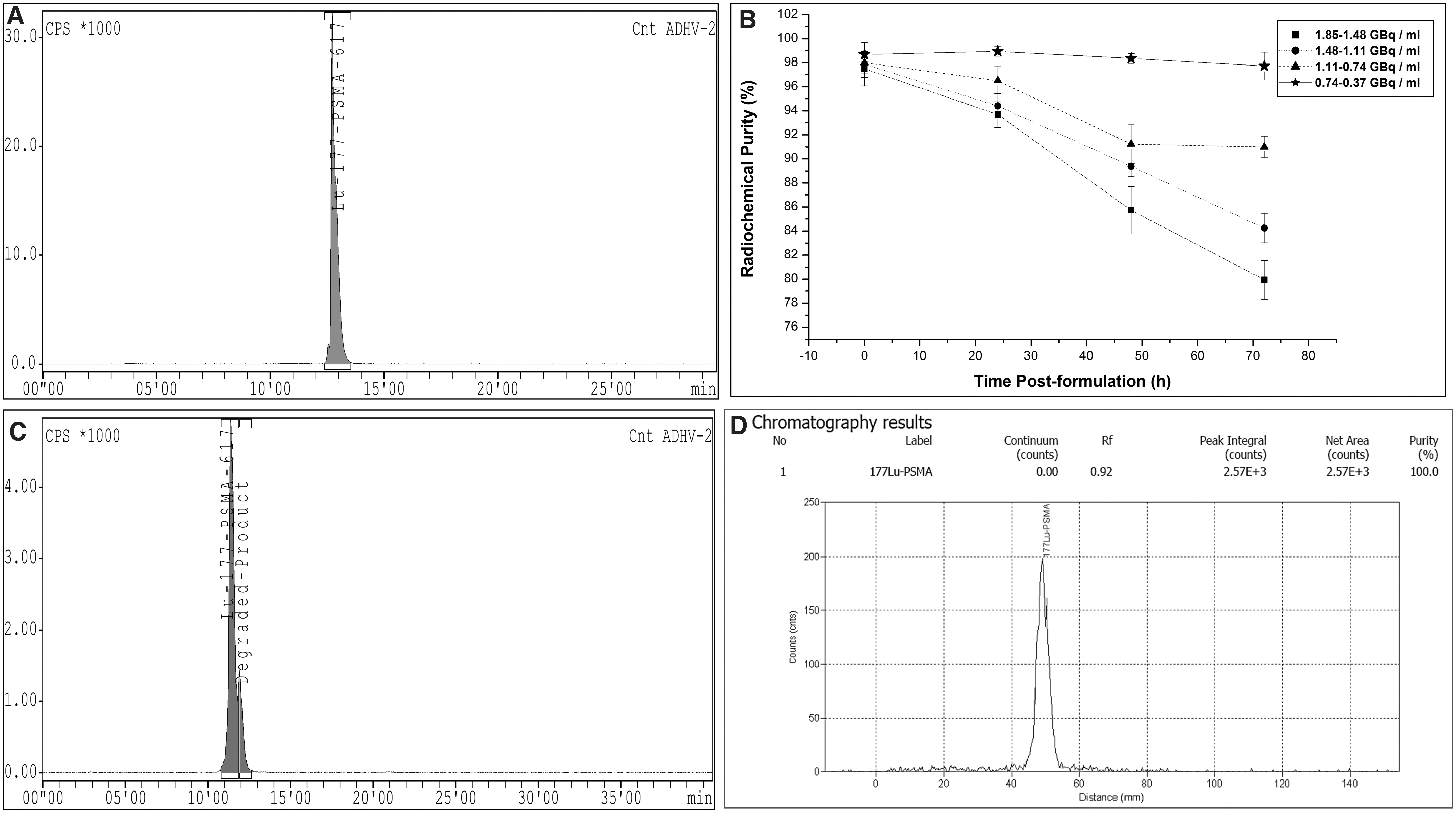
Radio-TLC chromatogram (Fig. 3D) ascertains the in vitro serum stability of [177Lu]Lu-PSMA-617 (n = 4) (RAC: 0.37–0.74 GBq/mL) with RCP of 98.23% ± 0.34% upon 24-h postincubation (37°C) and further storage for 72 h at −20°C.
Table 1 provides the physicochemical and biological quality control results of the formulated [177Lu]Lu-PSMA-617.
Quality Control Results of [177Lu]Lu-Prostate-Specific Membrane Antigen-617
EU, endotoxin unit; HPLC, high-performance liquid chromatography; PSMA, prostate-specific membrane antigen; RCP, radiochemical purity; TLC, thin layer chromatography.
In vitro cell binding and cell internalization of 177Lu-PSMA-617
In vitro cell-binding studies carried out in LNCaP cells showed a rapid and specific binding of [177Lu]Lu-PSMA-617 (0.5 nM) at different time points and reached 17.4% ± 2.4% at 60 min. To distinguish between the internalized activity and the total cellular uptake (cell surface and internalized activity), washing with ice-cold buffer (4°C) containing 10 μM PSMA-617 was carried out for removal of the cell-surface-bound [177Lu]Lu-PSMA-617. The LNCaP cells showed maximum internalization of (13.04 ± 1.57) % in 60 min, when incubated with 1 nM of the radioligand (prepared in bulk scale) in the presence of 10 μM of cold PSMA-617, indicating the unaltered specificity of [177Lu]Lu-PSMA-617 for PSMA antigen. The nonspecific cell binding and internalization were found to be in the range of 2.4% ± 0.45% and 1.2% ± 0.20%, respectively, as observed in PC3 cell line, which has low PSMA expression as per earlier reports. 15 The nonspecific cell binding was found to be 1.98% ± 0.37% in MCF7 cell line, which has low PSMA expression (data not shown).
The total cellular uptake and internalization kinetics of [177Lu]Lu-PSMA-617 in LNCaP cell line, at different incubation times, is given in Figure 4A.
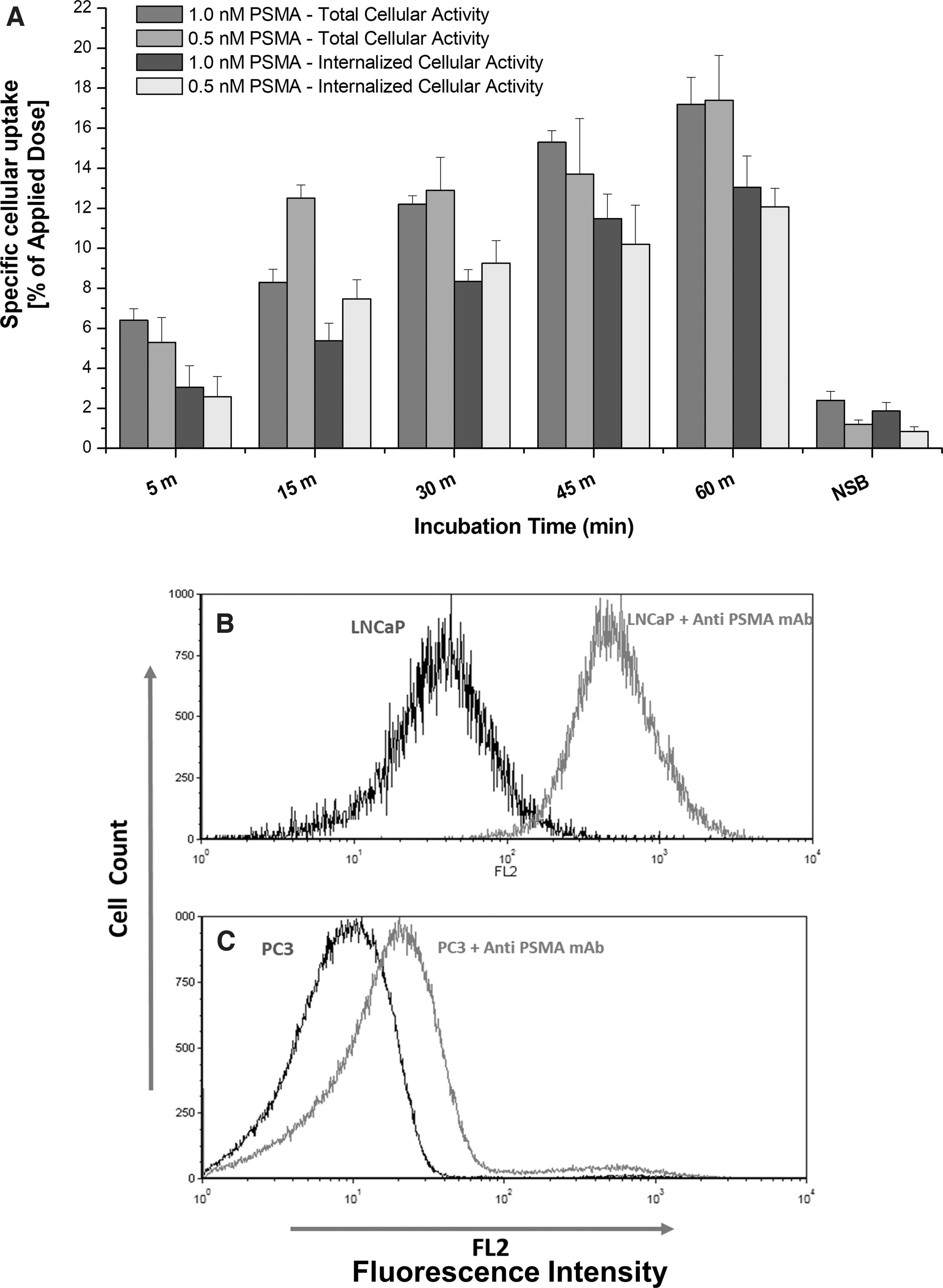
Flow cytometry studies were performed to determine PSMA expression levels of LNCaP and PC3 cells as given in Figure 4B. Significantly strong PE fluorescent signals were observable for LNCaP cell lines (Fig. 4B), whereas no significant signals were observed in PC3 cell line (Fig. 4C).
Biodistribution studies of [177Lu]Lu-PSMA-617 in male nude mice
Biodistribution profiles of [177Lu]Lu-PSMA-617 in male athymic NCR nude mice (sp/sp, CrTac:NCr-Foxn1nu ) carrying LNCaP prostate adenocarcinoma xenograft tumor are given in Figure 5A, B. In the biodistribution study, the uptake in the ∼1.5–2.0 cm3 volume tumor was 7.5 ± 2.6% ID/g at 24 h postinjection of [177Lu]Lu-PSMA-617. The observed uptake was found to be ∼20.28-fold higher than that of blood (0.22 ± 0.02% ID/g), which is statistically significant (p < 0.01). 16
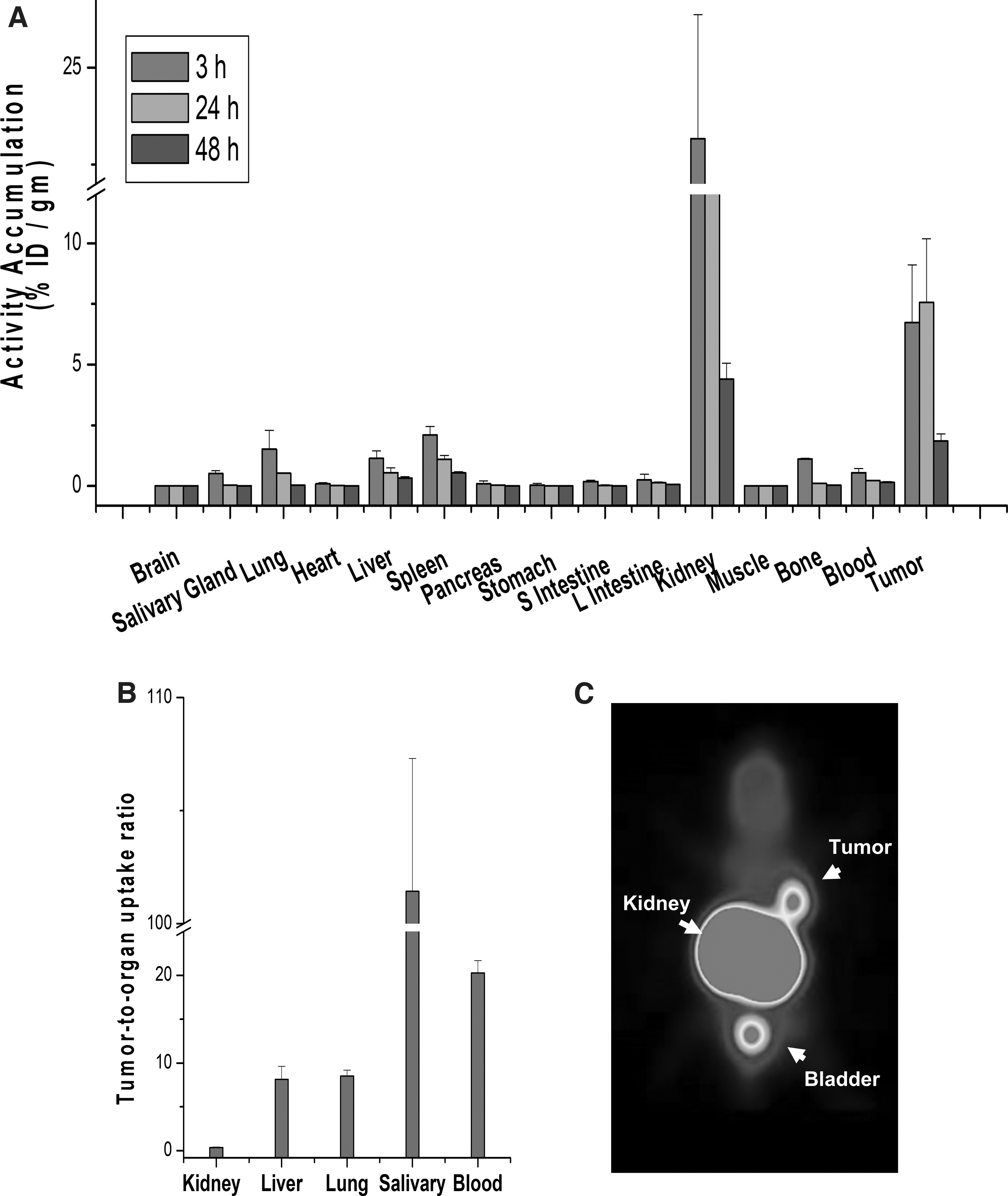
[177Lu]Lu-PSMA-617 was found to be cleared predominantly by the renal route, with 23.1 ± 3.2% ID/g in kidney after 1 h postinjection. Radioactivity in the blood and most of the organs decreased after 24 h postinjection. High uptake and long-term retention of radioactivity were found in the kidney (12.9 ± 2.1% ID/g) at 24 h postinjection. The accumulation of [177Lu]Lu-PSMA-617 in target organs including kidney and LNCaP xenograft tumor bearing NCR nude mice was further ascertained by [68Ga]Ga-PSMA-11 PET-CT imaging (Fig. 5C). Corresponding semiquantitative evaluation (SUVAvg) of tumor-to-kidney ratio shows ∼4.98% and 3.3% uptake at 1 and 3 h, respectively.
Clinical efficacy of [177Lu]Lu-PSMA-617
The primary aim of the study was to prepare a stable mutidose formulation of [177Lu]Lu-PSMA-617. Toward this, to evaluate the therapeutic outcome of using this formulation, representative post-therapy SPECT image of [177Lu]Lu-PSMA-617 (Fig. 6A) in a patient was recorded. Patient evaluation studies showed excellent localization of the radiotracer at the diseased mCRPC site as per the expected distribution. The post-therapy images confirm the excellent therapeutic response.

Discussion
In this report, we presented a cost-effective multidose formulation of [177Lu]Lu-PSMA-617 using a medium specific activity of [177Lu]LuCl3 produced from a domestic reactor. In our radiolabeling protocol, 0.1 N CH3COONH4 buffer solution is equivalent to three times the desired volume of [177Lu]LuCl3 and PSMA-617 taken together. Typically, during multidose formulations, radiolabeling is carried out using 29.6–37 GBq of [177Lu]LuCl3. While carrying out radiochelation with high activity of [177Lu]LuCl3, the two factors that are very critical are (1) the pH of reaction mixture and (2) the stability during radiolabeling process to prevent radiolysis.
Using 2.2–2.5 mL 0.1 M CH3COONH4 buffer containing 43–48 μg/mCi gentisic acid (equivalent to three times the desired volume of radionuclide and peptide precursor), it was possible to maintain pH of the reaction mixture at ∼4.0–4.5 and prevent any degradation of [177Lu]Lu-PSMA-617 owing to radiolysis during the heating process (95°C, 45 min).
In a typical hospital radiopharmacy setup of a high-volume nuclear medicine center, being able to achieve reproducible and consistent multidose formulation of [177Lu]Lu-PSMA-617, using carrier added [177Lu]LuCl3 (specific activity ∼0.74 GBq/μg) with RCP >95%, requires extensive optimization of the radiolabeling protocol with respect to metal [177Lu]:ligand (PSMA-617) ratios (M:L ratios) and incubation temperature.
Initial radiolabeling experiments were carried out at four different M:L ratios ranging from 1.1.5, 1:2, 1:2.5, and 1:3 using low to medium specific activity [177Lu]LuCl3 at 95°C for 45 min at pH ∼4.0–4.5. From Figure 7A, we concluded that at 1:1.5 and 1:2 M:L ratio, the RCY and RCP of the product [177Lu]Lu-PSMA-617 were <90% and <95%, respectively. Hence, for increasing the RCP to >95%, tC18 or C18 solid-phase extraction (SPE)-based purification became essential. However, when the M:L ratio is increased to 1:2.5, RCY and RCP improves to >97% and 98%, respectively. Further increasing the M:L ratio to 1:3 did not result in increase in RCY and RCP.
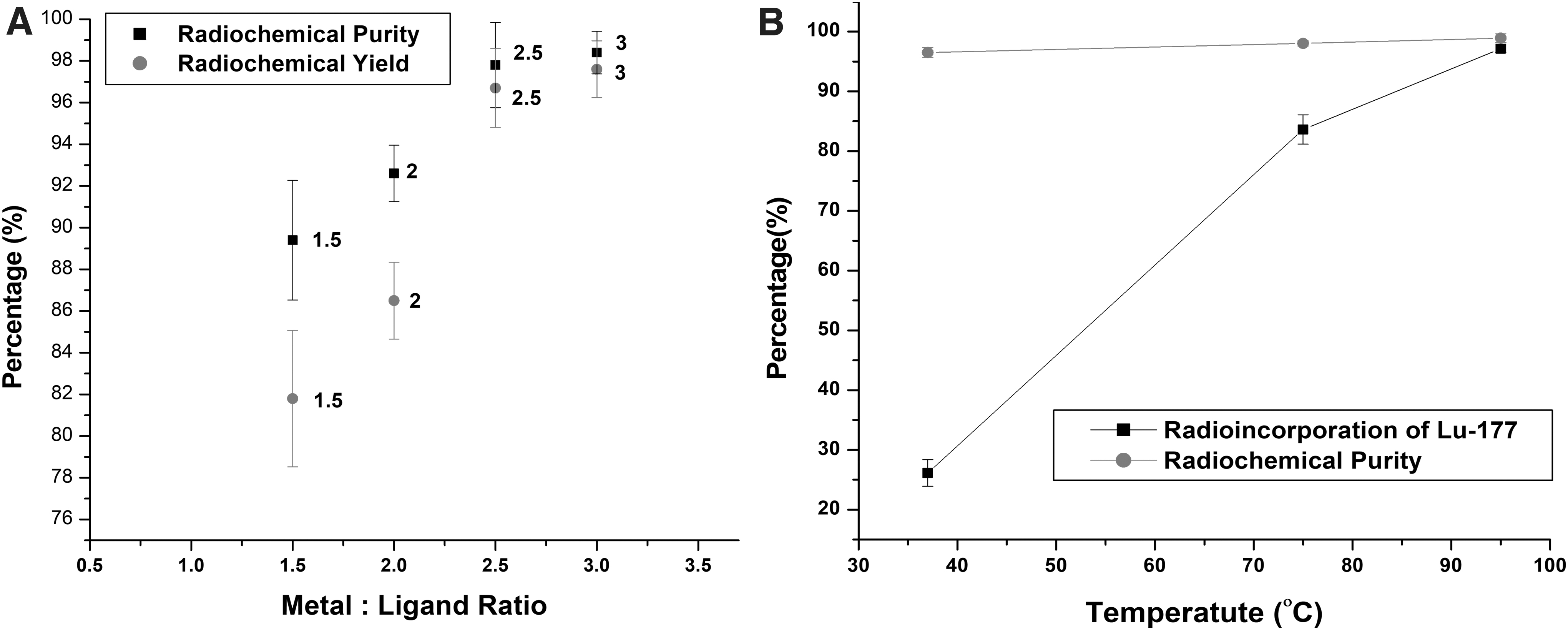
The effect of temperature on complexation of PSMA-617 with [177Lu]Lu3+ were studied at 1: 2.5 M:L ratio. During the standardization process, radiolabeling was carried out at three different temperatures, that is, 37°C, 75°C, and 95°C. PSMA-617 were incubated with carrier-added [177Lu]LuCl3 at each of the above temperatures, for 45 min, at pH 4.0–4.5. From Figure 7B it is observable that at 37°C, chelation of [177Lu]Lu3+ with PSMA-617 was <30%, and became <85% at 75°C. However RCP of the [177Lu]Lu-PSMA-617 (incubated at 37°C and 75°C) was found to be >95% only after C-18 SPE purification, postradiolabeling. PSMA-617 showed temperature-dependent chelation of [177Lu]Lu3+, reaching >95% at 95°C. The DOTA chelator in PSMA-617 requires minimal physical manipulation during coordination with [177Lu]Lu3+ as owing to its inherently constrained geometry, which decreases the entropic loss during [177Lu]Lu3+ coordination. The complex formation using DOTA chelator moieties requires heating at elevated temperatures (95°C), whereas the small molecular weight PSMA-617 moiety remains stable.
In the present radiolabeling method, we have restricted tC18 or C18 SPE purification, postradiolabeling, because tC18 or C18 totally removes the gentisic acid added in the reaction mixture. The removal of gentisic acid from the final product, especially during bulk dose formulations (29.6–31.4 GBq) was a major concern as the [177Lu]Lu-PSMA-617 starts undergoing radiolysis immediately after its formulation whereby the RCP reduces to <95%.
From this study, another parameter that was found to be affected by tC18 or C18 SPE purification, postradiolabeling was the RCY. The RCY of the [177Lu]Lu-PSMA-617 decreases by 10%–12% on SPE purification, which is almost equivalent to 3/4th of a single patient dose (4.81 GBq). In general, in SPE purification, ethanol is used as eluent. Restricting the use of tC18 or C18 purification, the final product was free from residual ethanol content. By adhering strictly to our radiolabeling protocol, we could obviate the tC18 or C18 SPE purification step while maintaining the RCP of the [177Lu]Lu-PSMA-617 between 98% and 99%.
Post-2013, European Association of Nuclear Medicine (EANM) and Society of Nuclear Medicine and Molecular Imaging (SNMMI) has documented practical guidelines on peptide receptor radionuclide therapy (PRRT), in neuroendocrine tumors using [177Lu]Lu-DOTA-TATE/[177Lu]Lu-DOTA-TOC and [90Y]Y-DOTA-TATE/[90Y]Y-DOTA-TOC. Regulatory aspects regarding radiopharmaceutical quality control and its administration to patients were documented in the guidelines. Subsequently in 2019, EANM has specified guidelines on PRLT in prostate cancer using 177Lu-labeled PSMA, wherein the PSMA-PRLT procedure guidelines were very much similar to PRRT, with respect to the quality control parameters and its administration.
Focusing on quality control aspects and administration of [177Lu]Lu-PSMA-617, the EANM guidelines address the following factors: (1) specific activity of carrier added [177Lu]LuCl3 177LuCl3 used for radiolabeling (>10 mCi/μg or 0.37 GBq/μg), (2) RCP of [177Lu]Lu-PSMA-617 (>98%), (3) specific activity of 177Lu-PSMA-617 (1–2 mCi of 177Lu/μg of PSMA-617, and (4) mass of PSMA-617 in radiolabeled product per patient dose (100–200 μg of PSMA-617/patient dose). 17,18
It is imperative to keep in mind the above requirements while formulating therapeutic doses of 177Lu-PSMA-617. Typically, considering a single patient dose to be 4.81 GBq, for radiolabeling of six patient doses 28.86 GBq of the agent needs to be prepared. In the present protocol, we used carrier added [177Lu]LuCl3 (29.6 GBq in 445 μL) with specific activity 0.74 GBq/μg. The starting [177Lu]LuCl3 activity was taken as 29.6 GBq, because the RCY in our optimized protocol was 97%–97.5%. Radiolabeling was carried out by maintaining ligand (PSMA-617) to metal (177Lu3+) molar ratio ∼2.5:1.
In the present radiolabeling protocol for multidose formulations, 40 μg of [177Lu]Lu3+ (0.226 μmol, 29.6 GBq) and 588 μg of PSMA-617 (0.564 μmol) were taken. Postfiltration 28.86 GBq of [177Lu]Lu-PSMA-617 in 15 mL of saline was obtained because the concentration of peptide (PSMA-617) in the final product was 40 μg/mL and the final concentration of [177Lu]Lu3+/μg of peptide amounts to ∼1.32 mCi or 0.048 GBq.
The above data translate to a patient receiving 100 μg of peptide postadministration of [177Lu]Lu-PSMA-617 (4.81 GBq in 2.5 mL), which is in accordance with EANM guidelines of PSMA-PRLT. In principal, specific activity of the carrier added [177Lu]LuCl3 and metal/ligand molar ratio determines the amount of peptide received by a patient after single cycle of [177Lu]Lu-PSMA-617 therapy.
The stability of [177Lu]Lu-PSMA-617 on storage at −20°C was determined with a view to adjudging its suitability for patient administration, after 24 h postformulation Toward this, the RAC of [177Lu]Lu-PSMA-617 plays a critical role in maintaining the stability of the product.
As per the present radiolabeling protocol, the RAC is maintained between 1.48 and 1.85 GBq/mL on the day of formulation. The [177Lu]Lu-PSMA-617 with RAC between 1.48 and 1.85 GBq/mL when stored at −20°C for 24 h, results in the RCP becoming <95%. Since postradiolabeling, RAC of the final formulation is an important factor that determines the radiolytic degradation of the product during storage at −20°C.
To circumvent this problem, the RAC of remaining doses of [177Lu]Lu-PSMA-617 were reduced to 0.37–0.74 GBq/mL by aseptically adding sterile pyrogen free 0.9% NaCl before storage at −20°C. On the successive days (24, 48, and 72 h postradiolabeling), the RCP of [177Lu]Lu-PSMA-617 was found to be between 98% and 99%.
The volume of [177Lu]Lu-PSMA-617 (RAC 0.55–0.74 GBq/mL) were further adjusted with sterile saline just before administration, such that each patient undergoing PRLT receives the required 100–200 μg of PSMA-617 postsingle cycle of PRLT.
In vitro cell-binding studies carried out in LNCaP cells showed high specificity of [177Lu]Lu-PSMA-617 for PSMA. In the in vitro experiments, the extent of cell binding was 17.4% when [177Lu]Lu-PSMA-617 was prepared with moderate specific activity (0.74 GBq/μg) [177Lu]LuCl3. The results of cell binding were comparable with those obtained with [177Lu]Lu-PSMA-617 prepared using high specific activity [177Lu]LuCl3. In vitro cell internalization and competition binding assays ascertain the specificity and efficacy of the radiopharmaceutical. Bioevaluation studies in LNCaP xenograft tumor-bearing nude mice further confirmed the specificity of the product. High uptake in the 1.5–2.0 cm3 volume tumors and long retention thereof ascertains the effective dose delivery to the prostate cancer tissue.
There was excellent symptomatic response with improved good health-related quality-of-life (HRQoL) scores and reduced levels of serum PSA in a sizeable fraction of mCRPC patients, thereby confirming excellent therapeutic efficacy of the [177Lu]Lu-PSMA-617. Desirable localization of [177Lu]Lu-PSMA-617 in lesions observed in post-therapy SPECT scan (24 and 72 h) of the patient proves the excellent in vivo stability of the formulations.
Conclusions
An optimized protocol for multidose formulation of ready-to-use [177Lu]Lu-PSMA-617 for therapeutic use in mCRPC patients was successfully accomplished using moderate specific activity 177Lu3+ produced by direct neutron activation [176Lu(nγ)177Lu], in a medium flux research reactor. The stability of the formulation was ascertained up to 72 h postformulation on storage at −20°C with RAC between 0.37 and 0.74 GBq/mL. The bioefficacy of the product was established by in vitro cell-binding studies in androgen-sensitive human prostate adenocarcinoma LNCaP cells and by in vivo biodistribution profile in male nude mice bearing LNCaP prostate carcinoma xenograft tumor. The [177Lu]Lu-PSMA-617 has also been confirmed to be clinically efficacious with respect to post-therapeutic responses in mCRPC patients (reduced levels of serum PSA, good HRQoL scores, and excellent localization in tumors). The results document the relevance of preparing multidose formulations of [177Lu]Lu-PSMA-617 in a hospital radiopharmacy set up toward offering high-volume, cost-effective PRLT in patients with mCRPC, on a routine basis.
Footnotes
Acknowledgments
The authors thank the staff of the Scintigraphy Section, Clinical Nuclear Medicine Section, Hospital Radiopharmacy Section and the animal house facility of RMC, BARC, for providing the facilities for carrying out the work. The authors also gratefully acknowledge the support and encouragement received from Director, Medical Group, BARC.
Disclosure Statement
There are no existing financial conflicts.
Funding Information
No funding was received for the work.