
Abstract
Abstract
Magnetic chitosan nanospheres immobilizing cobalt and manganese tetraphenylporphyrins were synthesized and employed as catalysts for the degradation of a model contaminant, methyl orange (MO) under irradiation with a high-pressure mercury lamp, an iodine tungsten lamp, or natural sunlight (NSL), respectively. Scanning electron microscopy showed that the prepared catalysts had a spherical uniformity with average diameters of ca. 300 nm. Catalytic results illustrated that these magnetic metalloporphyrin nanospheres were highly efficient and recyclable catalysts which can be used to degrade MO under NSL, and the nanospheres with Co(II) porphyrin were more active than those with Mn(III) porphyrin.
Introduction
E
In recent years, metalloporphyrins as a new catalyst started attracting environmental chemists' interests. Some metalloporphyrin complexes have been found to photocatalyze the organic substance oxidation by O2 under mild temperature and pressure (Xiang et al., 2011). Previously, we have researched the catalysis of cyclohexane with various metalloporphyrin magnetic nanospheres and found that these magnetic nanospheres could oxidate organic substance with a high efficiency and could be easily reused (Fu et al., 2008, 2009a, 2009b). This gives us the inspiration to resolve the azo dyes wastewater by metalloporphyrin magnetic nanospheres.
In the present work, we synthesized alternative catalysts based on magnetic chitosan-supported metalloporphyrin (M=Mn, Co) nanospheres, designed as MCSMnTPP and MCSCoTPP, respectively, and studied their photocatalysis behaviors to methyl orange (MO) solution, which is a representative azo dye. The recovery and reuse of these nanospheres were comparatively researched.
Experimental Protocol
Materials
Chitosan (molecular weight: 161.16 monomer, deacetylation degree 95%) was commercially purchased from Sigma. Methyl orange (MO; chemical structure shown in Fig. 1) was of analytical grade and was used without further purification. Ammonia, FeCl3·6H2O, FeCl2·4H2O, and glutaraldehyde were commercially obtained from Guangzhou Reagent Company and of analytical or reagent grade. All the solutions were prepared with double-distilled water.

Chemical structure of methyl orange (MO).
Preparation of catalysts
Preparation of Fe3O4 particles
The magnetic fluid used in this work, Fe3O4 particles, was prepared by chemical coprecipitation of ferrous chloride and ferric chloride. 250 mL 4% (v/v) fresh ammonia was refluxed in a 500 mL round flask at 80°C. 13.6 g FeCl3.6H2O and 5.0 g FeCl2.4H2O dissolved in 60 mL distilled water were added dropwise, and the mixture was then refluxed for 2 h. After cooling the reaction mixture to room temperature, the products were separated in a magnetic field, washed with H2O to neutrality, and then redispersed in distilled water. The X-ray diffraction (XRD) pattern of the Fe3O4 particles is shown in Fig. 2. The average diameter of the Fe3O4 particles obtained from XRD is ∼8.96 nm by the Scherrer formula. (Khorsand Zak et al., 2012).

X-ray diffraction pattern of Fe3O4 particles.
Preparation of metalloporphyrins
Metalloporphyrins, manganese tetraphenylporphyrins (MnTPP), and cobalt tetraphenylporphyrins (CoTPP) were prepared, purified, and characterized according to a previous report (Liu et al., 2007). A typical reaction was conducted as follows. Benzaldehyde was dissolved in propionic acid and then, pyrrole was added dropwise. The mixture was refluxed for 1 h at 120°C and then cooled to 80°C. Ethanol was added to the cooled mixture and filtered. The purple filter cake was tetraphenylporphyrins (TPP). TPP was metalized by mixing Mn(Ac)2 or Co(Ac)2 and TPP in HAc solution. The mixture was stirred at 65°C for 8 h. After washing the mixture to neutrality with H2O, drying over anhydrous Na2SO4, and concentrating via rotary evaporation, the residue was chromatographed on a silica gel column using CHCl3 as eluent.
Preparation of magnetic chitosan-supported metalloporphyrins
In a 250 mL round-bottomed flask equipped with a reflux condenser, a total of 3.0 g magnetic fluid, 5.0 g chitosan, and 40 mg metalloporphyrins were mixed in a mixture of 20 mL distilled water and 10 mL glacial acetic acid. The mixture was stirred at room temperature for 20 min. Then, 1.5 mL glutaraldehyde was slowly added dropwise, and the mixture was stirred at 70°C for 20–24 h under an N2 atmosphere. The product was washed with 1 M HCl solution to remove the unenclosed Fe3O4 and then with chloroform to remove the residual metalloporphyrin until no porphyrin could be detected in the chloroform, as measured on a ultraviolet–visible light (UV-vis) spectrophotometer. The final product was separated by applying an additional magnetic field (0.42 T) and dried at 65°C for 24 h in vacuum. Blank magnetic chitosan nanospheres were similarly obtained, without adding metalloporphyrins.
Apparatus
XRD patterns with diffraction intensity versus 2θ were recorded using an X-ray diffractometer (XRD, D8 Advance) using nickel-filtered copper radiation (Cu-Kα, λ=0.15418 nm) over the 2θ range 10–90° at room temperature. Scanning electron microscopy (SEM) analyses were performed with a JSM-6330F field emission scanning electron microscope. Transmission electron microscope (TEM) analyses were performed with a JEM-2010HR transmission electron microscope. The specimens for SEM and TEM were prepared by dispersing the final powders in ethanol with 20 min ultrasonication and placing a drop of this mixture on carbon-copper grids. UV-Vis spectra were recorded on a Shimadzu UV-3150 spectrophotometer. An Iris high-resolution (HR) inductively coupled plasma (ICP)–atomic emission spectrometer was used to determine the contents of metalloporphyrins and Fe3O4. Magnetic measurements were performed by using an XL-7 magnetic property measurement system.
Degradation rates of MO solutions were periodically scanned by Perkin-Elmer-Lambda-850. Catalysis of MO was carried out in a specially constructed reaction vessel (Fig. 3). A cylindrical quartz photochemical reactor with a volume of 50 mL was utilized for photocatalytic reaction. The lamp and the reactor maintained a distance of about 10 cm. Cooling water was used to keep the reaction at room temperature. Irradiation was carried out with a 125 W high-pressure mercury lamp (HPML, Guangzhou Bang Wo Electronics Co., Ltd.) or a 300 W iodine tungsten lamp (ITL, Guangzhou Bang Wo Electronics Co., Ltd.). The experiments under natural sunlight (NSL) were performed in May 2012 at Zhongshan, located at 113o53′ E, 22o11′ N. The supported metalloporphyrins were recovered from the catalytic system by magnetic settlement and then reused under identical conditions.
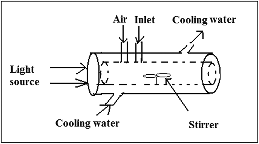
Illustration of the reaction apparatus.
Recommended procedures
Photocatalytic oxidative degradation of MO using magnetic chitosan-supported metalloporphyrins as catalysts in aqueous solutions was studied under room temperature and normal atmosphere pressure. The catalytic system consisted of corresponding catalysts and MO aqueous solution, which were added into the quartz reactor and then stirred with a magnetic stirrer before irradiation with different light sources (HPML, ITL, or NSL). A simultaneous air flow rate of 0.8 L/min was continuously sparged into the solutions with the intention of adding sufficient oxygen and retaining a stable reaction. The absorbance alternatives of the samples were standing aside for 5 min and were measured at 465 nm with glass cells. The degradation rate of MO in the reaction process could be calculated by the following formula:
where At is the absorbance of MO measured at 465 nm at time t, and A0 is the initial absorbance before the reaction.
Results and Discussion
Characterization
The morphology of the magnetic chitosan-supported metalloporphyrins was obtained by SEM, with a satisfying spherical uniformity and an average diameter of ∼300 nm. Meanwhile, the internal structures of these nanospheres were given by TEM, from which one can clearly find the Fe3O4 magnetic core embodied in the chitosan shells. A negligible difference was found between the nanospheres with different metalloporphyrins. Figure 4 gives the SEM and TEM images of MCSCoTPP.
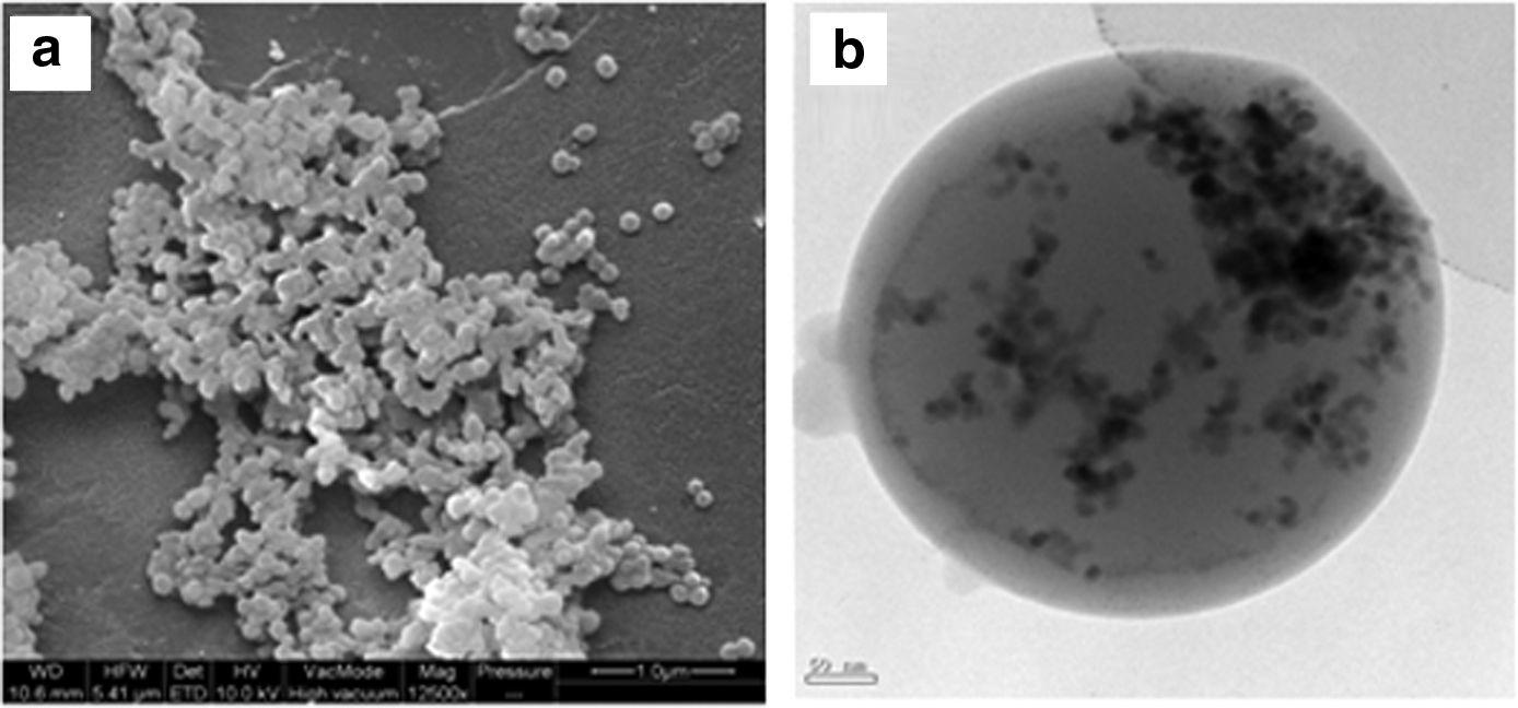
Scanning electron microscope image
The presence of metalloporphyrins in the microsphere support was confirmed by solid-state UV-vis spectra as well as by ICP results of the catalysts. No substantial ultraviolet spectra were observed with the blank magnetic chitosan nanosperes. However, typical absorption spectra of Mn(III) porphyrin and Co(II) porphyrin with Soret bands centered at∼480 and ∼428 nm, respectively, were observed in the solid-state UV-vis spectra of MCSMnTPP and MCSCoTPP (Fig. 5). The solid-state UV-vis results ambiguously evidenced the existence of metalloporphyrin in the nanospheres. Furthermore, the contents of metalloporphyrins in the magnetic nanospheres were calculated from the contents of Co or Mn, which can be obtained using ICP. The contents of metalloporphyrins in MCSMnTPP and MCSCoTPP were 0.43% and 0.52%, respectively. The ICP results further confirmed the presence of metalloporphyrins in the nanospheres. Meanwhile, the contents of magnetic cores (Fe3O4) in the nanospheres were similarly calculated to be 12.27% and 10.06% for MCSMnTPP and MCSCoTPP, respectively.

Solid-state UV-vis absorption spectra of
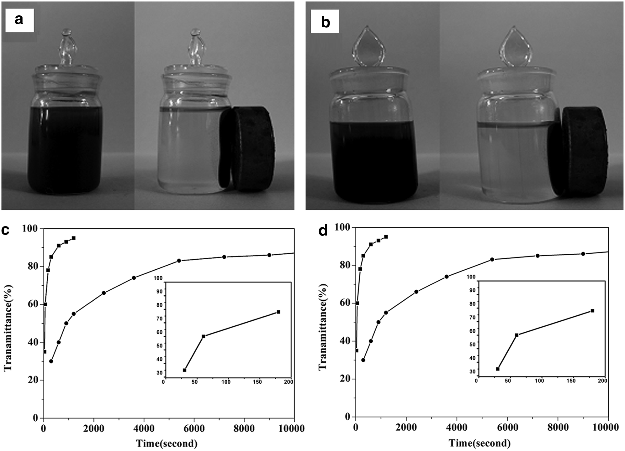
Since these nanospheres have magnetic cores, an important characteristic of the prepared catalysts is that they have excellent magnetic responsiveness which plays an essential role in their effortless recovery. Magnetization curves (Fig. 6) show that all of these magnetic nanospheres exhibit superparamagnetic behavior with zero coercivity and remanence, because the diameter of the magnetic Fe3O4 fluid used in the preparation of these nanospheres is 8.96 nm, which is smaller than the critical particle size of Fe3O4 particles (Zampronio et al., 2005). The saturation magnetizations of MCSMnTPP (3.41 emu/g) and MCSCoTPP (5.37 emu/g) are lower than those of Fe3O4 nanoparticles, which are due to the copolymer coating of the Fe3O4 nanoparticles in nanospheres (Huang et al., 1996; Santra et al., 2001). What is more, it was experimentally observed that these nanospheres dispersed in water are rapidly attracted by a conventional magnet which is placed close to the reaction vessel while the sedimentation in natural field was much more difficult (Fig. 7a, b). We monitored the transmittance change of MCSMnTPP and MCSCoTPP emulsion in natural and magnetic fields. It was found that the transmittance of emulsion in magnetic fields costs almost 1200 s to approach 100%, while that in natural fields costs almost 2×10⁴ s (Fig. 7c,d). This result evidenced the efficacy of magnetic separation.
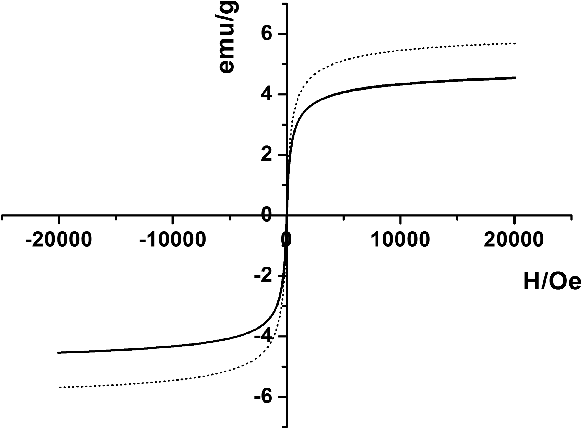
Magnetic hysteresis loops of MCSMnTPP (black) and MCSCoTPP (gray).
Catalysis
UV-Vis spectra evolution of MO solution
Figure 8 illustrates the evolution of UV-vis spectra of the solution as a function of irradiation time in the present of catalysts. The bands at wavelengths of 463 nm and 272 nm were attributed to the conjugated structure constructed via azo bond and the benzene structures of MO. These two bands decreased during the irradiation, indicating the decomposition of MO and the decoloration of the solution.

UV-vis spectra evolution of MO solution as a function of natural sunlight (NSL) irradiation time in the presence of MCSCoTPP (initial MO concentration: 30 mg/L, catalyst concentration: 200 mg/L).
Effect of light sources
The degradation of MO with MCSCoTPP under HPML, ITL, or NSL was comparatively investigated, and the results are depicted in Fig. 9. It is well known that the emission spectra of HPML mainly focused on the UV band, and the spectrum of ITL mostly shifted to visual light (Sansonetti et al., 1996). However, the spectrum of NSL was continuously alternated from ultraviolet to near infrared. As shown in Fig. 9, the degradation rate of MO irradiated with NSL was higher than that with HPML or ITL.
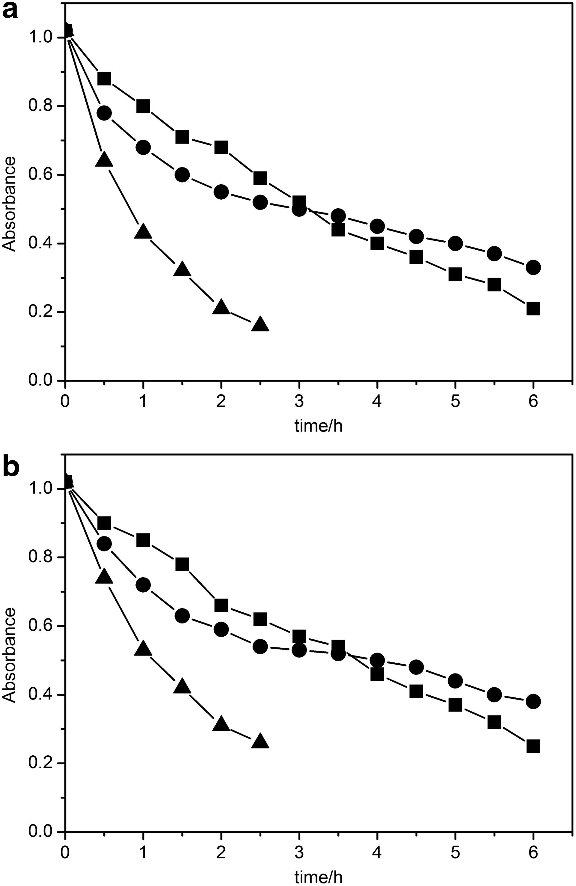
Degradation of MO under HPML (●), ITL (■), or NSL (▲) using MCSCoTPP
Since the main emission spectra of HPML concentrated in the ultraviolet region (Sansonetti et al., 1996), it was believed that the photocatalyst absorbed high energy from the ultraviolet light and existed at an excitation level, which then transmitted the energy to the substrate and itself came back to the ground state. The substrate at the excitation level was degraded by O2 and other oxidated species (Chen et al., 1999). On the other hand, the binding of dioxygen to the low valent metalloporphyrins, which is a key step in metalloporphyrins' photocatalytic behaviors, takes place under visible light. Thus, the degradation rate by visible light from ITL seemed swifter for the last 2 h.
NSL proved to be the most efficient light source in the photocatalytic process, as the degradation was rapid and complete under NSL. NSL with a continuous spectrum takes advantage of both ultraviolet and visible light and, thus, shows the best photocatalytic efficiency. For practical application in the water-treatment procedure, NSL should be chosen as an ideal source for irradiation.
Effect of pH
After adding catalysts and irradiating with NSL, MO photodegradation was conducted in the pH range from 3 to 10 by the previous adjustment with 5% NaOH or HCl solution. From the experimental results shown in Fig. 10, it was evidenced that the lower pH value was favorable to the degradation of MO by the catalysts in aqueous solutions. This could be attributed to the OH and NH2 groups on the surface of these chitosan-supported nanospheres. In an acidic environment, the OH and NH2 groups will change to OH2+ and NH3+ (Dotto and Pinto, 2011), which could attract the SO3− group of MO molecules, and, thus, the catalysts have higher photocatalytic efficiency. However, in the alkali environment, the lone-pair electrons on OH and NH2 will repulse the SO3− groups, and therefore alkali solution is unfavorable for the photocatalytic reaction.
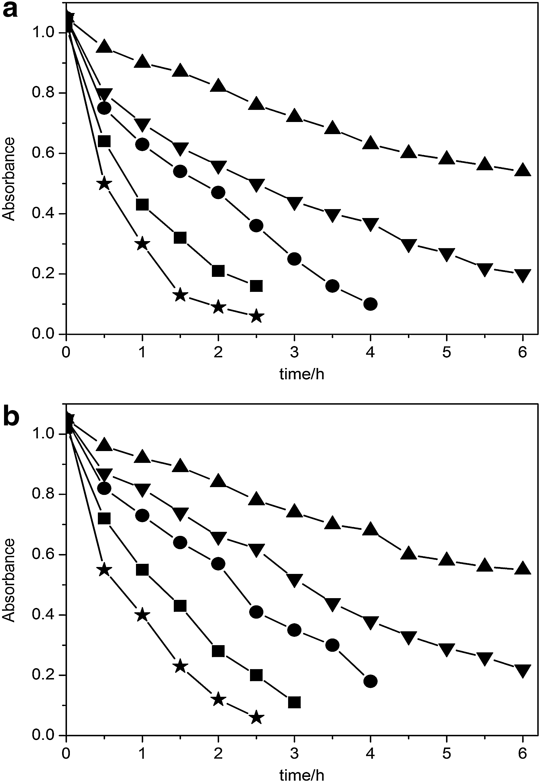
Effects of pH on photodegradation of MO solution using MCSCoTPP
Nevertheless, since chitosan is unstable in solution with a low pH value (Liu et al., 2010), it is preferred to carry out the photocatalytic reaction in a neutral or weak acidic environment.
Effect of catalysts
The photocatalytic degradation of MO with different catalysts in aqueous solutions was investigated, and the results are shown in Fig. 11. MO solution could be stably reserved in the dark, and it could exist for a long period of time with the irradiation of NSL (Fig. 11). The concentration of MO decreased swiftly in the first stage when magnetic chitosan nanospheres without porphyrins were added but the decrease became much slower after 1 h. This could be explained by the well-known fact that chitosan spheres could adsorb dyes in water. This process is a saturable absorption and is irreversible. Thus, after 1 h stirring with the blank chitosan nanospheres, the MO concentration could not be further decreased, as the chitosan was saturated at that timepoint. However, after stirring with catalysts under the irradiation of NSL, it was evident that MO degraded dramatically with irradiation time, and the reducing quantity reached 81% and 93% with MCSMnTPP and MCSCoTPP, respectively.
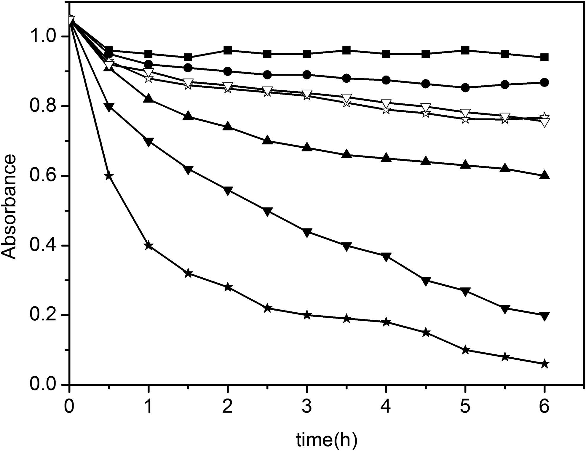
Photodegradation of MO solution under various conditions: no catalyst in dark (■); no catalyst in NSL (●); with MnTPP (▽); with CoTPP (✩); with blank magnetic chitosan nanospheres in NSL (▲); with MCSMnTPP in NSL (▼); and with MCSCoTPP in NSL (★). (15 mg/L MO was dissolved in 50 mL solution, and the metalloporphyrins in the nanospheres are 0.09 μmol, pH=5.00.)
In the reaction, the oxygen molecule possibly activated readily into O2− through electron transformation from metal ions, then abstracted the hydrogen atom of the substrate, and induced the photochemical reaction. Correspondingly, here MO was photodegraded into smaller molecular products at last. On the basis of the earlier studies (Chen et al., 1999; Fu et al., 2008), the photodegradation mechanisms of MO may follow processes such as
MCSMn3+TPP employs a similar mechanism as MCSCo2+TPP. However, it is well known that the cobalt TPP exists in the form of TPP Co(II), while manganese TPP exists in the form of TPPMn(III). It was reported that chloro (tetraphenylporphyrinato) metal (III) [TPPMIIICl] loses a chlorine radical and forms (tetraphenylporphyrinato) metal (II) [TPPMII], which then combines with an oxygen molecule and forms an activated radical species (TPPMIIIO•). (Tetraphenylporphyrinato) metal (II) is a key intermediate in the reaction with O2. Usually, cobalt TPP, which exists in the form of TPPCoII, favors the activation of oxygen. However, manganese (III) TPP are probably first changed to the forms TPPMnIICl, in contrast to Co, for which TPPCoII is not needed. It is probable that the more slowly TPPMnIIICl change into the form TPPMII, the more slowly they activate the oxygen molecule (Huang et al., 2007). Hence, the catalytic activity of TPPCoII was greater than that of TPPMnIIICl, and, thus, the MCSCoTPP had higher catalytic efficiency than MCSMnTPP.
Figure 11 also illustrates that under mild conditions, nonsupported metalloporphyrins (CoTPP and MnTPP) have limited catalytic activities compared with the chitosan-supported metalloporphyrin nanospheres. Earlier studies have indicated that metalloporphyrins would be dimerized, which would reduce the activity in the photocatalytic process (Chen et al., 1999; Fu et al., 2008, 2009a, 2009b). The chitosan support was believed to effectively prevent the dimerization of the single metalloporphyrin and, thus, enhance the photocatalytic activity. Moreover, the chitosan shell in these nanospheres provides a suitable microenvironment for the “accommodation” of porphyrin photocatalytic centers. Since chitosan could adsorb MO molecules in the solution, the substrate concentration of the photocatalytic reaction was remarkably increased around the metalloporphyrins. Thus, the catalytic capability of the chitosan-supported metalloporphyrins could remarkably enhance the catalytic capability of metalloporphyrin.
Reuse of the catalysts
Table 1 gives the photocatalytic results of these catalysts that were recovered and reused. From Table 1, one can clearly observe that the magnetic chitosan-supported metalloporphyrin nanospheres, which are reused for as many as five times through recovery from the reaction media by effortless magnetic separation, still show relatively high activity in the photocatalytic process. There is no loss of magnetic responsiveness or morphological change after recycling. Meanwhile, the UV-vis and infrared spectra of these recovered nanospheres did not show any substantial change compared with those of the fresh ones. Meanwhile, the spectra of the filtrates isolated from the reaction mixtures did not show any band related to metalloporphyrins, indicating no leaching of the complex from the support during reaction. The decreased catalytic activity of the reused catalysts could be attributed to the undegraded MO molecules adsorbed on the catalysts. The earlier results evidenced the expended catalytic life of these magnetic chitosan-supported metalloporphyrin nanospheres. These new catalysts exhibited distinct advantages than traditional chitosan in azo wastewater treatment.
The metalloporphyrins in the nanospheres are 0.09 μmol. 15 mg/L MO was dissolved in 50 mL solution.
MCSMnTPP, magnetic chitosan-supported manganese tetraphenylporphyrins; MCSCoTPP, magnetic chitosan-supported cobalt tetraphenylporphyrins.
Conclusion
This article presents a relatively facile technology that can be used to prepare magnetic chitosan-supported metalloporphyrin nanospheres and investigated their photocatalytic behaviors to MO solution. It is found that these catalysts have enhanced catalytic capabilities and an extended catalytic life than traditional chitosan or nonsupported metalloporphyrins. Metal ions are observed to affect the catalytic efficiencies of these catalysts, and Co(II) porphyrins show more efficient catalytic abilities than Mn(III) porphyrin. These catalysts may facilitate the research of new highly efficient and cost-saving treatment of dye wastewater.
Footnotes
Acknowledgments
This work was financially supported by the National Nature Science Foundation of China (21101033) and the Innovative Project for College Students of Guangdong (1057310041).
Author Disclosure Statement
No competing financial interests exist.