
Abstract
Waterborne pathogens are a leading cause of disease and death worldwide. Ultraviolet (UV) irradiation is an effective method of drinking water treatment through the destruction of cells at the molecular level. Throughput capacity per unit volume of the UV unit is a major driver of the treatment process. In this study, we developed a microscale-based reactor in combination with UV radiation to determine the inactivation efficacy of bacteriophage MS2 and Escherichia coli. Using the microscale-based reactor, we achieved inactivation of more than 7 log colony forming units per mL of E. coli by 8 s and removal of more than 7.5 log plaque forming units per mL of bacteriophage MS2 by 8 min residence times. These values meet and surpass the standards set by the U.S.-Environmental Protection Agency and the World Health Organization for drinking water treatment technologies. Results from mathematical modeling suggest that the best fit model is obtained when we assume that more than a single photon directly damages E. coli cells and allows a transitional damaged state or further inactivation by additional photons. The successful application of a point-of-use UV water disinfection device to inactivate pathogens creates new opportunities for household and commercial water treatment globally.
Introduction
Approximately 829,000 people die annually from waterborne pathogens, of which 297,000 are children under the age of 5 (WHO, 2019). Advances in sanitation, vaccination, and therapeutics have made improvements to the detriments caused by waterborne pathogens, but still two billion people globally use a drinking water source with fecal contamination (WHO, 2019). Chlorination of drinking water is effective at inactivating most pathogenic bacteria and viruses by breaking chemical bonds and disrupting enzymatic function (NRC, 1980). Chlorination, however, is highly ineffective at inactivating protozoa, such as Cryptosporidium, and some viruses, including bacteriophage MS2, due to their robust nature (Beck et al., 2016). In addition, chlorination processes lead to the formation of disinfection byproducts such as trihalomethanes and haloacetic acids, which are known to be carcinogenic and mutagenic (CDC, 2016). Proven methods of disinfection require high-intensity ultraviolet (UV) light for the destruction of protozoa and viruses at the molecular level and to inhibit their replication (Oguma et al., 2001; Beck et al., 2016). UV disinfection utilizes high-energy ultraviolet-C (UVC) photons (primarily at 254 nm) emitted from inert gas lamps (mercury, argon, etc.) that are absorbed by nucleic acids (EPA, 2016). Once absorbed, the UV radiation impedes reproduction by altering the cell DNA (Beck et al., 2016).
UV treatment of water is highly effective for a wide range of contaminants, both biological and chemical. Recently, the inactivation of SARS-CoV-2 (the virus responsible for the COVID-19 pandemic) in aqueous solutions by UV irradiation has been demonstrated (Ma et al., 2021; Robinson et al., 2021). UV disinfection efficacy is a function of exposure time, irradiation intensity, and general water quality parameters such as turbidity, suspended solids, and organic matter (EPA, 2016). Depending on the water treatment application, the cost of equipment, operation, and maintenance can be significant inhibitors to the adoption of UV treatment technologies. The cost of operation for these treatments is a function of the process's energy consumption or energy efficiency, which primarily consists of powering the UV light sources. Throughput capacity per unit volume of the treatment device can impact the equipment cost. In addition, systems that require longer residence times inherently require a larger internal flow volume per processing capacity. Residence time is also impacted by the ability to efficiently utilize the photons from the light source. Ultimately, there is a need for new design approaches to UV water treatment devices that provide flexibility for application scale, improved energy efficiency, and tighter process control.
Small-scale, energy-efficient, economically feasible, and rapid methods of disinfection utilizing UV can be beneficial in households as a point-of-use system for drinking water or in a diverse range of industries such as medical practices and restaurants. Recently, the application of activated carbon filters and carbon nanotube composite membranes in water disinfection point-of-use systems have been studied (Oh et al., 2019; Mulhern et al., 2021). Montenegro-Ayo and colleagues (2020) demonstrated waterborne pathogens inactivation in a portable photoelectric point-of-use device that combined filtration, UV, and titanium dioxide (TiO2) nanotubes. In another recent study, commercial stainless steel bottles were fitted with UVC-emitting caps for portable water disinfection applications (Mariita et al., 2021). The use of microscale-based reactors has been shown to improve process efficiency by leveraging intensified heat and mass transfer and process modularization (De Sa et al., 2016). Water treatment can benefit from the controlled laminar flow behavior resulting from low Reynolds number within micro- and milliscale channel architecture, which provides a more consistent residence time distribution profile. Laminar dispersion does occur and can be balanced by the short characteristic path length of the shallow channel depth allowing a species to “sample” an average of the laminar velocity profile. The shallow channel depth also reduces the effect of light attenuation as it passes through the fluid, especially at higher turbidity conditions. This results in uniform and consistent exposure of the fluid to the UV light and high utilization of the photons from the light sources. This in turn enables the reduction of the device footprint per equivalent throughput capacity when compared with conventional shell and tube configurations. In addition, microscale-based chemical processing technologies are highly suited for a numbering-up approach for scale-up to reach targeted capacities, which ensures performance behavior is maintained regardless of application scale.
Our group selected a microscale-based design approach as an opportunity to develop and demonstrate an excitingly new water treatment technology. There are several intrinsic advantages that microscale-based technologies bring into technical development. First, all microscale-based technologies are central to process intensification, as recently suggested by Jovanovic et al. (2021a, 2021b). Process intensification is the latest technical “emblem” of an integrally refined and optimized process technology (Stankiewicz et al., 2019). Superior rates and controllability of all transport phenomena (i.e., mass, heat, and momentum) are just a few of the collateral benefits of the microscale-based design approach. Second, achieving industrially relevant capacities with microscale-based technologies is effectively realized through the numbering-up approach, rather than the classic and often-unreliable scale-up approach. Third, thanks to the numbering-up approach, the optimal operating conditions demonstrated at the laboratory research scale are precisely preserved in the industrial scale devices, that is, no need for further process development. And finally, the capacity scale of microscale-based devices is infinitely flexible since adding or removing any number of elemental (i.e., microchannel) units is a trivial task.
Scales pertinent to a personal or industrial scale capacity have been demonstrated (O'Connor et al., 2021). On the techno-economic side, microscale-based technologies bring even more inherent advantages. Since the capacity range of microscale-based devices is infinitely adjustable, the development of modular technologies, and thus distributed water production is entirely feasible and economical. The capacity cost of microscale-based technologies is reaching new lows every day. Moreover, additive manufacturing (e.g., three-dimensional printing, laser-directed energy deposition, and laser powder bed fusion) enables a sharp reduction in manufacturing costs. In addition, a shorter timeline from laboratory to industrial-scale technology development reduces development costs. And finally, the overall capital investment and operating cost per unit capacity of microscale-based technologies are lower than comparable costs in classic technologies. Thus, the reduction of the investment risk, usually associated with a centralized economy of scale, is feasible (O'Connor et al., 2021).
In our group, we have developed a microscale-based reactor combined with UV irradiation that can provide novel applications to conventional drinking water treatment. In this article, we evaluated the design, development, and implementation of a microscale reactor utilizing UV radiation to determine proof-of-concept log-removal inactivation of bacteriophage MS2 and Escherichia coli at varying residence times and constant concentration in drinking water. The successful application of a point-of-use photolytic device to inactivate pathogens creates new possibilities for household and commercial water treatment globally.
Materials and Methods
Reactor design and fabrication
The reactor consists of a serpentine flow channel (Fig. 1) recessed on a metal plate with the open top side enclosed by a 14.6 × 8.9 × 0.64 cm quartz window (Technical Glass Products, Inc., Painesville, OH) allowing irradiation of the passing fluid by a UVC light source. The flow channel is 7.0 mm in width, 50 cm in length (through the centerline path), with a channel depth of 0.5 mm. The total irradiation area of the channel is 34.3 cm2. This flow channel was computer numerical control machined into the plate of aluminum alloy (6061 aluminum alloy).

(Left) Assembled reactor with flow channel visible through quartz window. The quartz window enables UVC irradiation of passing fluid. (Right) Diagram of serpentine flow channel on bottom plate of reactor. UVC, ultraviolet-C.
A top plate contains a recessed pocket to align and compress the quartz window against the bottom flow channel plate. The reactor was electroless nickel plated to prevent corrosion (CG Industries, Albany, OR). The flow channel is hermetically sealed against the quartz window through high-temperature silicone O-rings. The top and bottom metal plates are clamped together using 14 screws around the perimeter to provide evenly distributed compression. Water is fed to and from the flow channel by thru-holes in the bottom plate to avoid obstructing visible and irradiative access to the channels.
The thru-holes terminate on the reactor sides where 1/4–28 flat bottom fittings connect to the flow test loop. A syringe pump (Harvard Apparatus PHD 2000 Dual Syringe Pump, Holliston, MA) is programmed to deliver the liquid feed through high-performance liquid chromatography tubing with Luer-Lock connectors to the inlet port of the reactor. The outlet port is connected to a collection flask with an arm equipped with a high-efficiency particulate air filter (Vacushield Vent Device, Pall Laboratory). A 40 W 254 nm mercury germicidal lamp (Aqua Ultraviolet UV Sterilizer, Temecula, CA) with a constant photon output is placed at a 2.5 cm distance to irradiate the reactor. The lamp was sheathed in a polyvinyl chloride housing with a cut-out window approximately the size of the microscale reactor. The lamp was kept on during the testing period to ensure consistent UVC output.
Testing procedure
Before each experiment, the reactor and all the components were disassembled and sterilized. After reassembly, the UV flux (2.0 mW/cm2) was quantified with a UV-VIS AvaSpec-2648 spectrophotometer (Avantes BV, Apeldoorn, The Netherlands). Using a dual syringe setup, the reactor was primed and leak tested with autoclaved deionized water through a 60-mL syringe to purge any air bubbles for three residence times (Fig. 2). The influent sample containing a known concentration of the target contaminant was injected into the system through a second 60-mL syringe. The influent moved through the inlet, through the body of the reactor, and to the outlet. The treated effluent was collected into a vessel. For each test, ∼1 mL of the influent and effluent was collected, stored at 4°C, and analyzed within 12 h.

(Left) Diagram of the UV Reactor Test Loop. Water loaded with the microorganism is pumped through syringe pumps to the reactor where the fluid is then exposed to a UVC light source. The fluid exits the reactor and a UV containment or shield before it is collected in a vessel. (Right) Cross-section of reactor channel (Z = 0.5 mm). UV, ultraviolet.
Microbial enumeration
Target microorganisms were enumerated before and after photolytic inactivation in the reactor. E. coli strain ATCC 25922 was used as an indicator of fecal coliforms. E. coli culture was prepared in Luria broth (Criterion™; Hardy Diagnostics, Santa Maria, CA). Undiluted and 10-fold serial dilutions in phosphate-buffered saline (80 g/L NaCl, 2 g/L KCl, 14.4 g/L Na2HPO4, and 2.4 g/L KH2HPO4; Criterion, Hardy Diagnostics) of the influent and effluent were spread plated onto MacConkey agar with 4-methylumbelliferyl-beta-D-glucuronide (MUG) (Criterion; Hardy Diagnostics) and incubated at 37°C for 18–24 h. Pink colonies that fluoresced blue-green under UV light (366 nm) were identified and quantified as presumptive E. coli. Results were reported as colony forming units (CFU) per mL and the lower limit of detection was 14.3 CFU/mL.
Bacteriophage MS2 was used as a surrogate for enteric viruses. Following ISO 10705-3:2003, bacteriophage MS2 strain ATCC 15597-B1 was propagated and quantified through the double agar overlay method. In brief, the host culture of E. coli strain ATCC 700891 grown to stationary phase was added to 0.7% tryptic soy agar (TSA; Hardy Diagnostics, Santa Maria, CA) while molten, and poured on top of a 1.5% TSA plate. A sample of 100 μL was spotted on the agar, covered, and allowed to sit for 10 min to absorb. A method blank was performed between each dilution. Plates were incubated at 36°C for 16–24 h. Bacteriophage MS2 was quantified as plaque forming units (PFU) per mL. The lower limit of detection was 1 PFU/mL.
Mathematical model
Mathematical models for the photolytic degradation of E. coli and bacteriophage MS2 were developed using elementary chemical kinetics and the first principal transport equations. The chemical kinetics contained the concentration of target microorganisms (i.e., E. coli or bacteriophage MS2), the flux of light on the target microorganism's surface area, and the assumption of a repair mechanism (as evidenced by Oguma et al., 2001 and Nyangaresi et al., 2018 for E. coli and Radman, 1975 and Kadavy et al., 2000 for bacteriophages).
Several inactivation mechanisms were modeled. The initial model describes a single photon strike causing total inactivation (mechanism 1). The second model details a single photon causing inactivation, but with the additional reversible path for repair (mechanism 2). Whereas the third model incorporated a single photon inactivation allowing a transitional damaged state. The damaged state can undergo repair or, if hit by additional photons, will fully inactivate (mechanism 3). The reaction mechanisms across three stages of increasingly complex representation are as follows:

where Cinitial is the concentration of E. coli or bacteriophage MS2 in influent water entering the reactor, Cinactivated is the concentration of inactivated E. coli or bacteriophage MS2 in effluent treated water exiting the reactor, Cdamaged represents E. coli or bacteriophage MS2 cells with damaged DNA that still maintain the capability to repair, I represents the flux of light onto the surface of cells, k1 represents the reaction rate constant of photons hitting undamaged cells, k–1 represents the reaction rate constant of DNA repair mechanism triggering, and k2 represents the reaction rate constant of photons hitting damaged cells. The flux of light into the microscale channel was ∼2.4 × 10⁻⁵ [molphotons/m2 E. coli cells —s].
This value is determined from the flux of the light into the channel, the volume of the channel, the surface area to volume ratio of E. coli cells, and the mean residence time (Levin and Angert, 2015). Based on calculations using Beer–Lambert law, light attenuation through media in the 500 μm deep channel is negligible (>99.9% transmission) and, therefore, photon flux is assumed to be constant.
A schematic of the model geometry and boundary conditions is shown in Fig. 3. The fluid was Newtonian and flow through the reactor was laminar; therefore, the model used the following Navier–Stokes equations in the Cartesian coordinate system:
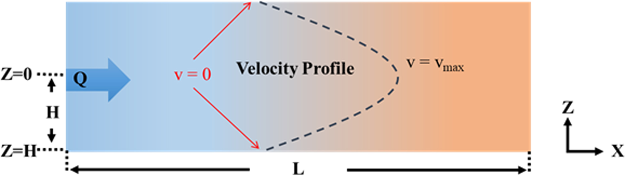
Schematic of the model geometry and boundary conditions for the flow channels in the microscale reactor.
No-slip condition (u = 0) at the channel walls and maximum velocity at the center of the channel were assumed. The material balance of the system was maintained through the convection-diffusion equations for each microorganism, as shown hereunder:

A diffusion coefficient (Dib) of 10–5 [cm2/s] was used (Berg and Turner, 1990). The subscript i represents the target microorganism (i.e., E. coli or bacteriophage MS2) and subscript b represents the continuous aqueous phase. The reaction terms were derived from the rate equations for each target microorganism and differed depending on microbial species and reaction mechanisms.
Numerical simulation and optimization
The numerical simulation of the model was solved in COMSOL Multiphysics 5.4 (COMSOL, Inc., Burlington, MA) as a rectangular channel with dimensions matching the experimental reactor in height, width, length, and photon flux. The model was initially fit to the averaged experimental data using arbitrary rate constants and subsequently ran through a MATLAB optimization program to find a best-fit line. The objective function for the optimization is the sum of the squared error (SSE) between the model and the experimental data. SSE was calculated from the error of the natural log of the concentration.
Results
Flow characteristics
The flow characteristics, including the volumetric flowrates, the average velocities, and the Reynolds numbers, within the microscale reactor during different residence times are shown in Table 1. The average volumetric flow rates ranged between 0.43 and 12.90 mL/min resulting in average velocities between 2 and 61 mm/s. The flow was laminar throughout the study with very low Reynold numbers (≤64).
Flow Characteristics Within the Microscale Reactor During Treatment
Inactivation of fecal indicators
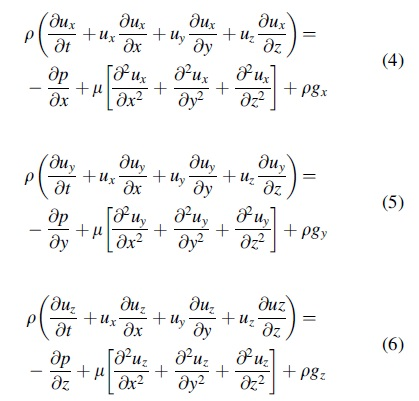
The microscale reactor was evaluated for the inactivation capabilities of E. coli and bacteriophage MS2 in water during UV irradiation (Fig. 4). The initial E. coli concentration in the inoculated untreated influent was 8.85 ± 0.08 (average ± standard error) log CFU/mL. Low E. coli inactivation in the microscale reactor was observed in the first 5 s of exposure to the UV irradiation (<3 log CFU/mL reduction). However, by 8 s residence time, E. coli concentration was reduced by more than 7 log CFU/mL and the treated water effluent levels were at or just above the limit of detection (i.e., 1.15 log CFU/mL). For bacteriophage MS2, the initial concentration in the inoculated influent water was 8.30 ± 0.22 log PFU/mL. The UV irradiation in the microscale reactor resulted in bacteriophage MS2 inactivation by 2.56 ± 0.73 logs PFU/mL within 30 s residence time, by 2 min the removal rates reached 4.03 ± 0.38 logs PFU/mL, and more than 7.5 logs PFU/mL bacteriophage MS2 were removed by 8 min.
Concentration of
Model results
Figure 4 shows the best fit model for the three reaction mechanisms in comparison with the experimental data. The mechanism 1 model, in which a single photon caused total inactivation of E. coli cells, illustrates poor agreement with the experimental data (Fig. 4a). The model fitted the initial slope of the experimental data but could not match the slope of the subsequent longer residence time data. The best fit value for the mechanism 1 model had an SSE of 830 (Table 2). The mechanism 2 model, where a single photon caused inactivation while having an additional reversible path for cell repair, showed improved fit with the initial slope approaching or nearly matching the experimental values.
Optimized Reaction Rate Constants and the Sum of Squared Errors for the Three Modeled Mechanisms Applied to the Inactivation of Escherichia coli and Bacteriophage MS2
SSE, sum of the squared error.
The subsequent slope did not match the slow decline in E. coli concentration, as the model slope went to zero. The best fit value for the mechanism 2 model was 19.3 for SSE (Table 2). Finally, the mechanism 3 model, where we incorporated a single photon inactivation while allowing a transitional damaged state or further inactivation by additional photons, showed the best agreement with the experimental data. In the mechanism 3 model, both the initial and final experimental slopes were reasonably modeled, and the final SSE value was 17.4.
The mechanism 3 model reached our experimental limit of detection (1.15 log CFU/mL) at 100 s residence time. One major note of interest is that the rate constant for the repair mechanism (k–1) is several orders of magnitude smaller than the other rate constants (k1 and k2). This agrees well that the concept of the repair mechanism is slower than the damage mechanism (Oguma et al., 2001; Rastogi et al., 2010). Considering the three modeled mechanisms, the best fit model is obtained when we assume that more than a single photon directly damages E. coli cells. The rate constants and SSEs for the three modeled mechanisms are shown in Table 2.
Modeling results for bacteriophage MS2 followed a similar trend as E. coli (Fig. 4b). Mechanism 1 model failed to align with inactivation levels associated with longer residence times. Mechanism 2 model performed better, accounting for the plateau in inactivation performance, and mechanism 3 model was able to be optimized to a high fit to the experimental results. Mechanism 3 model in fact had a stronger fit to the bacteriophage MS2 results than E. coli, most likely due to lower variance in the shorter residence time measurements. The rate constants and SSE for all three modeled mechanisms are listed in Table 2. Again, we see that k−1, the repair mechanism, is much slower than k1 and k2.
Discussion
Microscale-based chemical processing is rapidly growing in chemical, biochemical, and pharmaceutical manufacturing as industries desire step-change advancement resulting from process intensification and modularization. These benefits move beyond the limitations of traditional unit operations that have held the industry captive for over a century. Applications range from thermocatalytic microreactors to two-phase membraneless separation technologies and modular platforms that access a range of distributed feedstocks. Recently, researchers have investigated augmenting UVC water disinfection systems with photocatalytic reactions, such as TiO2 or silver nanoparticles, within microfluidic reactors.
Prabhakar et al. (2020) embedded a microfluidic disinfection device with silver nanoparticles to demonstrate a synergistic bactericidal effect. Further back, researchers have looked into augmenting UV disinfection with low-frequency ultrasound (Bazyar Lakeh et al., 2013) and improving flow characteristics through modified surface roughness (Sultan and Cho, 2016). There is also significant interest in implementing UV-light-emitting diodes (LEDs) instead of conventional mercury lamps. Using a modified tubular flow-through reactor, Wang et al. (2021) demonstrated strong inactivation, up to ∼6.5 Log inactivation, in their compact device. The authors also noted the importance of achieving high irradiance uniformity to maximize inactivation efficiency. Early study by Wurtele et al. (2011) tested a continuous flow, milli-scale reactor that resembled many of the microscale-based reactors seen in different areas of chemical engineering research. In our presented study, we have approached improving or intensifying UVC water disinfection through the microscale-based design of the fluid flow geometry. This design approach attempts to improve light uniformity, whereas reducing light attenuation and variance in fluid mean residence time, essentially targeting a key tenet of process intensification: “give all molecules or species the same experience” (Stankiewicz et al., 2019). The microscale-based reactor integrates well with augmentation methods and UV-LED implementations proposed in previous studies.
Results of this study on the inactivation of E. coli and bacteriophage MS2 during UV irradiation in the microscale-based reactor comply with the U.S.-Environmental Protection Agency (EPA; <1 CFU E. coli/100 mL and >4 logs removal of viruses; EPA, 2014) and the World Health Organization (≥4 logs removal of bacteria and ≥5 logs removal of viruses; WHO, 2019). When using bacteriophage MS2 as the challenge organism that has lower UV resistance than the human adenovirus, at least 7.4 logs inactivation of bacteriophage MS2 should be achieved to demonstrate the equivalent ability to achieve 4-log removal of adenovirus (Yates et al., 2006).
In our study, by 8 min UV irradiation within the microscale reactor, we achieved 7.54 ± 0.41 logs PFU/mL removal of bacteriophage MS2. Moreover, to achieve a 4-log reduction of viruses U.S.-EPA recommends a UV dose of 186 mJ/cm2 (EPA, 2016). In our study, with the UV flux of 2.0 mW/cm2 in the microscale reactor, we achieved 4-log PFU/mL inactivation of bacteriophage MS2 by 78 s, which translates to 156 mJ/cm2. In addition to bacteria and viruses, water treatment technologies undergoing validation should meet requirements for the inactivation of protozoan parasites such as Cryptosporidium parvum (EPA, 2016).
Since the inactivation mechanism of C. parvum by UV irradiation is similar to those of E. coli and bacteriophage MS2 (Oguma et al., 2001), effective removal of viruses (e.g., 6-log reduction of bacteriophage MS2) qualifies the system for meeting C. parvum removal (EPA, 2016). Therefore, under the conditions discussed in this study, 8-min UV irradiation in the microscale reactor will meet the requirements for bacteria, viruses, and protozoan parasites. This residence time can be reduced further through increased light flux into the reactor flow channel.
UV treatment of water is highly effective for a wide range of contaminants, both chemical and biological. Depending on the water treatment application, cost of equipment, cost of operation, and maintenance requirements can be significant inhibitors to adoption. Cost of operation is a function of the energy consumption or energy efficiency of the process, which primarily consists of powering the UV light sources. Throughput capacity per unit volume of the treatment device can impact the cost of the equipment. Systems that require longer residence times inherently require a larger internal flow volume per processing capacity.
Residence time is also impacted by the ability to efficiently utilize the photons from the light source. Ultimately, the application of UV irradiation in the microscale reactor provides flexibility for application scale, improved energy efficiency, and tighter process control. Finally, LED light sources that emit in the UVC range have experienced a significant increase in availability and thus a decrease in unit cost due to the demand for air purification systems created by the ongoing COVID-19 pandemic. Microscale-based reactors with their flat or two-dimensional architecture and compact footprint could be an ideal platform to integrate UVC LED technology to increase irradiation intensity while also improving energy efficiency.
Conclusions
Economical and effective treatment of drinking water for pathogens inactivation is a global necessity. UV irradiation is an effective method for the destruction of pathogens. To achieve desired levels of pathogen removal, the cost of equipment and operation, as well as maintenance requirements can be significant barriers to the implementation of the technology. In this study, we demonstrate a proof-of-concept study that removal of E. coli and bacteriophage MS2 (i.e., microbial pathogens indicators) can be achieved in <8 and 78 s, respectively. Results from mathematical modeling demonstrated that the best fit model is obtained with the assumption that more than a single photon directly damages E. coli cells, where the model allows a transitional damaged state or further inactivation by additional photons. The application of UV irradiation in the microscale reactor provides flexibility for application scale, improved energy efficiency, and improved process control.
Footnotes
Acknowledgment
We thank Sue Yee Yiu for the help and support in the project.
Author Disclosure Statement
No competing financial interests exist.
Funding Information
This study received funding support from the Oregon State University start-up funds.