



Abstract
Background
Concern about radiation exposure is leading to an increasing interest in low-concentration contrast medium administration.
Purpose
To evaluate the image quality and safety profile after administration of iodixanol 270 mg I/mL at 100-kVp tube voltage with iterative reconstruction in subjects undergoing computed tomography angiography (CTA).
Material and Methods
Patients who completed CTA examination using iodixanol 270 mg I/mL and 100-kVp tube voltage along with iterative reconstruction for coronary, aortic, head and neck, renal, or pulmonary arteries were included. Image quality was qualitatively and quantitatively evaluated. Incidence of adverse events (AEs) and adverse drug reactions (ADRs) within seven days and radiation dose were also analyzed.
Results
A total of 4513 individuals in 42 centers in China were enrolled, among which 4367 were included in efficacy analysis. The mean image quality score was 4.8 ± 0.45 across all arteries (all above 4.6) and 99.7% of the individuals’ images were classified as evaluable. The CT attenuation, signal-to-noise ratio (SNR), and contrast-to-noise ratio (CNR) in the regions of interest (ROIs) were 431.79 ± 99.018, 18.29 ± 11.947, and 28.21 ± 19.535 HU, respectively. Of all the participants, 68 (1.5%) and 65 (1.4%) experienced AEs and ADRs, respectively. No serious AEs or AEs leading to discontinuation occurred. The average effective radiation dose was 3.13 ± 2.550 mSv.
Conclusion
Iodixanol 270 mg I/mL in combination with 100-kVp tube voltage and iterative reconstruction could be safely applied in CTA and yield high-quality and evaluable images with reduced radiation dose.
Introduction
Ever since the application of computed tomography (CT) in clinical practice, there has been a growing concern on the consequence of radiation exposure on patients (1,2). Initial efforts to decrease radiation have mainly focused on lowering the tube current and reducing scan time and range (3,4). Increasing evidence demonstrates the benefits of applying low tube voltage, while not affecting CT image quality and contrast enhancement, and/or reducing the amount of CM administered (5–7).
In clinical practice, a tube voltage of 120 kVp was routinely used in CT examination. The commonly used iodine concentrations are 300 mg I/mL, 320 mg I/mL, 350 mg I/mL, 370 mg I/mL, and 400 mg I/mL in contrast-enhanced CT examinations (8–12). A few studies suggested that good diagnostic quality of CTA is acquired with an iodine concentration as low as 270 mg I/mL (iodixanol) (13,14). Furthermore, the feasibility of combining low iodine concentration, low tube voltage, and iterative reconstruction in contrast-enhanced CT scans, including CTA, to yield diagnostic quality exams has been shown in several studies (14,15). However, most of these single-center, small sample-size studies used a single CT scanner or single iterative reconstruction algorithm, which limits the wide application of the protocol.
The objective of this study was to assess image quality and safety using low iodine concentration (iodixanol 270 mg I/mL) and low tube voltage (100 kVp) scanning protocol with iterative reconstruction for coronary, aortic, head and neck, renal, or pulmonary arteries in a large, multicenter cohort.
Material and Methods
Study design
This was an open-label, prospective, and non-interventional study conducted at 42 centers in China. A total of 4532 individuals were screened and 4513 individuals were enrolled in the study from August 2013 to March 2015. The study design was in accordance with the Declaration of Helsinki and approved by the Institutional Ethics Committees. Written informed consent was obtained from all participants.
Study population
Demographic information, relevant medical/surgical history, concurrent medications, and pre-treatment events of all participants were recorded before the study began. Patients were consecutively included in this study if they: were aged ≥ 18 years; underwent a CTA examination with iodixanol 270 mgI/mL (Visipaque, GE Healthcare Ireland, Cork, Ireland) for coronary, aortic, head and neck, renal, or pulmonary arteries as part of their routine clinical care; and provided a written informed consent. Patients were excluded if they met one or more of the following exclusion criteria: (i) allergic to iodinated contrast agent; (ii) atrial fibrillation/flutter or any irregular heart rhythm considered to interfere with temporal acquisition of cardiac CT images (only applicable for coronary CTA); (iii) a resting heart rate > 100 beats per minute (bpm) and/or a resting diastolic blood pressure > 100 mmHg, or a resting heart rate > 70 bpm and beta-blocker therapy was contraindicated (only applicable for coronary CTA); (iv) a prior coronary artery bypass graft (CABG) procedure; (v) a prior pacemaker or internal defibrillator lead implantation (only applicable for coronary CTA); (vi) extensive vessel calcifications identified by non-contrast images (only applicable for coronary CTA); (vii) being unwilling or unable to discontinue metformin or metformin containing products; (viii) manifest thyrotoxicosis; (ix) previous enrollment in this study; and (x) suffering from a terminal, serious, or life-threatening disease, any medical or psychiatric condition, or any condition in which the study participation might compromised the management of the participant, or other reason that in the judgment of the investigator made the individual unsuitable for participation in the study.
Study process and CTA protocols
The study process is summarized in the study flowchart (Fig. 1). CTA scanning was performed using iodixanol 270 mg I/mL at a tube voltage of 100 kVp using a 64-slice or greater CT scanner. The images were reconstructed using iterative noise-reducing reconstruction software, such as adaptive statistical iterative reconstruction (ASIR) by GE Healthcare, Sinogram Affirmed Iterative Reconstruction (SAFIRE) by Siemens, iDose or iDose4 by Philips or comparable approved iterative reconstruction software provided by the CT manufacturer. The tube currents and contrast-injection protocols used in the four most commonly used scanners are listed in Table 1. The amount of the contrast medium was adjusted to the individual body weight of each patient (1 mL/kg body weight) and injected at a flow rate as indicated in Table 2 followed by ≥ 30 mL saline solution.
Study flowchart. Tube currents and contrast-injection protocols of different scanners. The amount of the CM and flow rate.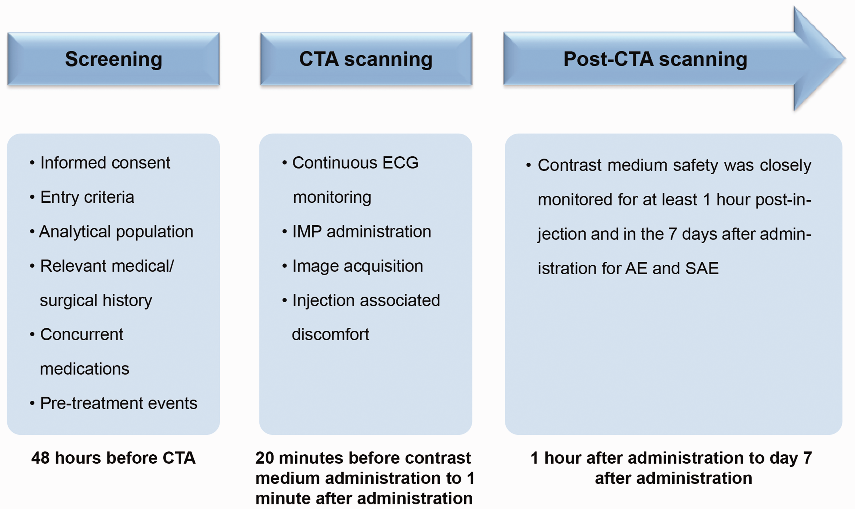


Efficacy assessment
Efficacy assessment included both qualitative and quantitative analyses of images. Four independent reading centers (IRCs) were selected to perform central reading of the scans and two blinded readers independently assessed the image quality at each IRC.
The primary efficacy of CTA was determined in terms of overall image quality, which was assessed using a 5-point scale depending on the contrast enhancement (brightness and uniformity), edge sharpness, and presence of image noise and beam-hardening/motion artefacts. A score of 5, 4, 3, 2, and 1 corresponded to excellent, good, adequate, sub-optimal, and unacceptable or poor vessel delineation, respectively. Specifically, excellent vessel delineation was defined as excellent contrast enhancement and minimal or no image noise or no artefacts; good as good or excellent contrast enhancement and less than average noise, presence or absence of artefacts not affecting image interpretation; adequate as acceptable or good contrast enhancement and average or less than average noise, or presence of artefacts but not affecting image interpretation; sub-optimal as less contrast enhancement and/or above average image noise, or presence of artefacts affecting image interpretation; and poor as unacceptable contrast enhancement and/or unacceptable image noise, or presence of artefacts affecting image interpretation. Based on this 5-point assessment scale, CTA images could be classified as: (i) evaluable including excellent, good, or adequate image quality; and (ii) non-evaluable including sub-optimal or poor image quality.
Quantitative measurements for contrast enhancement included CT attenuation values, SNR, and CNR. CT attenuation values were obtained at selected regions of interest (ROIs) and regions of comparison (ROCs) for each type of CTA images. In particular, coronary artery and aorta CTAs were investigated at the aortic root and the origins of left and right coronary arteries; head and neck artery CTAs at the origin of each carotid artery, each carotid artery bifurcation, and each distal internal carotid artery; pulmonary artery CTAs at the left and right main pulmonary arteries as well as the origins of lobular branches; renal artery CTAs at the renal artery level.
The SNR and CNR were calculated using the formula: SNR = AVA / BNA and CNR = (AVA–AVM) / BNM (AVA = attenuation value of ROI, AVM = attenuation value of ROC, BNA = SD value of ROI, BNM = SD value of ROC).
Overall participant safety
Safety assessments, including recording of adverse events (AEs) or treatment emergent adverse events (TEAEs), serious adverse event (SAEs), and adverse drug reactions (ADRs), were performed during the study procedure in a hospital setting at 1 h post injection, in-clinic evaluation, or telephone follow-up on day 1, and with a telephone follow-up on days 3 and 7. The participants were required to report in a timely manner any symptoms and signs to the study staff. In addition, injection-associated discomfort was assessed for up to 20 min after contrast administration using a standardized questionnaire which recorded the presence of discomfort as well as the intensity evaluated by a 0 - (none) to 10 - (the most intense sensation imaginable) point scale. The location was classified as peripheral, chest, abdomen, pelvis, any combination, or whole body. Radiation dose of each individual was obtained from the scanner dose report and the effective dose (ED) was calculated according to dose-length product (DLP) using the following formula: ED = k * DLP (k was a weighting factor dependent upon body regions and adopted by the International Commission on Radiological Protection (ICRP).
Sample size determination and statistical analysis
Based on a 10% drop-out rate and 1% rate of non-evaluable images, which were demonstrated from the interim analysis performed in October 2014, at least 1935 patients should be recruited to provide a 95% confidence interval (CI) with a margin of error of 0.5% (i.e. the length of the 95% CI is no wider than 1%). The efficacy cohort included individuals that received iodixanol and for who the CTA images were available, while the safety cohort included all participants who received iodixanol.
Descriptive statistics for continuous data included the number of participants, mean, standard deviation, median, and range (minimum–maximum). Descriptive statistics for categorical data included counts and percentages. Tabulations of summary statistics, graphical presentations, and statistical analyses were performed using SAS software, Version 9.2.
Results
Study population
Study population.

Efficacy results
Descriptive summary of image quality assessment.

SD, standard deviation.
Summary of quantitative measurements of contrast enhancement.

ROI, region of interest; ROC, region of comparison; SD, standard deviation; SNR, signal-to-noise ratio; CNR, contrast-to-noise ratio.

CTA for coronary artery in a 59-year-old woman with a BMI of 23 kg/m2, including an axial image (a), a curved multiplanar reformat (CPR) image (b), and a volume-rendering (VR) image (c).
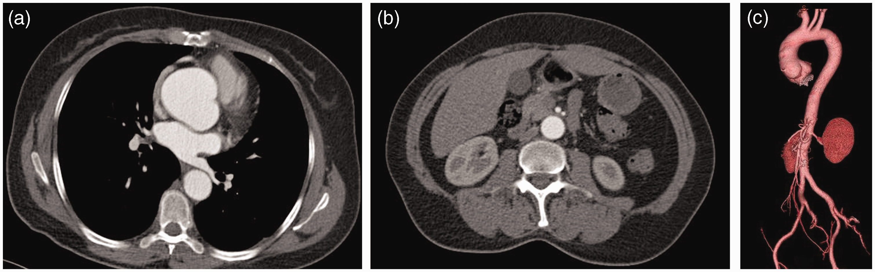
CTA for aorta in a 48-year-old woman with a BMI of 23 kg/m2, including axial images (a, b) and a VR image (c).
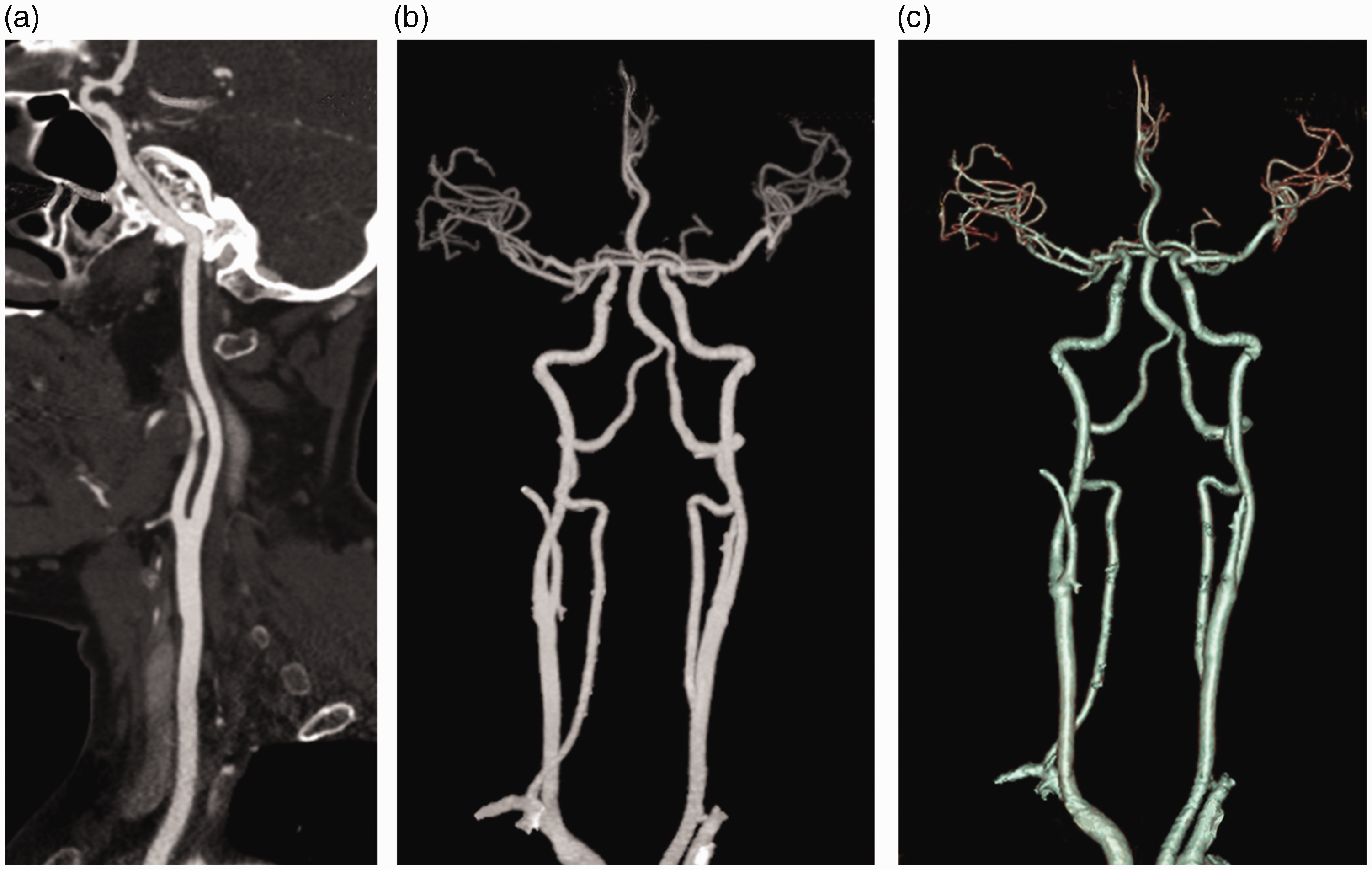
CTA for head and neck artery in a 61-year-old woman with a BMI of 24.1 kg/m2, including two CPR images (a, b) and a VR image (c).
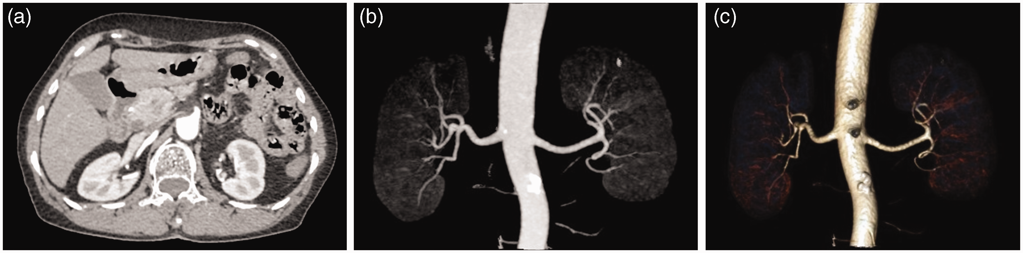
CTA for renal artery in a 66-year-old woman, including two maximum intensity projection (MIP) images (a, b) and a VR image (c).
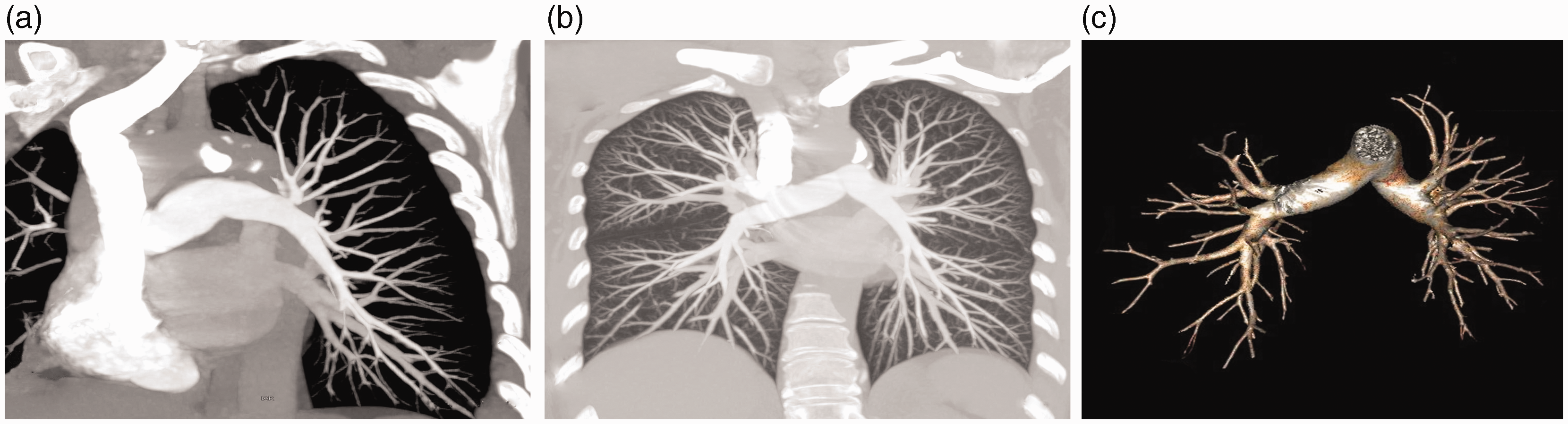
CTA for pulmonary artery in a 59-year-old patient with a BMI of 22.6 kg/m2, including an axial image (a), a MIP image (b), and a VR image (c).
There was no statistically difference in image quality assessment between the two readers except for the excellent and good vessel delineation. Assessment for the head and neck artery group showed the highest agreement percentage (97.1%), followed by the coronary artery group (67.4%), renal artery group (62.0%), aorta artery group, and pulmonary artery group (57.5%).
For quantitative analysis, the mean ± SD of ROI CT attenuation value, ROC CT attenuation value, SNR, and CNR were 431.8 ± 99.0 HU, 70.2 ± 29.9 HU, 18.3 ± 11.9, and 28.2 ± 20.0, respectively. The corresponding parameters in each of the above five groups are displayed in Table 5.
Safety results
In terms of safety analysis, the average dose of iodixanol was 60.9 ± 13.0 mL and the average flow rate was 4.9 ± 0.2 mL/s. In total, 68 (1.5%) participants experienced AEs, among which 65 (1.4%) were assessed as ADRs. The majority of AEs were mild (1.3%) and no SAEs or death occurred. There was no investigational medicinal product (IMP) discontinuation due to AEs. Nineteen (0.4%) participants experienced one or more early ADR that occurred within 1 h after iodixanol administration; the most commonly reported early ADRs were gastrointestinal disorders (0.2%) (10). Forty-seven (1.0%) participants experienced one or more late ADR that occurred in the period 1 h to one week after iodixanol administration; the most commonly reported late ADR was skin and subcutaneous tissue disorders (0.9%) (40). The most commonly reported AEs were skin and subcutaneous tissue disorders (1.0%) and rash (0.6%). The average DLP was 372.0 ± 242.4 mGy*cm and the mean ± SD of ED was 3.1 ± 2.6 mSv.
Discussion
In recent years, there has been an increasing interest in whether a low concentration of contrast medium can achieve satisfactory enhancement of diagnostic image quality due to the concern of the impact on radiation exposure and contrast safety in the patients (16,17). Contrast medium with a low concentration will result in a reduction of the iodine concentration in blood under the same infusion protocol and might even lower the CT attenuation, while the application of lower tube voltage may produce a higher CT attenuation value for iodinated structures and thus provide an opportunity to use low iodine concentration. Though a drawback of reduced tube voltage is that the noise of CTA images might increase and potentially compromise the diagnostic accuracy, the recent introduction of iterative reconstruction algorithm can dramatically reduce image noise and improve image quality (18,19).
According to the Society of Cardiovascular Computed Tomography (SCCT) guidelines on radiation dose and dose-optimization strategies in cardiovascular CT, a tube voltage of 80–100 kVp is suitable for individuals weighing ≤90 kg or with a BMI ≤ 30 kg/m2 (20). Most of the population in China are within this range (21). Therefore, we believe that iodixanol 270 mg I/mL with 100-kVp scanning protocol will be able to provide adequate contrast enhancement and image quality for disease detection and diagnosis in China.
The effects of either the low tube voltage or low tube current protocols on the dose reduction have been verified by many studies (22–25). The common limitations of these studies are the relatively small study population and less evidence for safety issues and/or diagnostic accuracy, which resulted in limited persuasiveness of the data. Another common limitation of these previous studies is that only a single iterative reconstruction algorithm has been evaluated in each study. Multicenter studies with commonly used CT scanners and iterative reconstruction algorithms are needed and the safety issues should be included as well.
Shen et al. (26) assessed the image quality and radiation dose of low-iodine concentration with low-voltage in the high-pitch dual-source CT (DSCT) thoracoabdominal angiography for detecting coronary arteries. Their results demonstrated that the “double-low” group (iodixanol 270 mg I/mL, 100 kVp) resulted in 34.3% less radiation (4.4 ± 0.5 mSv) compared with the other group (iopamidol 370 mg I/mL, 120 kVp; 6.7 ± 0.6 mSv) and 27.3% less iodine weight (20.36 ± 2.65 g) than the other group (28 ± 1.98 g). In the present study, iodixanol 270 mg I/mL and 100 kV were used as a double-low protocol. Results showed that the ED of radiation was 3.1 ± 2.6 mSv for the whole population which was relatively low.
In terms of image quality, our study included both qualitative and quantitative evaluation. In each CTA group, the mean image quality scores were all > 4.6 and a total of 99.7% participants’ images were classified as evaluable, confirming the efficacy performance of iodixanol in this study. Some studies have suggested that CT attenuation value of 350–500 HU may be the appropriate attenuation to observe arterial abnormalities (27,28). A study conducted by Zheng et al. (29) compared the vascular enhancement, image quality, and radiation dose of coronary CTA between a “double-low” protocol (iodixanol 270 mg I/mL, 80 or 100 kVp) with the conventional protocol (iopromide 370 mg I/mL, 100 or 120 kVp), which demonstrated that the contrast enhancement was still maintained without impairing image quality in the “double-low” group (ROI CT attenuation value =394.19 ± 68.09 vs. 383.72 ± 63.11 for patients with BMI ≥ 25 kg/m2; 576.63 ± 95.50 vs. 569.51 ± 118.93 for patients with BMI < 25 kg/m2). Another study focused on head-and-neck vessel CTA also showed significantly higher CNR in the “double-low” group (contrast media of 270 mg I/mL and 80 kVp) compared with a conventional scan (contrast media of 320 mg I/mL and 120 kVp) (30). In the present study, a mean arterial ROI CT attenuation value of 431.8 ± 99.0 HU was achieved, suggesting good visualization of arteries.
The safety analysis results showed that iodixanol 270 mg I/mL was well tolerated in this study. Sixty-eight (1.5%) participants experienced AEs, among which 65 (1.4%) were assessed as ADRs. As the only iso-osmolar contrast media (IOCM) available for intravascular injection, iodixanol is formulated to be isotonic to normal body fluids in all concentrations by the balanced addition of electrolytes, which makes it have less impact on the renal tubular function than other non-ionic media (31).
Some limitations need to be addressed. First, the diagnostic accuracy was not evaluated in this present study. Second, only 100 kVp was assessed in this study; future studies are needed to compare the efficacy of 70 or 80 kVp with 100 kVp for CTAs. Third, this was an observational study and no comparison was performed upon the conventional scanning and contrast infusion protocols.
In conclusion, the application of low iodine contrast iodixanol 270 mg I/mL in combination with a relatively low voltage of 100 kVp and iterative reconstruction in clinical CTA can be performed safely with very good image quality and reduced radiation dose.
Footnotes
Acknowledgments
The authors thank all investigators from the 42 involved centers who participated in this study. The scientific support from GE Healthcare is gratefully acknowledged.
Declaration of Conflicting Interests
The author(s) declared no potential conflicts of interest with respect to the research, authorship, and/or publication of this article.
Funding
The author(s) received no financial support for the research, authorship, and/or publication of this article.