



Abstract
Background
Alzheimer's disease (AD) is a progressive and irreversible neurological disorder that gradually deteriorates an individual's ability to carry out even the simplest tasks.
Objective
This study was undertaken to investigate the potential therapeutic efficacy of a novel bithiophene in a rat model of aluminum-induced AD pathology.
Methods
A total of 108 adult male albino rats weighing 160 ± 20 g, were randomly assigned to six groups: (1) a control group administered DMSO, (2) group receiving a high dose of bithiophene (1 mg/kg), (3) a model group received AlCl3 (100 mg/kg), those rats were then treated by either (4) bithiophene low dose (0.5 mg/kg), (5) high dose (1 mg/kg), or (6) memantine (20 mg/kg).
Results
Low dose bithiophene treatment was a promising strategy for mitigating oxidative stress and improving synaptic plasticity. This was demonstrated by reductions in malondialdehyde level, and increased activities of superoxide dismutase and catalase, and elevated glutathione content. Likewise, low dose bithiophene enhanced synaptic plasticity through a reduction in excitatory glutamate and norepinephrine levels, while increasing dopamine. Moreover, bithiophene significantly downregulated the expression of GSAP, GSK3-β, and p53, which are implicated in AD progression. This treatment also decreased caspase 3 and amyloid-β (Aβ1–42) accumulation in the hippocampus. Finally, behavioral assessments revealed that low dose bithiophene significantly enhanced learning abilities, as proved by Morris water maze.
Conclusions
Low dose bithiophene mitigated AD through ameliorating oxidative stress, promoting synaptic plasticity, inhibiting the Aβ accumulation, and enhancing the cognitive functions in a rat model.
Keywords
Introduction
Alzheimer's disease (AD) is a progressive and irreversible fatal brain ailment that gradually impairs one's capacity to perform even the most basic tasks. Now, the illness is the sixth most common cause of death in the United States. The most recent World Alzheimer Report of 2022 stated that an estimate of 55 million people worldwide was affected with dementia in 2020, with AD representing 60–70% of all the dementia cases. 1 This number is expected to double every 20 years, therefore, people suffering from AD are expected to reach 131.5 million by 2050. Taking all these facts together results in concluding that the incidence of AD is 1.5 times greater than the incidences of all cancer types combined. Additionally, the expenditures of treating AD are predicted to exceed $1.1 trillion by 2050, placing a significant burden on society. 2
AD is a complex disorder with many factors and hypotheses. The most prominent one being the amyloid cascade which explains the abnormal accumulation of amyloid-β (Aβ) plaques. 3 A second hypothesis is the tau hypothesis which addresses the role of tau tangles within neurons. 4 Other hypotheses are the neuroinflammation hypothesis which attributes AD to the chronic inflammation in the brain due to damaged neurons, 5 the cholinergic hypothesis which stresses the role of acetylcholine deficiency in the development of AD, 6 and the oxidative stress hypothesis. 7 The certain etiology of the disease is not known up to this very moment. However, aluminum, which is easily ingested by humans, is thought to be one of the factors responsible for the onset and progression of AD. Aluminum is a well-established neurotoxin, that has long been related to AD, possibly due to its ability to cause oxidative damage, 8 inhibition of protein phosphatase 2A (PP2A) activity, 9 hyperphosphorylation of tau, 9 and granulovacuolar degeneration 10
It is worth mentioning that no anti-Alzheimer's medicinal agents have been discovered yet. All the current treatments are just symptoms-alleviating. The alarming rate of new dementia diagnosis, exceeding 10 million annually, translates to approximately one new case every 3.2 s. This stark statistic underscores the critical and ever-increasing need for the development of effective treatment and preventive strategies. 11 Thiophenes are five-membered sulphur-containing heteroaromatic rings generally used as the pharmacophores of a wide pallet of agents with antimicrobial, 12 antimutagenic, 13 antiproliferative,14–17 and antioxidant.18–20 activities. Previous investigations have explored the effects of thiophenes through in silico and in vitro studies, revealing their modulation of amyloid beta and inhibition of the tau protein. 21 However, the present research work represents the first examination of the therapeutic effects of a new thiophene against AlCl3-induced Alzheimer's disease in the hippocampus of an animal model.
Methods
Animals and experimental design
A group of 108 adult healthy male rats (Rattus norvegicus), weighing 160 ± 20 g, were obtained from the Elmarg Animal House (Cairo, Egypt). The G power program was used to calculate the sample size. The rats were kept in an environment with temperature and humidity controls and a 12-h natural light/dark cycle. Food and tap water were freely available to all rats. Animals were handled humanely, and all procedures followed the ARRIVE principles. The Faculty of Science Ethics Committee on Animal Use accepted the research procedure (Code ASU-SCI/ZOOL/2023/6/1). The rats were split up into six groups of eighteen after two weeks of acclimation (Figure 1). Group one: DMSO was given orally to the naïve animals every day during the final 30 days of the 75-day experiment. Group two: animals were given a daily oral dose of bithiophene dissolved in DMSO (1 mg/kg, Figure 2) for 30 days every other day. 22 Group three: For 45 days in a row, the animals were given daily oral doses of AlCl3 (100 mg/kg, or 20.23 mg Al3+) which was reported to cause AD-like pathology. 23 Groups four and five: Bithiophene at low or high dosages (0.5 or 1 mg/kg, respectively) was given orally to animals with AD-like pathology every other day for 30 days (15 doses/month). Group six: memantine (20 mg/kg) in isotonic saline administered orally once a day for 30 days to animals exhibiting AD-like pathology. This medication is commonly used as a reference for AD. 24
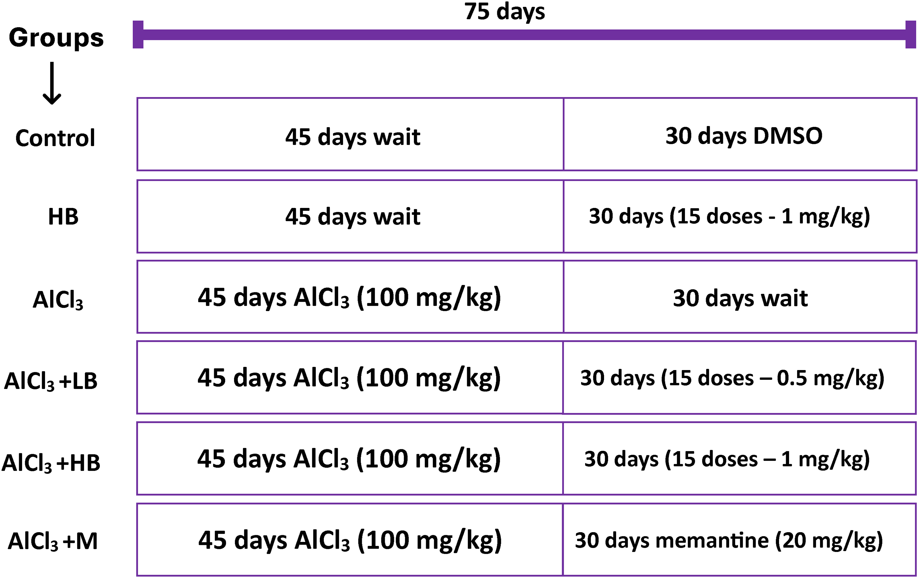
A scheme for experimental design.

Chemical structure of the new bithiophene derivative.
Drugs and chemicals
As previously reported, 4-((2,2′-bithiophen)-5-yl) benzamidine, or the derivative of bithiophene (Figure 2), was synthesized and characterized. 22 Just before the oral administration, it was dissolved in DMSO. Memantine hydrochloride and AlCl3 were purchased from Sigma (St Louis, Mo, USA). All other chemicals used were of analytical grade.
Morris water maze (MWM) behavioral test
After the final dose, the MWM test was conducted as described elsewhere. 25 The pool was divided (virtually) into four quadrants. It was filled with starch-turbid water to hide a 9-cm circular platform located in the target quadrant. Briefly the rats were trained four times a day for four days (learning trials). The distance cut by each rat to escape the pool was calculated. Then on the fifth day, the hidden platform was removed, and rats were allowed to freely explore the pool (Figure 3), and time spent in each quadrant was recorded (probe trial). Behavioral data were collected by recording all trials with a high-resolution Sony 20-megapixel camera. Subsequently, video recordings were analyzed using Python 3.9.8. The path taken by each rat from the drop-off point to the target zone during each trial was then visualized using Matplotlib Pyplot version 3.4.3.
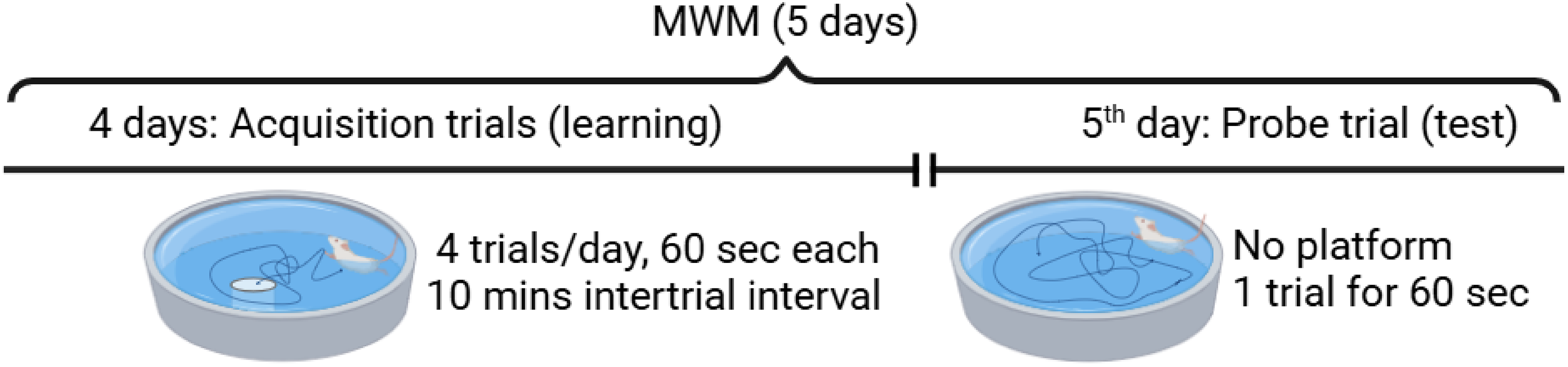
Timeline of Morris water maze behavioral test.
Tissue sampling and biochemical analyses
At the end of the experiment and after a 12-h fasting, all animals were weighed and sedated with isoflurane. Initially, the rats were quickly beheaded, and their brains were removed on ice and dissected. The hippocampi of the first six rats were weighed. Up until the execution of the biochemical measurements, all tissues were kept at −80°C. The tissues were homogenized in 10% (w/v) ice-cold phosphate buffer saline. The malondialdehyde (MDA), 26 reduced glutathione (GSH), 27 superoxide dismutase (SOD), 28 catalase, 29 total protein content, 30 monoamine oxidase activity, 31 and acetylcholinesterase activity, 32 were all measured using colorimetric assays, with absorbance measured spectrophotometrically as outlined in the associated references. The amyloid-β (Aβ1–42) was quantified using sandwich ELISA research kit (Biorbyt, UK. Cat# orb410690) using a microliter plate reader at 450 nm. The concentrations of Aβ1–42 were determined using a standard curve plotted on a graph.
The hippocampi removed from the remaining six rats were homogenized in either acidified butanol (for dopamine, norepinephrine, and serotonin) or 0.4 mol/L perchloric acid (for glutamate and GABA). The samples were then centrifuged at 10,000 rpm for 10 min at 4°C. After that, the supernatant was separated, aliquoted, and kept cold until it was examined. Colorimetric measurements were made of the biogenic amine neurotransmitters, which include serotonin, norepinephrine, and dopamine. 33 Glutamate and GABA, two neuroactive amino acid neurotransmitters, were evaluated using high-performance liquid chromatography (HPLC, Agilent 1260), as previously described. 34
Real-time PCR analysis
Using specific primers, the levels of gene expression for γ-secretase-activating protein (GSAP), glycogen synthase kinase 3-β (GSK3-β), p53, Bcl2, Caspase-9, and glyceraldehyde-3-phosphate dehydrogenase (GAPDH) as a housekeeping gene were assessed in the hippocampi from the last six animals. The primers listed in Table 1 were all purchased from Sigma (St Louis, MO, USA). Three halves of the hippocampi of these rats were used in the immunodetection of caspase 3.
Sequence of primers of the investigated genes and the house keeping gene.

Immunodetection of caspase 3
After the brains were rapidly dissected and washed in isotonic saline, they were fixed in 10% formalin for 24 h (Fischer, Cornell Lab, Egypt) and then dehydrated through an ascending series of ethanol concentrations. The samples were subsequently cleared in terpineol and embedded in paraffin wax (Leica, Wetzlar, Germany). The brain samples were sectioned to a thickness of 5 µm using a YiDi semi-automated microtome. The sections then underwent deparaffinization, hydration, and treatment with H2O2. Polyclonal rabbit anticaspase-3 (Neo Markers Laboratories, CA, USA) was used to stain the sections and a secondary antibody (goat antirabbit immunoglobulin antibody conjugated with peroxidase) was purchased from BioCare Medical (CA, USA). More details are given elsewhere. 35
Statistical analysis
The data are presented as mean ± SEM. The Kolmogorov-xSmirnov normality test was passed by every value, and One-Way Analysis of Variance (ANOVA) was employed. Subsequently, distinct groups were compared using Tukey's posttest for multiple comparisons. GraphPad Prism (8.02., 2020) for Windows was used for all analyses. At p < 0.05, the differences were deemed statistically significant.
Results
Oxidative stress analyses
Treating rats with AlCl3 resulted in a significant (p < 0.001) increase (283%) in the MDA hippocampal content. The low dose of bithiophene and memantine significantly lowered the MDA hippocampal levels (75.4%, p < 0.001 and 45%, p < 0.01, respectively) in the Al-intoxicated rats compared to the model group. The high dose bithiophene increased the MDA levels significantly (p < 0.01) by 46.9% compared to the AlCl3 group (Figure 4A). The aluminum treatment significantly (p < 0.001) increased the SOD by 1185%. Treating the Al-intoxicated rats with low or high dose of bithiophene, or memantine significantly (p < 0.001) lowered the SOD levels by 81%, 49% and 75% compared to AlCl3 group. Treating normal rats with a high dose of bithiophene significantly (p < 0.01) lowered the catalase levels by 50% in comparison to control rats (Figure 4B). Similarly, treating the normal rats with aluminum chloride decreased the catalase activity significantly (p < 0.001) by 78% in comparison to control rats. The Al-intoxicated rats showed the lowest catalase activity which was increased significantly upon treatment with low-dose, high-dose bithiophene, or memantine by 313%, 100%, and 350% respectively. The high dose bithiophene showed significantly lower catalase activity by 52% when compared to the low dose group (Figure 4C). Upon treating normal rats with a high dose of bithiophene, the GSH content decreased significantly (p < 0.001) by 42.8% in comparison to control rats. Also, treating the normal rats with AlCl3 caused the GSH to drop significantly (p < 0.001) by 86% in comparison to control rats. The Al-intoxicated rats showed the lowest GSH levels, these levels increased significantly (p < 0.001) when treated with low dose of bithiophene or memantine by 420% and 360%, respectively. The high dose bithiophene treatment showed significantly (p < 0.001) lower GSH levels (by 46%) when compared to the low dose of bithiophene, and insignificantly elevated the GSH level in the hippocampus compared to the AlCl3 group (Figure 4D).
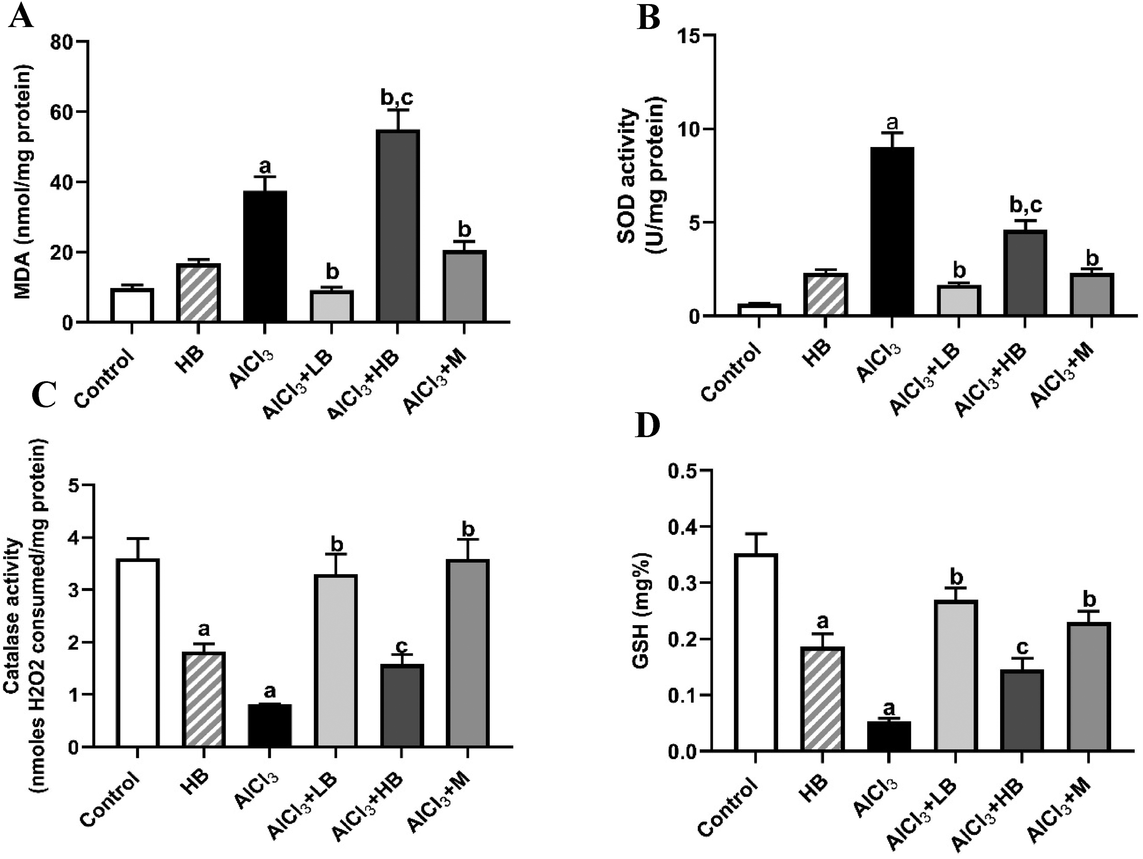
Effects of the different treatments on various oxidative stress parameters. (A) malondialdehyde (MDA), (B) superoxide dismutase (SOD), (C) catalase, and (D) reduced glutathione (GSH). The data are expressed as mean ± SEM, n = 6. aSignificant difference versus control. bSignificant difference versus AlCl3. cSignificant difference versus low dose bithiophene group. HB: high dose of bithiophene; LB: low dose of bithiophene; M: memantine.
Neurotransmitters and neurotransmitter catabolizing-enzymes
The aluminum insult caused a significant (p < 0.01) increase in the glutamate hippocampal content by 60% as compared to the control. This surge was significantly lowered upon treating the demented rats with either low dose of bithiophene or memantine by 48% (p < 0.001) and 29% (p < 0.05), respectively (Figure 5A). Al-insult caused no significant change in the GABA content in the hippocampus. Only the high dose of bithiophene caused a significant (p < 0.01) decrease in the hippocampal GABA content by 44% when compared to the model group (Figure 5B). The Al-insult also caused a significant (p < 0.01) increase in the level of norepinephrine (NE) by 98% in comparison to the control group. Then, treating the Al-insulted rats with low doses of bithiophene caused a significant (p < 0.05) reduction in the NE by 39% (Figure 5C). The Al-insult caused the dopamine levels to drop significantly (p < 0.05) by 36%. The low dose of bithiophene and the memantine caused significant (p < 0.001) surges in the levels of dopamine by 110% and 99%, respectively (Figure 5D). Treating normal rats with bithiophene high doses caused a surge (p < 0.001, 283%) in the serotonin levels compared to the control rats. While aluminum insult caused no significant change, memantine and high doses of bithiophene increased the serotonin levels significantly (p < 0.001) by 280% and 180%, respectively compared to the model group (Figure 5E).
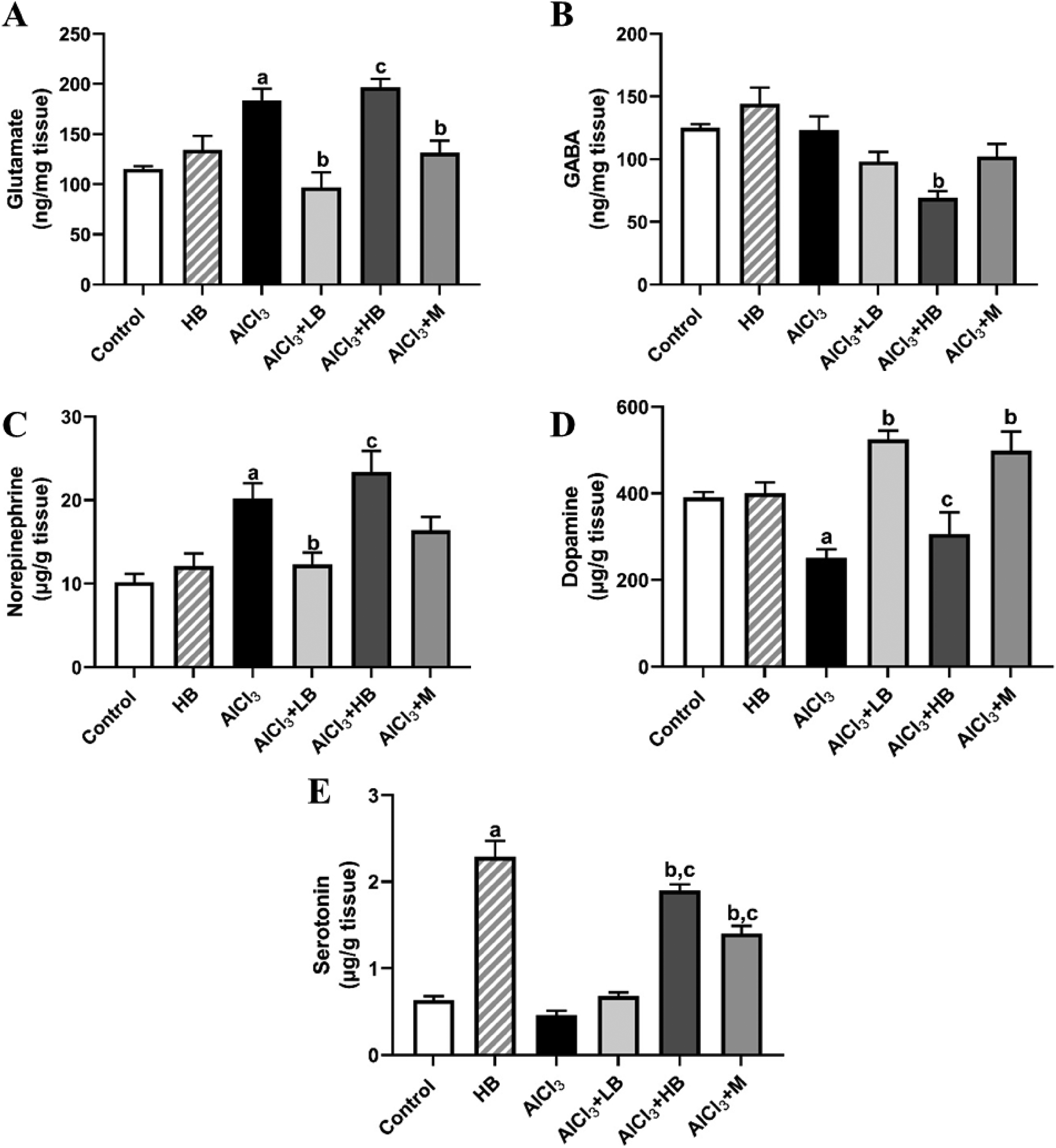
Effects of the different treatments on the neurotransmitters (A) glutamate, (B) GABA, (C) norepinephrine, (D) dopamine, and (E) serotonin. The data are expressed as mean ± SEM, n = 6. aSignificant difference versus control. bSignificant difference versus AlCl3. cSignificant difference versus the low dose bithiophene group. HB: high dose of bithiophene; LB: low dose of bithiophene; M: memantine.
The high dose of bithiophene conferred no significant change in acetylcholinesterase (AChE) or monoamine oxidase (MAO) activities in normal rats. However, aluminum treatment caused a significant (p < 0.05) increase in the enzyme activity by 43%. Only the high dose of bithiophene significantly (p < 0.01) lowered the AChE activity in the Al-intoxicated rats by 39% (Figure 6A). aluminum treatment caused a significant (p < 0.001) increase in the MAO activity by 120%. Treating the Al-intoxicated rats with low-, or high dose of bithiophene, or memantine caused significant reductions in the MAO activity by 76% (p < 0.001), 23% (p < 0.05), and 78% (p < 0.001), respectively. In lowering the MAO levels of the Al-intoxicated rats, the low dose bithiophene was significantly (p < 0.001) superior to the high dose by 230% (Figure 6B).
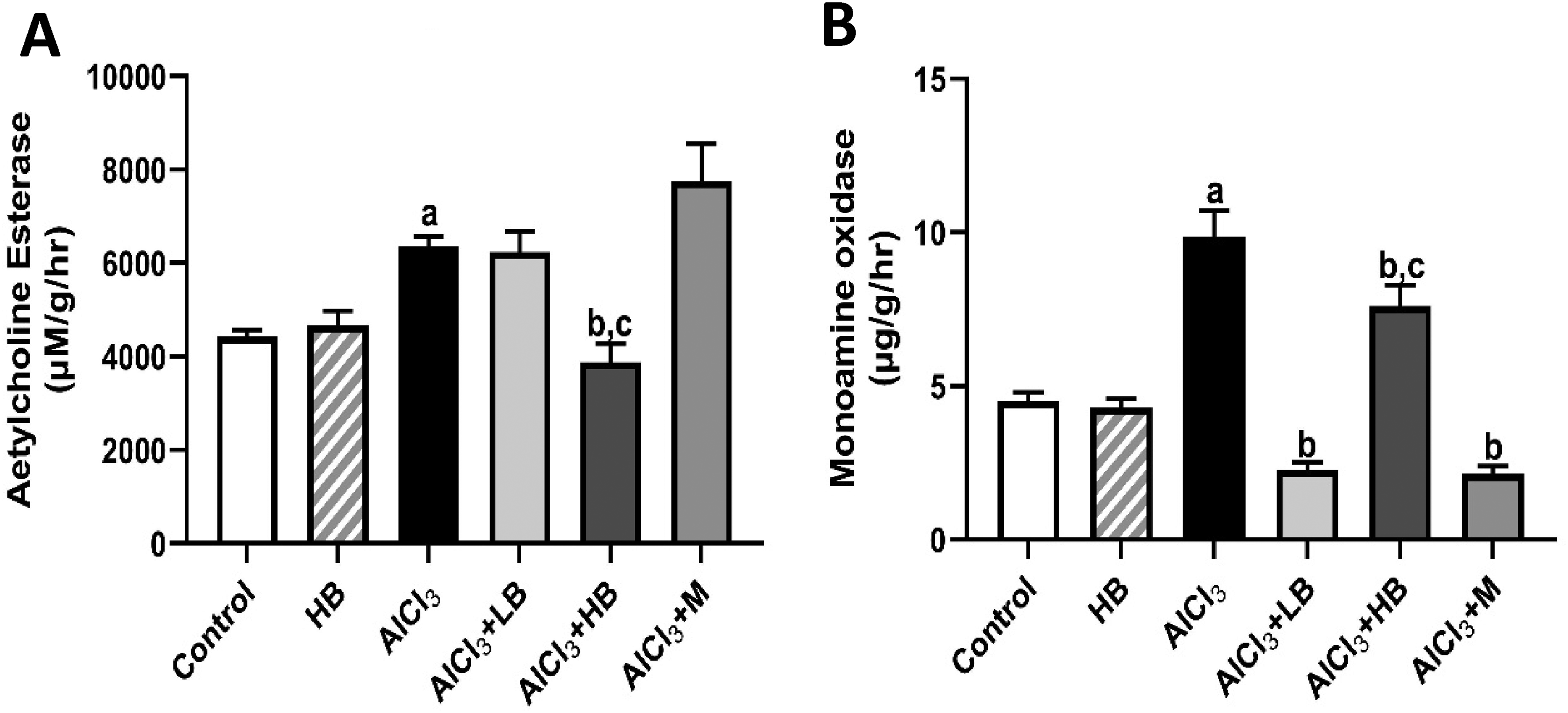
Effects of the different treatments on neurotransmitter catabolizing enzymes: (A) acetylcholine esterase (AChE), (B) monoamine oxidase (MAO). The data are expressed as mean ± SEM, n = 6. aSignificant difference versus control. bSignificant difference versus AlCl3. cSignificant difference versus the low dose bithiophene group. HB: high dose of bithiophene; LB: low dose of bithiophene; M: memantine.
Gene expression analysis
RT-PCR analyses showed that aluminum intoxication caused a significant (p < 0.001) 4-fold upregulation of the GSAP gene expression as compared to the control. This elevation was significantly (p < 0.001) downregulated by the low dose bithiophene (40 folds), high dose bithiophene (6.7 folds), and memantine (1.5 folds) when compared to the Al-intoxicated group (Figure 7A). Treating normal rats with bithiophene at the high dose caused a significant (p < 0.001) 16.6-folds downregulation of GSK3-β gene expression, while treating animals with aluminum caused a significant (p < 0.001) downregulation by only 2.2-fold compared to the control. Treating the Al-intoxicated rats with low dose of bithiophene or memantine caused a further significant (p < 0.001) downregulation by 3.8 and 3.1 folds, respectively (Figure 7B) compared to the Al-intoxicated animals. For Casp9, treating the normal rats with bithiophene high dose caused a significant (p < 0.001) 4.2-fold downregulation. aluminum treatment also caused a significant (p < 0.001) 3.2-folds downregulation. None of the treatments could reverse this downregulation (Figure 7C). Al3+ caused a significant (p < 0.001) 20-fold downregulation in the expression of p53. This gene was significantly (p < 0.05) upregulated, upon treating Al-intoxicated rats with bithiophene at low dose by 6.2 folds (Figure 7D). For Bcl2 gene, treating rats with bithiophene high dose or aluminum significantly (p < 0.001) downregulated the gene expression by ∼5-fold, and none of the treatments abated this down regulation (Figure 7E).
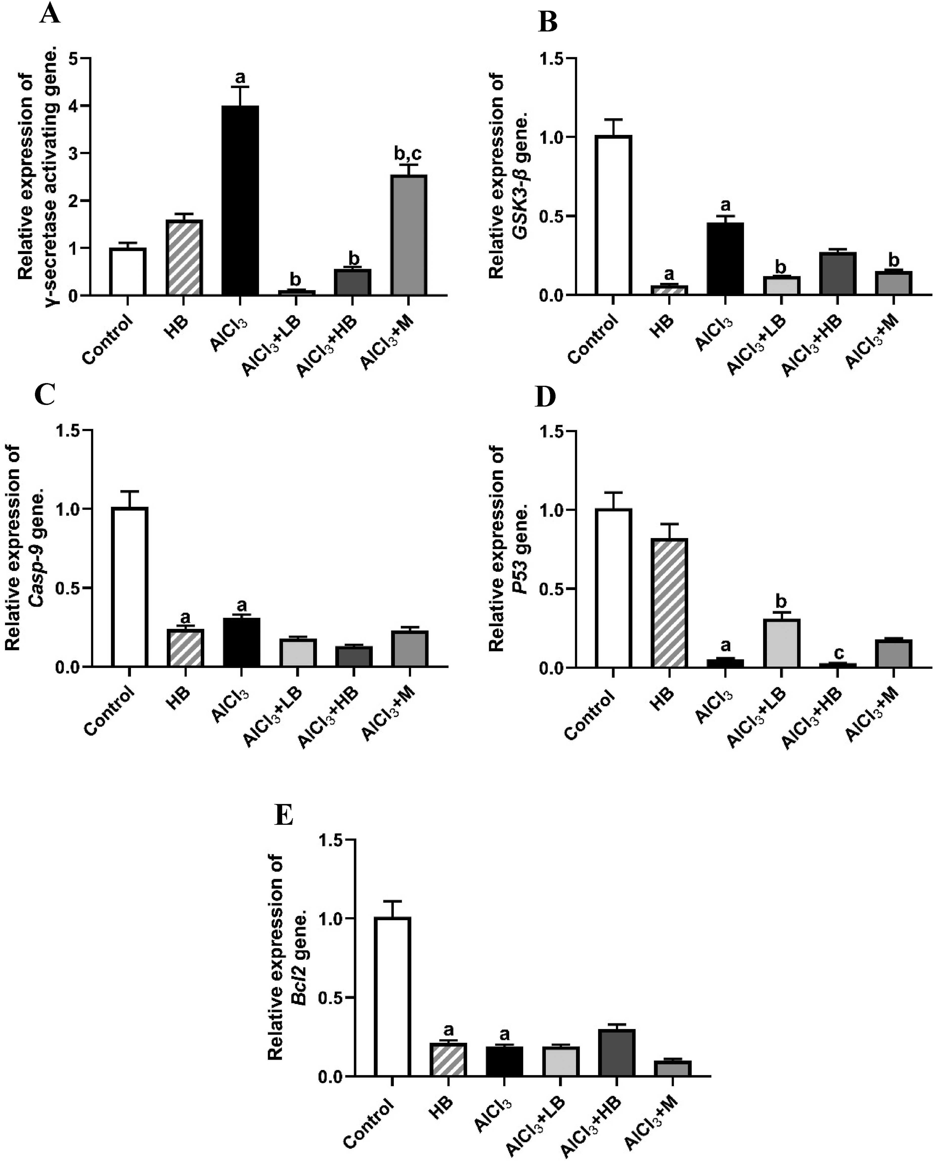
The effects of treatments on various genes expression: (A) γ–secretase activating protein, (B) GSK3-β, (C) Casp9, (D) p53; and (E) Bcl2. The data are expressed as mean ± SEM, n = 6, p < 0.05. aSignificant difference versus control. bSignificant difference versus model. cSignificant difference versus low dose bithiophene group.
Active cleaved caspase-3 analysis
The microscopic examination of the hippocampus for immunohistochemical expression of active cleaved caspase-3 in the different groups showed that the control group exhibited the faintest staining all through the hippocampal structure. Treating animals with bithiophene at high dose or Al3+ increased caspase-3 significantly by 200% (p < 0.05) and 500% (p < 0.001), respectively. While treating the Al-intoxicated rats with bithiophene low dose or memantine significantly reduced caspase-3 deposition by 72% (p < 0.001) and 51% (p < 0.01), respectively (Figure 8 and Table 2).

Photomicrographs of immunoreaction stain for caspase-3 in the hippocampi of rats, n = 3, (A) control group, (B) the bithiophene group, (C) Al-insulted group, (D) bithiophene low doses group, (E) bithiophene high dose group, and (F) the memantine group. Note that the control and the bithiophene groups show the least Casp-3 reactivity, and so the lightest stain, while the AlCl3 group shows the densest reaction. While the low dose of bithiophene and the memantine groups show moderate reactions.
Activated caspase 3 intensity score in the hippocampi.

The results are expressed as mean ± S.E.M of the relative area covered by the positive reaction in reference to the whole area, n = 3, aSignificant difference versus control. bSignificant difference versus AlCl3, cSignificant difference versus low dose bithiophene group at p < 0.05.
Amyloid-β (Aβ1–42) detection
Aluminum intoxication caused a significant (p < 0.001) increase in the Aβ deposition in rats’ hippocampi by 119%. Such heavy accumulation was significantly decreased by the bithiophene low dose (39%, p < 0.01), bithiophene high doses (29%, p < 0.01), or memantine (45%, p < 0.001) treatments (Figure 9).
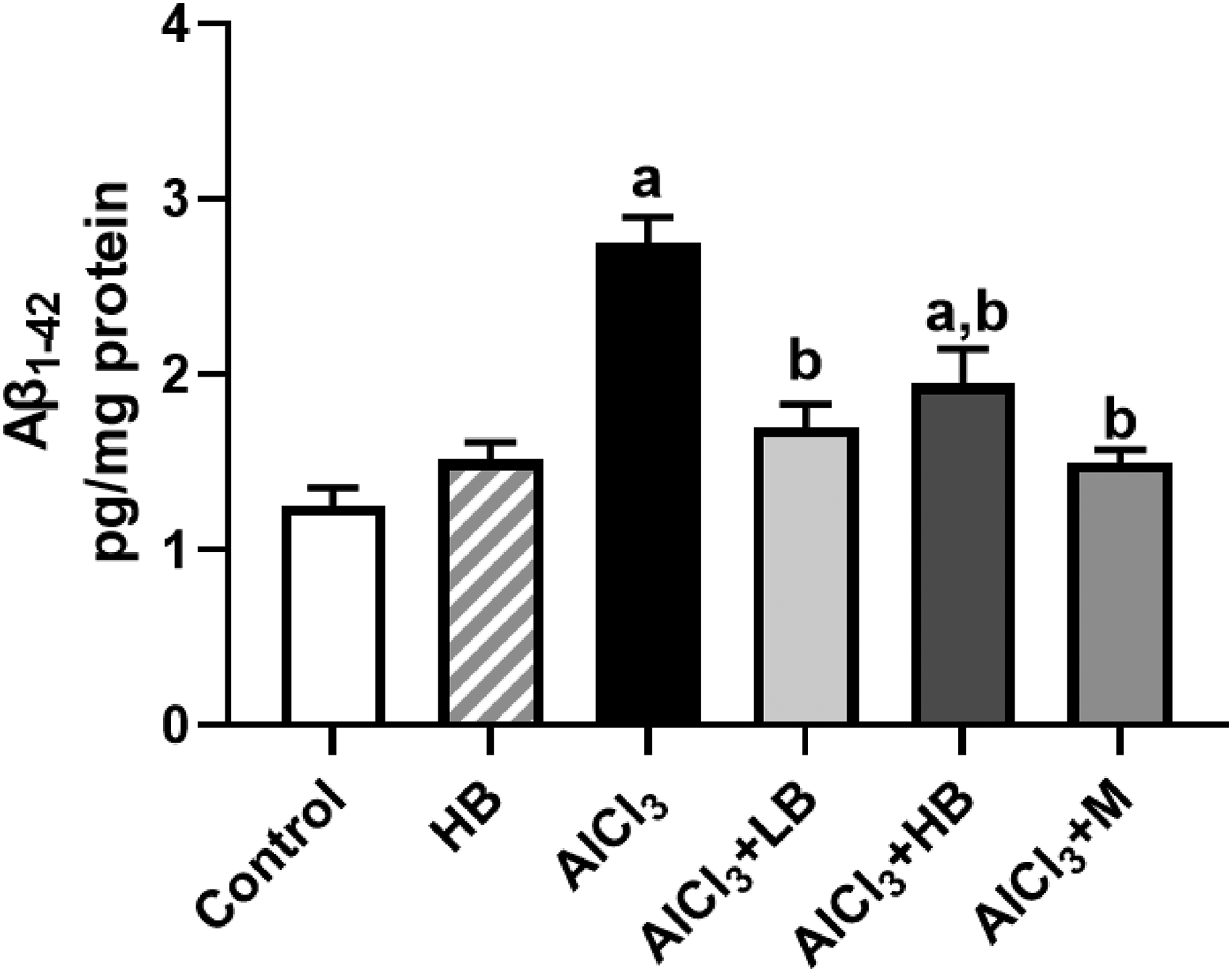
Effects of different treatments on Aβ1–42 concentration. The data are expressed as mean ± SEM, n = 6.
Behavioral analyses
Learning trials
In the control rats, the distance required for the rat to reach the platform decreased progressively as the training proceeded along the four consecutive days. The Al-insulted rats, however, consistently took significantly longer paths to reach the platform compared to the control group. This latency in the Al-insulted group was significantly shortened by high and low doses of bithiophene as well as memantine treatments. Low dose was more potent than the high dose and was equipotent to memantine (Figure 10A).

(A) Differences in the rate of learning among the different groups. (B) the time spent in the platform quadrant by the different groups. (C) Charts of the paths taken by the group type during the probe trials. The data are expressed as mean ± SEM. n = 6. aSignificant difference versus control. bSignificant difference versus AlCl3. cSignificant difference versus low dose bithiophene group. HB: high dose of bithiophene; LB: low dose of bithiophene; M: memantine; NW: Northwest; NE: Northeast; SW: Southwest; SE: Southeast; C1: Control; C2: high dose of bithiophene; C3: aluminum group; C4: rats with AD-like pathology treated with low doses of bithiophene; C5: rats with AD-like pathology treated with high doses of bithiophene; C6: rats with AD-like pathology treated with memantine.
Probe trials
The probe trial lasted for a minute and the time spent in each quadrant was divided by the whole minute and hence, the time share of each quadrant was calculated (Figure 10B). The control rats showed the highest platform quadrant (PQ) time, while the Al-insulted rats showed the lowest PQ time. This time was significantly increased by the treatment with low dose bithiophene or memantine (Figure 10B). The pathway charts for the different groups are shown in (Figure 10, C1-C6).
Discussion
The incidence of AD is rapidly increasing, without remedy currently available to halt its progression. While the precise mechanisms underlying AD development remain unclear. One of the factors that is thought to have a role in the initiation and development of the illness is exposure to aluminum. Aluminum can readily infiltrate the human body through ingestion. Moreover, studies have reported an association between aluminum exposure and increased blood-brain barrier permeability, potentially aggravating the pathogenesis of AD.11,36
Due to its high lipid content, considerable oxygen consumption, and low concentration of antioxidants, the brain is the most susceptible organ to oxidative stress. In the present study, aluminum intake caused a surge in MDA level and SOD activity in the hippocampal tissues in accordance with previous studies.26,37 It also caused a depletion in both catalase and reduced glutathione as previously reported. 38 Oxidative stress and lipid peroxidation can contribute to hippocampal dysfunction and neuronal damage observed in various neurological conditions including AD. 39 Production of reactive oxygen species (ROS), due to aluminum insult, caused a surge of SOD, the first line of defense. Enhanced SOD activity can lead to a paradoxical increase in H2O2 production which can then inhibit catalase activity and function. Apparently, the resulting oxidative stress and ROS overwhelmed the cellular antioxidant defense system leading to depletion of GSH through its reaction with ROS and subsequent formation of oxidized glutathione. 40 Nevertheless, low dose of bithiophene as well as memantine reversed the previous aluminum effects on MDA, SOD, catalase, and GSH with superiority of the bithiophene low dose over memantine in most cases. The bithiophene high dose failed to restore the levels of catalase and GSH and caused a further increase in MDA. The low dose bithiophene's antioxidative activity likely arises from its multifaceted mechanisms of action. Thiophene moieties within the bithiophene structure may function as metal chelators or as free radical scavengers by contributing electrons or hydrogen atoms to neutralize free radicals. 41 Furthermore, the bithiophene may modulate the activity of antioxidant enzymes as thiophene compounds have been shown to enhance SOD activity in a previous study. 42 Thiophenes have also been demonstrated to increase the cellular production of antioxidant molecules like glutathione improving the cellular oxidative milieu. 43
Another major hallmark of AD is the neural and synaptic loss, as cholinergic transmission plays a crucial role in memory and learning. AD is characterized by a cholinergic deficit, which can be attributed to either a decrease in acetylcholine (ACh) levels or increased AChE activity, the enzyme responsible for ACh degradation. AChE and MAO alterations are involved in the etiology of AD.38,44 Consistent with this established pathology, the present study reported a significant elevation in the hippocampal activity of AChE of the AD group, aligning with previous findings.45,46 Only high doses of bithiophene demonstrably reversed the Al-induced surge in the AChE activity. MAO, a mammalian flavoenzyme located in the outer mitochondrial membrane, has a vital function in the metabolism of monoamine neurotransmitters, which include norepinephrine, dopamine, and serotonin. Disruptions in the MAO activity have been implicated in various neuropsychiatric disorders such as depression or anxiety. 47 Al-intoxication enhanced oxidative stress which caused a surge in the MAO activity. 48 Again, together with memantine, the bithiophene low doses brought the surge of MAO caused by Al to the normal ranges.
Glutamate, the central nervous system main excitatory neurotransmitter, plays a crucial role in learning and memory. Beyond its well-established function in long term potentiation via N-methyl-D-aspartate receptor activation, Glutamate is essential for synaptic plasticity and memory consolidation. 49 In the present study Al-intoxication caused a significant increase in glutamate levels. Oxidative stress and ROS accumulation are well-known inducers of mitochondrial dysfunction, which can further exacerbate glutamate production, leading to glutamate excitotoxicity. 50 Both the low dose of bithiophene and the memantine treatments significantly reduced glutamate levels. Their propensity to lower ROS and oxidative stress may be the cause of this impact. The main inhibitory neurotransmitter in the central nervous system is GABA. It has long been known that Al damages GABAergic neurons. 51 In the present study however, Al-intake did not affect the GABA content in the hippocampus. Interestingly, treatment of Al-intoxicated rats with high-dose bithiophene resulted in a significant decrease in GABA levels compared to the aluminum group. This finding necessitates further exploration of the mechanisms by which bithiophene affected the GABAergic system. The potential applications of bithiophenes’ effects in neurological diseases characterized by disturbances in GABA, such as anoxic-ischemic injury and schizophrenia, offer promising paths for future research and clinical exploration. NE level increased after Al-insult due to deteriorations in the cellular oxidative milieu or increased de-novo synthesis. The co-occurrence of norepinephrine disturbance with cholinergic dysfunction in AD 52 underscores the potential importance of targeting both neurotransmitters while searching an effective treatment. Only the low dose of bithiophene significantly reversed that increase in NE caused by aluminum probably due to its strong antioxidant activity. Aluminum is known to inhibit dopamine β-hydroxylase; the enzyme that synthesizes dopamine, 53 or it can boost the catabolizing enzyme MAO 54 as reported in the current study, which results in a decrease in dopamine content. ROS resulting from aluminum damages dopaminergic neurons and interferes with dopamine release and signaling. In the current investigation, memantine and low dose bithiophene both effectively restored the dopamine depletion caused by aluminum in the hippocampal regions. It has been demonstrated that several licensed medications for AD, including rivastigmine, donepezil, and memantine, increase dopamine levels in the brain tissues. 55 Serotonin is a monoamine neurotransmitter that regulates several physiological processes, including mood, food, emotions, sleep, and memory. 56 The current investigation found no significant change in serotonin levels due to aluminum intoxication. The high dose of bithiophene or memantine administered to the rats that were exposed to Al dramatically raised the levels of serotonin in the hippocampal tissue. The capacity of memantine and bithiophene high dose to reduce MAO activity or to lessen oxidative stress is thought to be responsible for the increase in serotonin that results from these agents. However, the low dose bithiophene lowered the MAO activity and exhibited a strong antioxidant activity but did not affect the serotonin level.
The gamma-secretase complex, which regulates the cleavage of the amyloid precursor protein and the production of Aβ peptides that clump together to form the dangerous plaques essential to the pathogenesis of AD, is mostly regulated by γ-secretase activating protein (GSAP). 57 Al upregulated the GSAP expression in this study. The effects of aluminum exposure on this gene have not been studied before. However, Al-intoxication is known to increase the expression of the γ-secretase gene. 58 Bithiophene low and high doses as well as memantine significantly reversed the Al-induced upregulation of GSAP. The amelioration of this gene could be attributed to the antioxidative capacity of bithiophene and memantine, but more investigation is necessary.
Al-intoxication unexpectedly resulted in a significant downregulation of GSK-3β which is a constitutively active serine/threonine kinase engaged in brain development, metabolism, and regulation of cell cycle. 59 This was very confusing as elevated level of GSK-3β has been associated with reduced ACh synthesis and increased esterification, contributing to the cholinergic deficit observed in AD. Notably, both the low dose of bithiophene and the memantine treatments caused further reductions in the expression of this gene. This finding aligns with previous reports linking GSK-3β upregulation in AD to hyperphosphorylation of tau and subsequent learning impairment. 60 It is noteworthy that GSK-3β downregulation within the hippocampus has been observed in late stages of neurodegeneration, 61 potentially explaining the differences between our findings and established roles of GSK-3β in the early stages of AD. 62
In addition, caspase-9 upregulation and activation has been linked to increased expression of GSK-3β and elevation of GSK-3β-mediated hyperphosphorylation of tau. 63 Caspase-9, a cysteine protease belonging to the caspase family, critically regulates apoptosis, a central process in AD-associated neurodegeneration. Functioning as the intrinsic pathway's key initiator caspase, it is activated by factors like oxidative stress or mitochondrial dysfunction. 64 Al-intoxication caused a significant downregulation of casp-9, while none of the treatments reversed such effect. In AD, neuronal death occurs through apoptosis, and caspase 9 activation is a part of it. Yet, some research suggest caspase 9 reduction is more prominent in later stages of AD. 65 We can propose that aluminum reduced caspase 9 which in turn decreased the expression of GSK-3β indicating that the model we used in this study represents a late stage of AD-like pathology.
The tumor suppressor gene p53, plays a critical role in maintaining healthy cells by regulating cell division, repairing DNA damage, and triggering apoptosis when necessary. p53 dysfunction has been linked to AD. 66 Our study revealed that aluminum intoxication significantly reduced p53 expression in the hippocampus. This suggests that aluminum-induced apoptosis in our model might occur independently of the well-established p53 pathway. The decreased p53 expression led to decreased DNA repair allowing neurons to accumulate mutations and so increasing their liability to neurodegeneration. Interestingly, only the low-dose bithiophene treatment effectively restored p53 expression to near-normal levels, highlighting its potential role in mitigating the negative effects of aluminum exposure. Another key player in intrinsic apoptosis is caspase 3 which is the main executioner caspase. It plays a critical role in the final stages of apoptosis by initiating the dismantling of cellular components. 67 Caspase-3 activation has additionally been implicated in promoting tau hyperphosphorylation. 68 Aluminum treatment significantly increased caspase-3 activity compared to the control group. Notably, both low-dose bithiophene and memantine treatments effectively normalized caspase-3 levels, with bithiophene exhibiting superior efficacy. This enhanced effect of bithiophene might be attributed to its well-documented radical scavenging and potent antioxidant properties associated with thiophene moieties. Caspase 3 is known to be activated by initiator caspase 9 through p53 cascade when the damage is severe and exceeds the repair mechanisms. However, in the current study the expression of p53 and caspase9 was downregulated by aluminum. Many studies reported that caspase 3 could be activated and upregulated but not through the classical pathway of p53/caspase9. Caspase 3 was elevated without induction of caspase 9, 69 or not through p53. 70 Caspase 3 was even elevated with parallel downregulations in both caspase 9 and p53. 71 The molecular mechanism behind this elevation of caspase 3 activity needs further investigation. Bcl-2, a protein critical for cell survival, acts as a gatekeeper in apoptosis within cells. Studies suggest disruptions in Bcl-2 expression are associated with AD. 72 Aluminum intoxication significantly reduced Bcl-2 expression in the hippocampus, potentially due to the oxidative stress. This stress may activate pathways promoting cell death or disrupt mitochondria, both of which could contribute to Bcl-2 downregulation and further protein depletion, yet none of the treatments was able to reverse this effect.
The Aβ is normally secreted from neurons and degraded. However, in AD patients it is secreted and aggregated into insoluble plaques that are potentially neurotoxic. Prolonged administration of AlCl3 significantly increased Aβ1–42 levels due to either increased production or impaired clearance due to its ability to upregulate GSAP. 73 Bithiophene as well as memantine significantly reduced the Aβ1–42 levels to normal range. One possible explanation for such result is the ability of thiophenes to enhance the Aβ clearance from the brain by activating microglia. In our previous recent study, activated microglia were demonstrated in histological sections of bithiophene treatments. 74 We have also recently shown that aluminum exposure resulted in severe degeneration of both the histological and ultrastructural aspects of CA1 pyramidal cells, as well as the thickness of the stratum granulosum in the dentate gyrus. The low dose of bithiophene restored the normal histological and cytological structure of both cortical and hippocampal neurons affected by aluminum treatment. 74
We evaluated the spatial reference memory through the MWM test. 75 Successful navigation in the MWM task relies on two key pillars: the development of coping strategies and spatial learning. 76 The first involves the rodent adapting to the stressful environment by utilizing spatial working memory. The second pillar, spatial reference memory, comes into play as the animal learns the platform's fixed position within the maze. In the present study, AlCl3 daily oral intake caused Al-intoxicated rats to fail the MWM test, taking significantly longer times and distances in the learning trials and significantly lengthy paths in the probe trials. As well as the least amount of time in the target quadrant. These findings go in accordance with those of a previous study. 77 Bithiophene low dose treatment reversed the effects of Al on memory deficits by showing significantly shorter paths to reach the platform during the learning trials, even shorter than those treated with the memantine. It also showed the highest time shares in the platform quadrant. Only the bithiophene low dose and the memantine treatment showed significantly higher time share in the platform quadrant. In this behavioral investigation, we used the standard procedure of acquisition and extinction. The ability of acquisition (learning trials) and recall of memories was represented in the escape latency. In the Al-insulted group per se, dementia was observed as longer distances and times required by the Al- insulted rats to reach the escape platform compared to the control rats. In the extinction phase (probe trial) the Al-insulted rats also proved demented by significantly taking shorter times searching for the supposedly hidden platform in the target quadrant.
Conclusion
This study demonstrated the therapeutic potential of bithiophene in reversing the Al-induced neurodegeneration in a rat model of AD. Bithiophene achieved this action through ameliorating oxidative stress, improving cognitive functions, enhancing synaptic plasticity, preventing the Aβ accumulation, and reducing apoptosis. Taken together, these findings proved that bithiophene represents a promising candidate for AD that deserves further research.
Footnotes
Acknowledgments
The authors thank Dr Mohamed A Ismail, Professor of Organic Chemistry at Mansoura University, for supplying the bithiophene material.

Author contributions
Kholoud AbdElRaouf (Data curation; Formal analysis; Investigation; Methodology; Validation; Visualization; Writing – original draft); Hussein SH Farrag (Methodology; Supervision); Monir A El-Ganzuri (Supervision); Wael M El-Sayed (Conceptualization; Data curation; Formal analysis; Supervision; Validation; Visualization; Writing – review & editing).
Funding
The author(s) received no financial support for the research, authorship, and/or publication of this article.
Declaration of conflicting interests
The author(s) declared no potential conflicts of interest with respect to the research, authorship, and/or publication of this article.
Data availability
All data are presented in the manuscript.