



Abstract
Background
A disintegrin and metalloproteinase 17 (ADAM-17) has multiple pathophysiological functions in Alzheimer's disease (AD). However, the clinical relevance of ADAM-17 in AD is not clear yet.
Objective
This study aims to investigate the levels of circulating ADAM-17 and their association with AD.
Methods
This cross-sectional study recruited 40 normal cognition (NC) participants and 36 AD patients. Plasma ADAM-17 and biomarkers of neurodegeneration were determined. The association of plasma ADAM-17 with cognitive functions and biomarkers of neurodegeneration was analyzed.
Results
Plasma ADAM-17 levels were elevated in AD patients in comparison with NC subjects. Plasma ADAM-17 was positively associated with Clinical Dementia Rating (CDR) scores, but negatively associated with the Mini-Mental State Examination scores and Montreal Cognitive Assessment scores. Plasma ADAM-17 levels were positively associated with the levels of Aβ40, Aβ42, and p-Tau181.
Conclusions
These findings suggest a link between ADAM-17 and the pathogenesis of AD from a clinical perspective.
Introduction
Alzheimer's disease (AD) is currently the seventh leading cause of death worldwide and one of the main causes of disability and inability among older adults.1,2 The main pathological hallmarks of AD include extracellular senile plaque composed of amyloid-β (Aβ) and intracellular neurofibrillary tangles composed of hyperphosphorylated tau protein. 3
As an α-secretase enzyme of amyloid-β protein precursor, a disintegrin and metalloprotease 17 (ADAM-17) can compete with β-secretase, which could cleave Aβ precursor protein to generate Aβ,4–10 thus promoting the nonamyloidogenic pathway. Furthermore, loss-of-function mutation of ADAM-17 is associated with the onset of familial AD, suggesting a protective role of ADAM-17 in the AD brain. 11 However, ADAM-17 is also involved in inflammatory pathways in the brain.12–15 Therefore, ADAM-17 has complex pathophysiological functions in the brain.
Currently, there is no research on the clinical relevance of ADAM-17 in AD patients. Therefore, this study aimed to investigate the association between ADAM-17 and AD clinical severity and biomarkers of neurodegeneration. We examined the levels of ADAM-17 in AD patients and explored their association with clinical severity of AD and biomarkers of neurodegeneration.
Methods
Study population
This study recruited individuals with cognitive complaints who visited the memory clinic at the Department of Neurology, Daping Hospital between January 2022 and December 2022. Subjects with normal cognition were recruited from the Health Examination Center from the same hospital during the same period. A total of 40 subjects with normal cognition (NC) and 36 subjects diagnosed with AD were included in this study. The inclusion criteria included: (1) willingness to participate; (2) NC subjects should have no memory complaints and tested normal in cognitive assessment; (3) AD subjects should have cognitive impairment in cognitive screening. The exclusion criteria included: (1) had a coexisting disorder that may affect cognition, such as schizophrenia, infections of the central nervous system, Parkinson's disease, and autoimmune encephalitis; (3) had any type of tumor; (4) had a systemic inflammatory disease, chronic infectious disease, or acute infection during the past 4 weeks; (5) declined to participate or blood sampling. Written informed consent for participation and blood sampling was obtained from all participants or their legal guardians. The research protocol fulfills the criteria of the Ethics committee of Daping Hospital, Third Military Medical University.
Clinical assessments and diagnosis of AD
All participants of the study were recruited from a single cohort, the China Aging and Dementia Study (CADS) from the Daping Hospital, Chongqing, China. The CADS cohort was established as a research platform, following the 2011 National Institute on Aging and Alzheimer's Association (NIA-AA) recommended Aβ deposition pathologic tau, and neurodegeneration (ATN) research framework,16,17 to characterize neurodegeneration-related cognitive impairment diseases and NC individuals. The general cognitive functions were examined with the Mini-Mental State Examination (MMSE). Subjects with an MMSE score below 27 were further subjected to a battery of neuropsychological tests, including the Clinical Dementia Rating (CDR), Montreal Cognitive Assessment (MoCA), Activities of Daily Living (ADL), Hachinski Ischemic Scale (HIS), and Boston Naming Test.18–22 CDR was used to judge cognitively normal (CDR = 0), MCI (CDR = 0.5), and dementia (CDR ≥ 1). About 1/3 of all subjects with a CDR of 0.5 or above underwent an MRI scan, an Aβ-amyloid PET scan using 11C Pittsburgh Compound B (PiB) tracer, and an FDG-PET scan to confirm the cause of cognitive decline. Additionally, AD participants received apolipoprotein E (APOE) genotyping according to the established protocol in our laboratory.23,24
Measurements of plasma ADAM-17, Aβ40, Aβ42, t-Tau, and p-Tau181
Fasting blood was collected from all participants between 07:00 and 08:00 am. The blood samples were centrifuged within one hour after collection, and the ethylenediamine tetraacetic acid (EDTA) plasma was equally divided into 0.5 mL polypropylene test tubes and stored at −80°C until use. Written informed consent was obtained from all participants and their legal guardians before blood samples were collected.
After collecting the plasma samples from all participants, a human ADAM-17 combined with an immunosorbent assay (ELISA) kit (Jingbai Biology Co., Ltd, Hangzhou, China) was used to determine the levels of ADAM-17 in plasma. Plasma levels of Aβ40, Aβ42, and t-Tau were measured by the commercially available single molecular array (SIMOA) human neurological 3-Plex A test kit (Quanterix, USA) using automated SIMOA HD-1Z analyzer (Quanterix, USA). Plasma levels of p-Tau181 were measured by the commercially available SIMOA human p-Tau181 test kit (Quanterix, USA) using an automated SIMOA HD-1Z analyzer (Quanterix, USA).
Statistical analysis
For each plasma cytokine, the normality distribution was checked using the Shapiro-Wilk test. Since ADAM-17 levels did not exhibit a normal distribution, they were natural logarithmic (ln) transformed for analysis. Continuous variables were described as means ± standard deviations (SD) or median (IQR) where appropriate, while classified data were summarized as numbers and frequencies. The frequency differences of gender and APOE ε4 were evaluated by chi-square test. Differences in other demographic characteristics and ADAM-17 levels were tested by a two-sample independent t-test (or students’ t-test and Welch's correction if the F test showed significant differences between groups). We used partial correlation analysis to adjust for age, sex, and education levels when analyzing the correlation between ADAM-17 levels and Aβ and tau levels. The computations were performed with SPSS version 20.0 (SPSS Inc., United States).
Results
Characteristics of participants
The characteristics of the participants are shown in Table 1. There was no difference in age, sex, education level or frequencies of diabetes mellitus, hypertension, and cardiovascular disease between the AD group and the NC group. As expected, the mean MMSE scores of the AD dementia group were significantly lower than those of the NC group (Table 1).
The characteristics of all participants.

NC: normal cognition; AD: Alzheimer's disease; MMSE: Mini-Mental State Examination; APOE: apolipoprotein E; SD: standard deviation.
Plasma ADAM-17 is upregulated in AD patients
The overall levels of plasma ADAM-17 levels in AD subjects were higher than those in NC subjects (mean ± SD: 467.6 ± 682.8 versus 222.2 ± 184.7 pg/ml, p = 0.032; Figure 1(a)). Furthermore, the difference was even more significant between the APOE ε4 (+) AD patients and NC subjects (mean ± SD: 668.0 ± 833.8 versus 225.9 ± 191.7 pg/ml, p = 0.003; Figure 1(b)). After logarithmic transformed, levels of plasma ADAM-17 levels in AD subjects and NC subjects have no significant difference (mean ± SD: 5.543 ± 0.967 versus 5.220 ± 0.534 pg/ml, p = 0.072; Figure 1(c)), but the difference was significant between the APOE ε4 (+) AD patients and NC subjects (mean ± SD: 5.895 ± 1.088 versus 5.224 ± 0.555, p = 0.003; Figure 1(d)), and between the APOE ε4 (+) AD patients and the APOE ε4 (-) AD patients (mean ± SD: 5.895 ± 1.088 versus 5.151 ± 0.636, p = 0.019; Figure 1(d)).
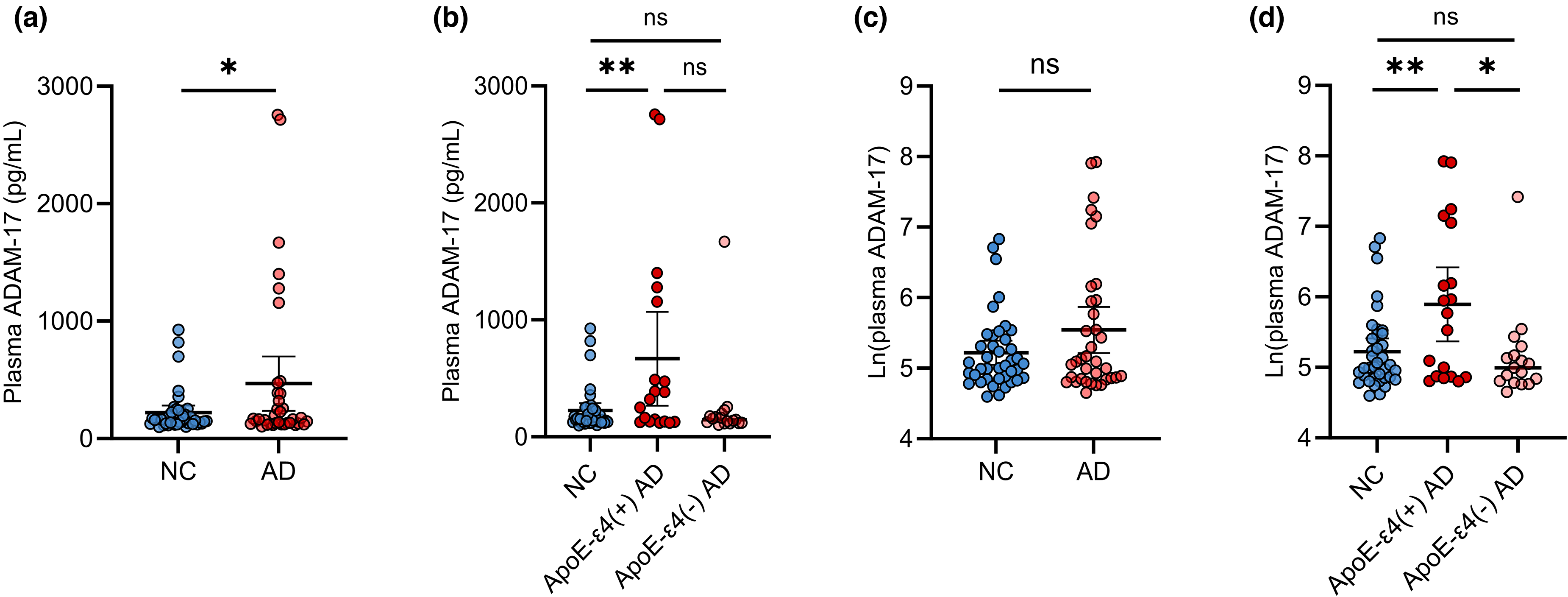
Comparison of plasma ADAM-17 levels and ln (plasma ADAM-17 levels) between different groups. (a) Comparison of plasma ADAM-17 levels between NC subjects and AD patients. (b) Comparison of plasma ADAM-17 levels between NC subjects, APOE ε4 (+) AD patients and APOE ε4 (-) AD patients. (c) Comparison of ln (plasma ADAM-17) between NC subjects and AD patients. (d) Comparison of ln (plasma ADAM-17) between NC subjects, APOE ε4 (+) AD patients and APOE ε4 (-) AD patients. ADAM-17: A disintegrin and metalloprotease 17; NC: normal cognition; AD: Alzheimer's disease; APOE: apolipoprotein E.
Plasma ADAM-17 is associated with the AD biomarkers
In AD patients, plasma ADAM-17 levels were positively correlated with Aβ40 (γ = 0.331, p = 0.049; Figure 2(a)), Aβ42 (γ = 0.359, p = 0.032; Figure 2(b)), and p-Tau181 levels (γ = 0.377, p = 0.024; Figure 2(e)). In NC subjects, there was no correlation between ADAM-17 levels and Aβ40 (γ = −0.040, p = 0.808; Figure 2(a)), Aβ42 (γ = 0.077, p = 0.636; Figure 2(b)), or p-Tau181 levels (γ = 0.022, p = 0.893; Figure 2(e)). Among NC subjects and AD patients, plasma ADAM-17 levels were not correlated with Aβ42/40 (Figure 2(c)) and t-Tau levels (Figure 2(d)).
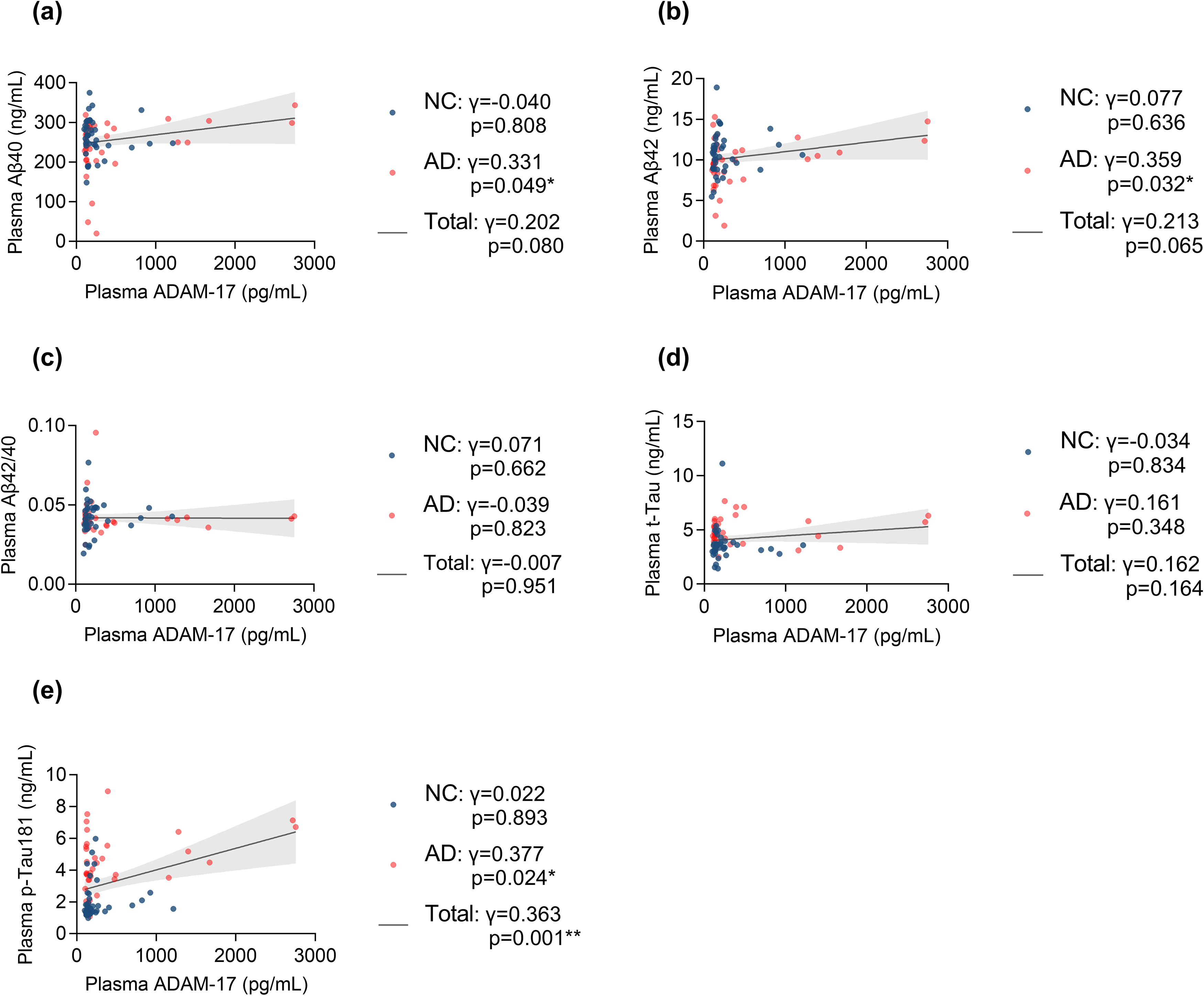
Correlation analysis between plasma ADAM-17 and plasma aβ, tau levels. (a) Correlation between ADAM-17 levels and Aβ40 levels. (b) Correlation between ADAM-17 levels and Aβ42 levels. (c) Correlation between ADAM-17 levels and Aβ42/40. (d) Correlation between ADAM-17 levels and t-Tau levels. (e) Correlation between ADAM-17 levels and p-Tau181 levels. ADAM-17: A disintegrin and metalloprotease 17; Aβ: amyloid-β; AD: Alzheimer's disease; NC: normal cognition.
Plasma ADAM-17 is associated with cognitive functions
In AD patients, plasma ADAM-17 levels were negatively correlated with MMSE scores (γ = −0.343, p = 0.040; Figure 3(a)) and MoCA scores (γ = −3651, p = 0.0285; Figure 3(b)), and positively correlated with CDR scores (γ = 0.4928, p = 0.0026; Figure 3(c)), but no correlation with ADL scores, HIS scores and Boston Naming Test scores. In NC subjects, there was no correlation between ADAM-17 levels and MMSE scores (γ = 0.057, p = 0.727; Figure 3(a)).
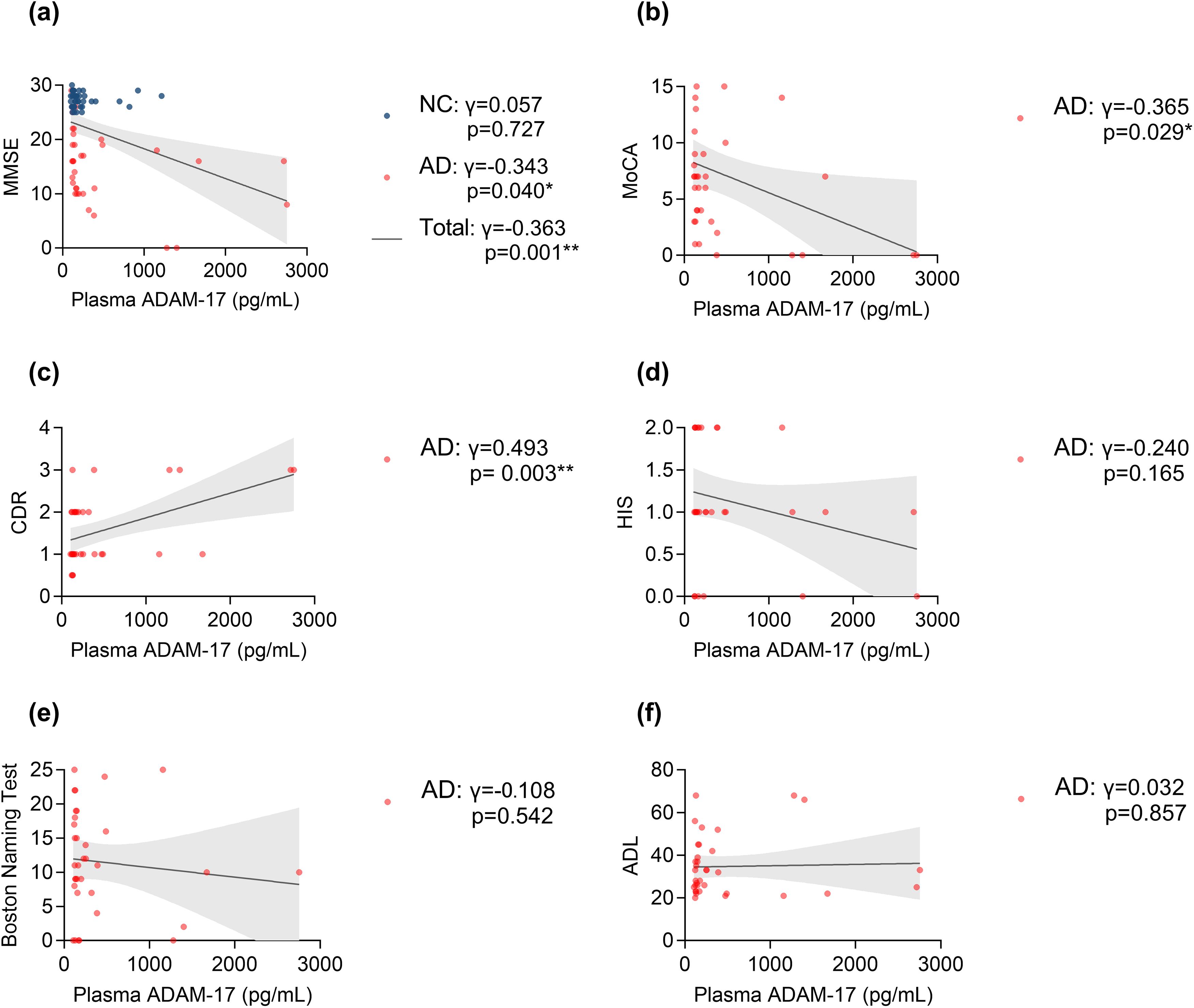
Correlation analysis between plasma ADAM-17 and cognitive function scores. (a) Correlation between ADAM-17 levels and MMSE scores. (b) Correlation between ADAM-17 levels and MoCA scores. (c) Correlation between ADAM-17 levels and CDR scores. (d) Correlation between ADAM-17 levels and HIS scores. (e) Correlation between ADAM-17 levels and Boston Naming Test scores. (f) Correlation between ADAM-17 levels and ADL scores. ADAM-17: A disintegrin and metalloprotease 17; AD: Alzheimer's disease; NC: normal cognition; TG: Total group; MMSE: Mini-Mental State Examination; MoCA: Montreal Cognitive Assessment; CDR: Clinical Dementia Rating; HIS: Hachinski Ischemic Scale; ADL: Activities of Daily Living.
Discussion
In this cross-sectional study, we investigated the clinical relevance of plasma ADAM-17 in AD in a Chinese cohort. Briefly, we found that plasma ADAM-17 is upregulated in AD patients. Furthermore, plasma ADAM-17 is associated with the severity of cognitive impairment and biomarkers of neurodegeneration.
The circulating levels of ADAM-17 have rarely been investigated in AD. A previous study reported that cerebrospinal fluid ADAM-17 activities are upregulated in AD patients. 25 Another study found upregulated activities of ADAM-17 in plasma in patients with mild cognitive impairment and AD dementia. 26 Consistently, a study conducted in the Chinese population also identified increased ADAM-17 activities in AD patients, 27 which is in accordance with our present findings. The upregulation of ADAM-17 as a consequence of AD or an initiation event in AD is not clear yet.
As an α-secretase, ADAM-17 has a physical protective role in the brain by inhibiting the production of Aβ.5,6,9,10 Consistently, a rare loss-of-function variant in ADAM-17 is linked to an increased risk of developing AD, 11 suggesting the complete loss of function of ADAM-17 is pathogenic in AD. However, there has been increasing evidence pointing to the opposite conclusion that ADAMs targeting cytokines and chemokines can actually cause inflammatory processes, and consequently neurodegeneration.12–15 Therefore, there are two possible mechanisms underlying the increase in ADAM-17 in AD patients. The first possibility is that the increase in this α-secretase is a consequence of AD, serving as an adaptive response to resist the accumulation of Aβ in the brain. The second possibility is that the increase in ADAM-17 is an upstream event of AD that contributes to the formation of the inflammatory environment in the brain. However, we could address this issue based on the current findings.
In the present study, we discovered that plasma ADAM-17 is associated with biomarkers of neurodegeneration, such as Aβ42, Aβ40, and p-Tau181. These biomarkers reflect brain amyloidosis and the severity of neuronal injury with high accuracy.28–30 Therefore, we believe that ADAM-17 might be a reliable indicator of brain neurodegeneration in AD patients. This notion is supported by the finding that APOE ε4 carriers had higher levels of ADAM-17 than noncarriers, with the general understanding that APOE ε4 carriers typically exhibit a higher burden of amyloid in the brain compared to noncarriers. 31 Previous research has shown that APOE ε4 gene promotes abnormalities in microglia, causing neurotoxic microglia-derived factor TNF-α. 32 The hydrolysis product sTNFR of the latter correlated strongly with ADAM-17 activity in AD subjects. 25 Furthermore, ADAM-17 could cleave TNFR2 signaling pathway, 33 which is a neuroprotective pathway. 34 So it is a reasonable supposition that APOE ε4 promotes the expression of TNF-α, thus increasing ADAM-17. Afterwards, they both promote neuroinflammation, tau phosphorylation and Aβ production.
This study has several limitations that should be noted. Firstly, the small sample size and cross-sectional nature may restrict the evaluation of its findings. Longitudinal cohort studies would be beneficial for establishing a causative relationship between ADAM-17 upregulation and the development of AD from a clinical perspective. Mechanistic studies are also necessary to address this issue. Secondly, we did not investigate the changes in ADAM-17 along the AD continuum, which is crucial for understanding the role of ADAM-17 in the progression of the disease. Thirdly, the proportion of APOE ε4 carriers among the healthy controls is not clearly specified, which leads to our lack of comprehensive research on APOE, and inability to be adjusted by APOE genotype. This issue needs to be further investigated in the future. Lastly, the correlation between circulating levels of ADAM-17 and its levels in the brain remains undetermined. Therefore, it is imperative to measure the levels of ADAM-17 in the cerebrospinal fluid as they may provide a more accurate reflection of the brain environment. However, this study provides valuable clinical evidence that supplements the findings of the increased activities of ADAM-17 in AD.
Footnotes
Acknowledgments
The authors have no acknowledgments to report.
ORCID iDs

Author contributions
Zu-Qi Chen (Conceptualization; Data curation; Investigation; Methodology; Resources; Software; Writing – original draft); Meng-Ting Wang (Methodology; Resources); Cheng-Rong Tan (Data curation; Resources); Shan Huang (Data curation; Resources); Fa-Ying Zhou (Data curation); Ying-Ying Shen (Data curation); Gui-Hua Zeng (Data curation); Dong-Yu Fan (Funding acquisition; Project administration; Supervision; Writing – review & editing); Yan-Jiang Wang (Funding acquisition; Project administration; Supervision; Writing – review & editing).
Declaration of conflicting interests
The authors declared no potential conflicts of interest with respect to the research, authorship, and/or publication of this article.
Funding
The authors disclosed receipt of the following financial support for the research, authorship, and/or publication of this article: This study was supported by the Chongqing Science and Health Project (No. 2024GGXM003 to WYJ), the Key Special Project of National Key Research and Development Programme (No. 2023YFC3605400 to WYJ), and the National Natural Science Foundation of China (No. 82201585 to FDY).
Data availability
The raw data supporting the conclusions of this article will be made available by the authors, without undue reservation.