



Abstract
Background
Cognitive impairment (CI) is a complex condition, and older adults with CI are several times more likely to develop Alzheimer's disease (AD) than their cognitively normal peers. The gut–brain axis plays a crucial role in neurodegeneration, with gut microbiota potentially affecting cognition via autophagy regulation.
Objective
This study aims to elucidate the association between Ruminococcus gnavus and CI in older adults, dissect the autophagy-related mechanisms potentially involved in this association and provide new insights and evidence for the role of R. gnavus in AD-related CI.
Methods
Fecal 16S rRNA profiles from 30 elders with CI and 30 matched controls were compared, then pseudo-germ-free aged mice were monocolonized with R. gnavus. Cognitive performance (Morris water maze), autophagy markers (quantitative PCR, western blot analysis, histology and ELISA) and untargeted metabolomics were evaluated to identify autophagy-related pathways.
Results
R. gnavus was enriched in patients with CI and inversely correlated with Mini-Mental State Examination scores. In aged pseudo-germ-free mice monocolonized with R. gnavus, learning and memory declined and autophagy was suppressed: the mRNA levels of Atg2a, Atg5, Atg7, Atg9a, Atg14, Atg16, Beclin-1, Lc3b and Ulk1 decreased, whereas P62 increased. Consistent protein changes, decreased LC3B and Beclin-1 levels with elevated P62 and immunohistochemistry indicated reduced LC3B-positive plaques in the CA1, CA3 and cortex. Metabolomics revealed disrupted glycerophospholipid metabolism and enriched autophagy-related pathways.
Conclusions
The enrichment of R. gnavus is associated with CI, with autophagy potentially mediating its effects on cognition via the gut–brain axis. Causality remains to be established through longitudinal studies.
Keywords
Introduction
Cognitive impairment (CI) is a chronic, progressive condition associated with ageing, affecting memory, learning, orientation and comprehension. 1 With the intensification of population ageing, the prevalence of CI has become increasingly common. According to data from the Chinese Longitudinal Healthy Longevity Survey (2002–2018), the prevalence of CI amongst the elderly in China reached 24.9% in 2018, making China the country with the largest population of elderly individuals affected by CI worldwide. Elderly individuals with CI have a 4- to 10-fold higher risk of developing Alzheimer's disease (AD) compared with healthy elderly individuals 2 and therefore pose a substantial societal burden.
Traditional pathology has identified amyloid-β (Aβ) deposition, tau protein hyperphosphorylation, chronic neuroinflammation and oxidative stress as core drivers of CI. However, treatment options for CI remain limited, with most current strategies aimed only at alleviating symptoms. A growing body of evidence now points to a critical role of gut microbiota in the pathophysiology of CI. The gut microbiome influences neurodevelopment and cognitive function via interconnected pathways involving the vagus nerve, immune–endocrine signaling and microbiota-derived metabolites.3,4 The disruption of microbial homeostasis (dysbiosis) can lead to cognitive deterioration. 5 For example, rats experiencing gut dysbiosis exhibit anxiety-like behavior and spatial memory deficits. 6 Similarly, dysbiotic mouse models demonstrate increased systemic inflammation, cerebral Aβ deposition and cognitive decline. 7 Fecal microbiota transplantation (FMT) can partially reverse these impairments. For example, transferring microbiota from healthy donors to AD mice markedly reduced cerebral Aβ plaques and neurofibrillary tangles, thereby improving behavioral performance. 8 However, a fatal incident involving toxin-producing bacterial overgrowth following FMT, reported in the New England Journal of Medicine in 2019, 9 led the U.S. FDA to halt related clinical trials and mandate the functional characterization of individual bacterial strains prior to therapeutic application. 10 Consequently, defining strain-specific roles and toxicities to enable precise microbiota interventions has become a central focus of gut–brain axis research.
Our preliminary analyses of fecal samples from elderly individuals, conducted as part of the current study, identified a remarkable enrichment of the gut bacterium Ruminococcus gnavus in patients with CI, and we further noted a significant negative correlation between its abundance and Mini-Mental State Examination (MMSE) scores. These preliminary findings prompted us to investigate existing evidence regarding R. gnavus. R. gnavus, a member of the genus Ruminococcus within the phylum Firmicutes, has repeatedly been linked in previous studies to chronic inflammatory and metabolic conditions, including metabolic syndrome, type 2 diabetes, and Crohn's disease.11–13 Limited epidemiological and experimental data have also suggested a potential link between R. gnavus abundance and CI. For instance, in a porcine model, elevated R. gnavus levels raised serum cortisol concentrations, reduced brain N-acetylaspartate levels and induced neuronal acidification, 14 a mechanism implicated in AD pathogenesis. 15 Additionally, R. gnavus enrichment has been observed in mice exhibiting anesthesia-induced cognitive deficits. 16 Human studies have similarly associated high R. gnavus abundance with poor cognitive scores in healthy children 17 and patients with poststroke CI. 18 An elderly cohort study further confirmed increased R. gnavus levels in individuals at high risk for neurocognitive disorders. 19 Conversely, several 16S rRNA sequencing studies and systematic reviews have reported decreased R. gnavus levels in mild CI and AD populations.20–22 These conflicting findings, which are likely attributable to differences in study design, ethnicity, diet, geography, sequencing regions and diagnostic criteria, suggest that R. gnavus abundance may be an environmentally modulated marker rather than a definitive causal factor.
Autophagic dysfunction is a recognized pathological feature of AD and other forms of CI. 23 The gut microbiota can influence central and peripheral autophagic processes through diverse microbial metabolites. Specifically, microbiota-derived metabolites have been shown to activate autophagy, enhance toxic protein clearance and improve cognitive function. 24 Probiotic supplementation enhances hippocampal autophagy, subsequently improving cognitive outcomes in animal models, 25 whereas FMT from healthy individuals induces intestinal mucosal autophagy, facilitating the clearance of toxic proteins and inflammatory mediators and reducing gut barrier damage. 26
To date, no studies have directly investigated whether R. gnavus or its metabolites influence CI via autophagy. Although Coletto et al. 27 reported that R. gnavus modulates cerebral gene expression, the specific autophagy-related mechanisms involved in this modulation remain unclear. We aim to address the above gap by (i) determining the statistical association between R. gnavus abundance and CI through an elderly case–control study and (ii) assessing the bacterium's effect on cognitive phenotypes and autophagic activity by using a pseudo-germ-free mouse monocolonization model. Our findings will substantially enhance our understanding of microbiota–host interactions and their relationship with CI.
Methods
Study population
Our study included elderly individuals aged 65 years and above who participated in health check-ups in Baiyun District, Guangzhou, in 2022. For inclusion, participants were required to complete a questionnaire survey, undergo biochemical tests and provide stool samples. Exclusion criteria were carefully selected to minimize confounding effects from inflammatory or infectious conditions known to influence gut microbiota composition independently. Specifically, we excluded individuals with (a) digestive system diseases (e.g., gastroenteritis, appendicitis, inflammatory bowel disease, or other chronic gastrointestinal conditions); (b) infectious diseases associated with systemic inflammation, including active tuberculosis and other chronic infections; (c) severe conditions known to affect cognitive function (e.g., dementia, stroke and traumatic brain injury); (d) recent lifestyle or medical interventions likely to alter gut microbiota composition, such as dietary changes within the past six months or the use of antibiotics or other microbiota-altering medications within the past three months. All participants provided written informed consent. Following screening, 598 eligible elderly individuals remained. We employed propensity score matching (PSM) based on gender, age, educational level, hypertension, diabetes and lipid profiles to mitigate potential confounding from demographic and clinical factors. Gut microbiota analyses were subsequently performed on 30 individuals in the CI group and 30 individuals in the cognitively normal control (NC) group.
Questionnaires and biochemical measurements
Standardized questionnaires were administered face-to-face by trained interviewers. The questionnaires captured demographic characteristics; lifestyle factors; educational attainment; and medical history, with a particular focus on comorbidities likely influencing gut microbiota composition or cognitive function—such as hypertension, type 2 diabetes, coronary heart disease, prior stroke and chronic respiratory diseases—as well as current medication usage. Dietary intake was evaluated by using a food-frequency questionnaire adapted from instruments developed by Liu 28 and Wang. 29 Participants were instructed to avoid antibiotics, probiotics and prebiotics for two weeks prior to sample collection. On the day of the health examination, fasting venous blood samples were collected and transported within 2 h to the laboratory of a local community hospital for biochemical analyses, including the analyses of serum total cholesterol, high-density lipoprotein cholesterol, low-density lipoprotein cholesterol, triglycerides and fasting blood glucose. Resting blood pressure was measured three times by using an automated sphygmomanometer after a seated rest of at least 5 min, and mean systolic and diastolic values were utilized for analysis. These variables were subsequently applied to exclude participants with potentially confounding conditions and inform PSM, ensuring that the reported microbiota–cognition associations were not confounded by differences in comorbidities, medication usage, or dietary habits.
Cognitive function assessment
Cognitive function was assessed by using the Chinese version of the MMSE, which evaluates five dimensions, namely, orientation, attention and calculation, memory, language ability and visual–spatial skills, with a maximum score of 30 points. The MMSE sets cutoff scores on the basis of years of education, with 17 points for less than 1 year, 20 points for 1–6 years and 24 points for ≥7 years. Scores below these thresholds are considered indicative of CI. The MMSE is widely used in dementia epidemiological research and has shown high validity and reliability.30–32
Fecal sample collection and 16s rRNA gene sequencing
Fecal samples were collected in 5 mL sterile fecal collection tubes and immediately placed in a low-temperature resistant box for transport to the laboratory. A 1000 mg stool sample, extracted from the middle layer of the feces by using sterile samplers, was placed in a 2 mL sterile preservation tube and stored at −80°C. All samples were collected and stored within 2 h of defecation. Genomic DNA was extracted from stool samples by using the CTAB method. DNA concentration and purity were assessed on 1% agarose gels. DNA was diluted to 1 ng/μL with sterile water on the basis of concentration. The 16S rRNA gene was amplified by employing the V4 region–specific primer 515F (GTGCCAGCMGCCGCGGTAA) and 806R (GGACTACHVGGGTWTCTAAT) with barcodes and Phusion® High-Fidelity PCR Master Mix (New England, Biolabs). DNA purity was evaluated through 1.0% agarose gel electrophoresis and Thermo NanoDrop One spectrophotometer (Thermo Scientific, the USA). Sequencing libraries were conducted following the standard procedure of TruSeq® DNA PCR-Free Sample Preparation Kit (Illumina, the USA) using purified samples. Library quality was assessed by applying a Qubit@ 2.0 fluorometer (Thermo Scientific,the USA) and Agilent Bioanalyzer 2100 system. Libraries were sequenced on the Illumina NovaSeq platform to generate 250 bp paired-end reads.
Animal experiment design
Twenty healthy aged specific pathogen–free (SPF) female mice (11 months old, 25–30 g) were obtained from the Laboratory Animal Centre of Southern Medical University and randomly allocated into the control and R. gnavus groups (n = 10 per group). Mice were housed under barrier conditions (four per cage) with free access to sterile water and standard chow (AIN-93M diet containing 14% protein, 10% fat, 5% fiber and essential vitamins and minerals). The diet composition was formulated to influence gut microbiota or autophagy minimally, thereby reducing experimental variability. Mice were orally administered an antibiotic cocktail for five consecutive days, then subjected to a two-day antibiotic-free washout period, to establish pseudo-germ-free conditions. The cocktail comprised vancomycin (100 mg/kg/day), ampicillin (200 mg/kg/day), metronidazole (200 mg/kg/day) and gentamicin (40 mg/kg/day). This antibiotic combination targets Gram-positive bacteria (vancomycin and ampicillin), Gram-negative bacteria (ampicillin and gentamicin/neomycin) and anaerobic organisms (metronidazole), effectively eliminating most intestinal microbes.33–36 Short-term antibiotic treatment is well tolerated without observable intestinal pathology, 34 creating a suitable pseudo-germ-free environment for recolonisation. 37 The antibacterial mechanisms of these antibiotics are well characterised,38,39 and their efficacy and safety have been validated in previous studies.39–41 Stable monocolonization with R. gnavus under comparable conditions has been previously reported. 42 In this study, gentamicin was used to replace neomycin due to its similar antimicrobial spectrum and minimal intestinal absorption, enabling effective luminal decontamination. Some protocols employ both antibiotics concurrently to maximize gut sterilisation. 43 After the establishment of pseudo-germ-free conditions, aged mice in the R. gnavus group were gavaged with 200 µL of R. gnavus suspension (109 CFU/mL; GDMCC 1.2727, ATCC 29149) every 72 h for 12 weeks, whereas control mice received an equal volume of sterile water. All animal procedures adhered to institutional ethical guidelines (approval number SMUL2022010).
Metabolomics analysis
At the conclusion of the animal experiment, plasma samples were collected from mice for untargeted metabolomics analysis. Plasma was rapidly frozen in liquid nitrogen and stored at −80°C until analysis. Samples were retrieved from the −80°C freezer and thawed on ice until no ice remained in the samples, with all subsequent operations conducted on ice. After being thawed, the samples were vortexed for 10 s, and 50 µL aliquots were transferred to labelled centrifuge tubes. Subsequently, the samples were added with 300 µL of the 20% acetonitrile–methanol internal standard extraction solvent (methanol and acetonitrile, Merck, Germany), vortexed for 3 min and centrifuged at 12,000 rpm for 10 min at 4°C. The resulting supernatant (200 µL) was transferred to a labelled centrifuge tube and stored at −20°C for 30 min. After this process, the samples were centrifuged again at 12,000 rpm for 3 min at 4°C, and 180 µL of the supernatant was transferred to a corresponding injection vial liner for instrumental analysis. Samples were analyzed through liquid chromatography–tandem mass spectrometry. Chromatographic analysis was performed with a Waters ACQUITY Premier HSS T3 Column (1.8 µm, 2.1 mm × 100 mm). The mobile phase consisted of 0.1% formic acid (Aladdin, China) in water and 0.1% formic acid in acetonitrile. The column temperature was maintained at 40°C, with a flow rate of 0.4 mL/min and an injection volume of 4 µL. Mass spectrometry was conducted by using a TripleTOF 6600 + mass spectrometer (Foster City, CA, USA), and high-performance liquid chromatography was performed with an LC-30A system (Japan).
Statistical methods
Categorical variables were described as frequencies and percentages and compared by using the χ² test. Continuous variables were tested for normality through the Shapiro–Wilk test. Variables following a normal distribution were compared by employing t-tests and expressed as mean ± standard deviation or standard error of the mean, whereas nonnormally distributed variables were analyzed through the Wilcoxon rank–sum test and presented as the median with interquartile range. All statistical analyses were performed by utilizing STATA/SE 17.0 software, and two-sided p < 0.05 was considered statistically significant.
For 16S rDNA sequencing analysis, α-diversity differences between groups were evaluated through t-tests and the Wilcoxon rank–sum test. β-Diversity indices were analyzed by using two nonparametric methods: analysis of similarity (ANOSIM) and ADONIS (PERMANOVA). Differences in taxonomic abundance between groups were assessed by employing the linear discriminant analysis effect size (LEfSe) method. Linear discriminant analysis (LDA) was performed to rank species by their effect size, with a threshold LDA score set at 4. The correlation between differentially abundant gut microbiota and MMSE scores across different cognitive dimensions was assessed through Spearman correlation analysis. Statistical significance was set at p < 0.05 or FDR-corrected p < 0.05 (two-sided). Metabolite analysis was performed by utilizing MetaboAnalyst 4.0, with statistical significance set at p < 0.05.
Results
Comparison of NC and CI group characteristics before and after PSM
Before matching, the mean MMSE score was 25.17 in the NC group and 13.89 in the CI group. Compared with the NC group, the CI group was older, more male-dominated and had fewer individuals with junior high school education or higher, with these differences being statistically significant (p < 0.05). After matching, the mean MMSE score was 25.03 in the NC group and 13.77 in the CI group, and other characteristics were not significantly different between the two groups, indicating successful matching (Table 1).
Characteristics of participants before and after propensity score matching (n [%], median [IQR]).

Significant differentially abundant bacteria in elderly adults with CI relative to those in elderly adults with normal cognition
Species accumulation box plots (Figure 1A) indicated that curves flattened with increasing sample size, suggesting adequate sample size and species richness. α-Diversity analysis, utilizing the ACE, Chao1 and Shannon indices, showed no significant differences in species number, diversity, or evenness between the NC and CI groups (Figure 1B). β-Diversity analysis revealed a significant difference in microbial community composition between the two groups, with PCoA results indicating distinct community structures. ADONIS analysis confirmed significant differences (p = 0.001) contributing to 5.1% of the total variation (R2 = 0.051) (Figure 1C). ANOSIM demonstrated that intergroup differences were significantly greater than intragroup differences (R = 0.122, p = 0.001) (Figure 1D), supporting the significant differences in microbial community composition.
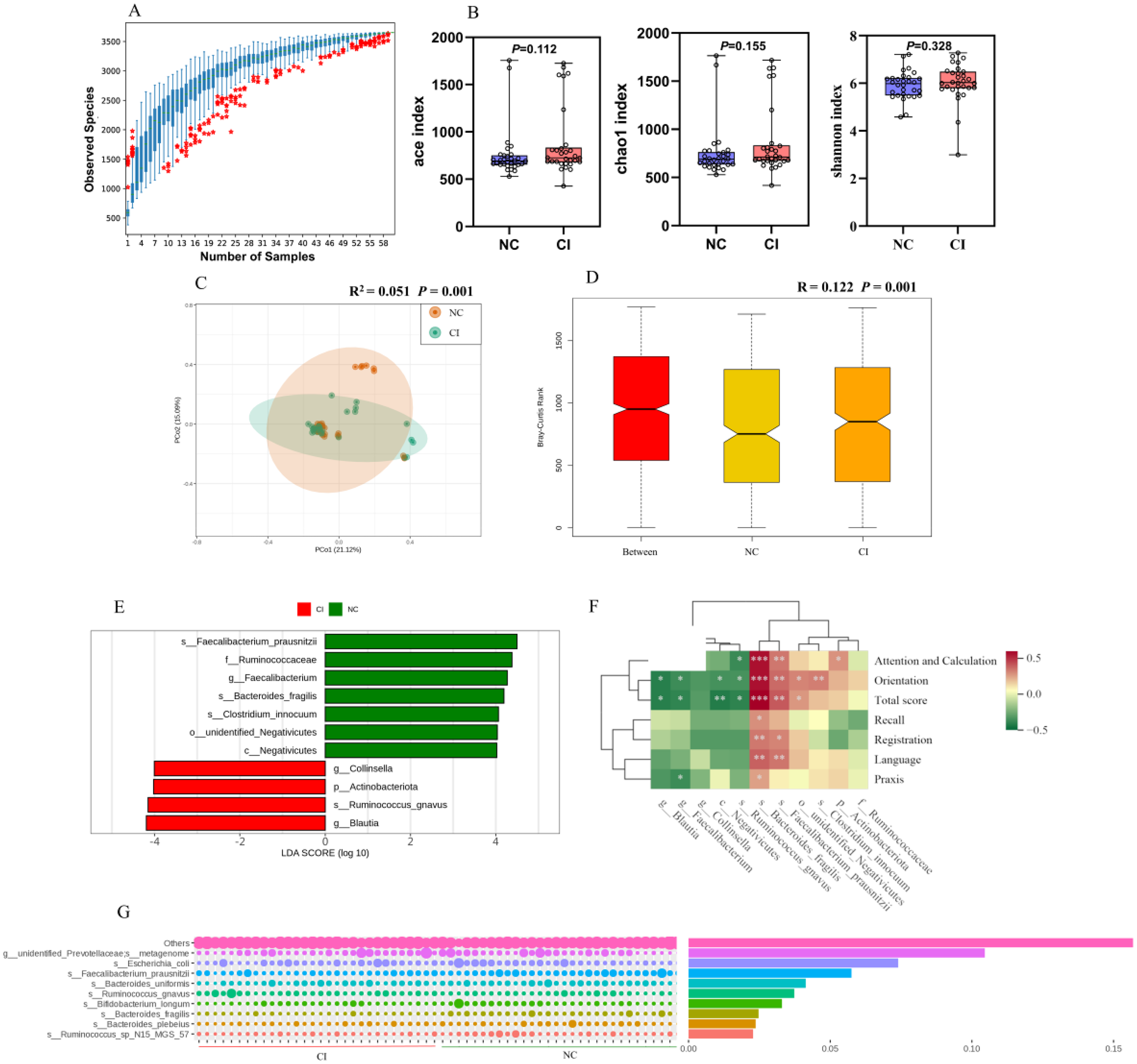
Analysis of gut microbiota diversity. (A) Species accumulation boxplot (n = 30 per group). (B) α-Diversity analyses between the cognitively normal control (NC) and cognitive impairment (CI) groups at the OTU level by using the ACE, Chao1, and Shannon indices. (C) Principal coordinates analysis based on unweighted UniFrac distance between the NC and CI groups at the OTU level. (D) Bray–Curtis distance between the NC and CI groups at the OTU level. (E) Differential species between NC and CI groups (linear discriminant analysis effect size). (F) Correlation between the relative abundances of different microbiota and total score of cognitive function and scores of various dimensions. (G) Top 10 differential species contribution and abundance between the NC and CI groups (Simper). *p < 0.05, **p < 0.01, ***p < 0.001.
LEfSe and Simper analyses revealed that the relative abundances of Coriobacteriia, Blautia, R. gnavus and Actinobacteria were higher in the CI group than in the NC group. By contrast, the NC group exhibited greater abundances of Faecalibacterium prausnitzii, Ruminococcaceae and Faecalibacterium (Figure 1E). The top 10 species contributing to intergroup differences included Prevotellaceae, Escherichia coli, F. prausnitzii, Bacteroides monomorphicis uniformis, R. gnavus, Bifidobacterium longum, Bacteroides fragilis, Bacteroides plebeius and Ruminococcus sp N15 MGS 57 (Figure 1G). Spearman correlation analysis indicated that the increased abundance in the CI group was negatively correlated with total cognitive scores and dimension-specific cognitive scores. Notably, the abundance of R. gnavus showed significant negative correlations with attention, computation ability, localization ability and the total MMSE score. Conversely, in the NC group, increased R. gnavus abundance was positively correlated with these scores. Importantly, R. gnavus was the only species that was significantly enriched in the CI group, ranking fifth in contribution to intergroup differences, indicating a strong association with CI.
R. gnavus colonization in mice
Fecal DNA was extracted on day 7 after five days of mixed antibiotic gavage and a two-day washout period. The PCR amplification of the 16S V4 region resulted in no detectable band on day 7 (Supplemental Figure 1), confirming the successful establishment of the pseudo-germ-free model. Multimodal in vivo imaging revealed that R. gnavus colonized the mouse intestine at 24, 48 and 72 h postgavage (Figure 2A), with the highest colonization at 24 h and some fluorescence still detectable at 72 h (Figure 2B). After 12 weeks, quantitative fluorescent PCR confirmed significantly higher R. gnavus expression in the Rg group feces relative to in control group feces, indicating successful colonization (Figure 2C).

R. gnavus colonization in mice after gavage. (A) Whole-body fluorescence images of mice after gavage with R. gnavus at 24, 48, and 72 h. (B) Fluorescence intensity analysis quantified by using the ROI autofeature of the instrument's software. (C) Relative expression of R. gnavus copies in the feces of mice. Data are presented as mean ± standard error of the mean (SEM) in each group, n = 10, *p < 0.05, **p < 0.01, ***p < 0.001.
R. gnavus induced CI in mice
After 12 weeks of monocolonization with R. gnavus, the spatial learning and memory abilities of aged mice were assessed by using the Morris water maze test. In the place navigation test, the latency to find the platform decreased with training days across all groups, indicating enhanced learning and memory abilities. ANOVA revealed significant differences in group effects (F = 7.752, p < 0.01) and time effects (F = 35.63, p < 0.01), suggesting that intervention and training time influenced spatial learning and memory in the mice. A t-test analysis of daily latency demonstrated that the Rg group exhibited significantly prolonged latency on days 2 and 3 compared with the control group, indicating impaired learning ability (Figure 3B). In the spatial exploration experiment, compared with those in the control group, the mice in the Rg group had a lower distance travelled, percentage of distance travelled and percentage of time spent in the target area and greater average distance close to the platform. All of these differences were statistically significant. These findings suggest that R. gnavus colonization induced learning and memory impairments in aged mice (Figure 3C–J).

R. gnavus colonization altered cognitive function and spatial learning in mice. (A) Representative traces of mice in the Morris water maze. (B) Escape latency in the learning trial. (C) Path length proportion in the target quadrant in the probe trial. (D) Average path length to the platform in the probe trial. E. Platform crossing times in the probe trial. Data are presented as mean ± SEM in each group, n = 10, *p < 0.05, **p < 0.01.
R. gnavus attenuated autophagy levels in the mouse brain
At the gene level, the fluorescence quantitative PCR results of autophagy-related genes in the hippocampus of mice in each group showed that the expression levels of the autophagy-related genes Atg2a, Atg5, Atg7, Atg9a, Atg14, Atg16l1, Beclin-1, Lc3b, and Ulk1 reduced; those of the autophagy-related genes Atg13, P62, Parkin and inflammation-related cytokine TNF-α significantly elevated; and those of the autophagy-related genes Atg12, Wipi1 and Gabarap did not significantly differ in mice in the Rg group relative to those in mice in the control group. Overall, the monocolonization of R. gnavus caused a down-regulation of autophagy-related gene expression levels in the mouse hippocampus (Figure 4A–P).

R. gnavus colonization altered autophagy-related gene expression in the mice hippocampus (A–P). Levels of gene transcripts are shown as fold changes relative to the levels of the housekeeping gene β-actin. Data are presented as mean ± SEM in each group, n = 6, *p < 0.05, **p < 0.01, ***p < 0.001, ****p < 0.0001.
The autophagy markers LC3B and P62 and the reference protein β-actin were quantified at the protein level in the mouse hippocampus by using a fully automated protein detector. LC3B and P62 were normalized to β-actin, with results showing a significant reduction in LC3B and an increase in P62 in the Rg group relative to in the controls. This finding implies that R. gnavus significantly suppressed autophagy protein levels in the mouse hippocampus (Figure 5A). ELISA results uncovered decreased levels of reactive oxygen species and Beclin-1 protein in the Rg group (Figure 5B).

R. gnavus colonization altered the expression levels of autophagy-related proteins in the mouse hippocampus and cortex. (A) Relative expression levels of LC3B and P62 in the mouse hippocampus. (B) Reactive oxygen species levels and Beclin-1 protein expression in the mouse brain presented as mean ± SEM, n = 4. (C) Immunohistochemistry analysis for LC3B in hippocampal CA1 and CA3 regions (arrows indicate autophagy), scale bars: 200 (original) and 50 μm (magnified). (D) Staining for LC3B in the cerebral cortex (arrows indicate autophagy), same scale bars. (E–G) Mean analysis of the proportion of areas positive for LC3B immunohistochemical staining in mouse brain tissue in each group. Data are presented as mean ± SEM in each group, n = 4, *p < 0.05, **p < 0.01, ****p < 0.0001.
The immunohistochemical staining of brain cross-sections from each group of mice with the autophagy marker LC3B antibody revealed that LC3B-positive plaques in the CA1 and CA3 regions of the hippocampus in the Rg group had significantly reduced compared with those in the control group (Figure 5C). In the cerebral cortex, the area of LC3B-positive plaques reduced, whereas staining intensity increased (Figure 5D). The analysis of the positively stained areas using the Colour Deconvolution plugin in ImageJ software showed that the proportion of LC3B-positive staining in the CA1 and CA3 regions of the hippocampus (Figure 5E and F) and the cerebral cortex (Figure 5G) significantly decreased in the Rg group. This result indicates that autophagy levels in the CA1 and CA3 regions of the hippocampus and cerebral cortex were significantly lower in the Rg group than in the control group.
Metabolic changes in aged mice colonized with R. gnavus
Mass spectrometry revealed significant differences in plasma metabolite composition between the Rg and CN groups, as demonstrated by principal component analysis (Figure 6A). Orthogonal partial least squares discriminant analysis (OPLS-DA) was performed to validate these differences further, illustrating a clear metabolic distinction between the two groups (Supplemental Figure 2A). Model validation confirmed the robustness of OPLS-DA, with Q2 > 0.5 and R2Y = 0.994, indicating high stability and no overfitting (Supplemental Figure 2B). A total of 770 differential metabolites were identified on the basis of VIP > 1 and p < 0.05, with 617 metabolites significantly decreased and 153 metabolites significantly increased in the Rg group compared with those in the CN group (Figure 6B). Among the top 20 metabolites with the highest VIP values (Figure 6C), glycerophospholipids, including PA (18:1[9Z]/18:1[9Z]), 1-stearoyl-2-palmitoyl-sn-glycero-3-phosphocholine and 4,21-dehydrocorynantheine aldehyde, were significantly elevated in the Rg group. Additionally, amino acids and metabolites, such as methionine, and aldehydes, like 1-palmitoyl-2-oleoyl-sn-glycero-3-phosphate, were significantly higher in the Rg group than in the CN group. The hierarchical clustering analysis of differential metabolites identified key metabolic groups, including amino acids, phenolic compounds, organic acids and lipids (Figure 6D). KEGG pathway enrichment analysis revealed that the autophagy pathway was significantly enriched (Figure 6E). Other significantly enriched pathways included amino acid metabolism, glycerophospholipid metabolism and cofactor biosynthesis, as well as pathways related to energy metabolism, such as pentose and glucuronate interconversions and butanoate metabolism. Further analysis within the autophagy pathway identified 11 core metabolites, all of which were glycerophospholipids, representing 6.71% of the total differential metabolites in this pathway (Figure 6F). These results indicate that the autophagy pathway and its associated glycerophospholipid metabolites are central to the metabolic changes induced by R. gnavus monocolonization.

Metabolic differences between aged mice in the R. gnavus monocolonization and control groups (n = 6 per group). (A) Principal component analysis plot. (B) Volcano plot of differential metabolites. (C) Variable importance in projection score plot of differential metabolites. (D) Pathway enrichment analysis of differential metabolites. (E) Hierarchical clustering heatmap of differential metabolites. (F) Pathway classification of differential metabolites.
Discussion
In our case–control study, R. gnavus was significantly enriched in patients with CI, and its abundance was negatively correlated with MMSE scores. Similarly, the monocolonization of pseudo-germ-free aged mice with R. gnavus impaired learning and spatial memory and disrupted autophagic homeostasis in the hippocampus and cerebral cortex. This disruption was evidenced by the downregulated transcription of autophagy-related genes (Atg2a, Atg5, Atg7, Atg9a, Atg14, Atg16l1, Beclin-1, Lc3b, and Ulk1), decreased protein levels of LC3B and Beclin-1 and increased accumulation of P62, indicating impaired autophagic flux. Additionally, hippocampal TNF-α levels were elevated, signifying neuroinflammation activation.
Our findings suggest that R. gnavus overgrowth is closely associated with cognitive decline, supporting the hypothesis that gut dysbiosis contributes to neurocognitive disorders.44–46 Previous studies have reported elevated R. gnavus abundance in various neuropsychiatric conditions, such as generalized anxiety disorder, wherein microbial diversity decreased but R. gnavus increased; 47 in an elderly cohort at high neurocognitive risk; 19 and in a postoperative cognitive dysfunction mouse model. 16 Although these epidemiological data are not proof of causality, they consistently associate R. gnavus with poor cognitive performance.
Recent research has highlighted the potential neurotoxicity of R. gnavus. He et al. 48 identified a ∼10-fold enrichment in R. gnavus carrying the phenylalanine decarboxylase gene in patients and mouse models of hepatic encephalopathy. Colonization resulted in the abnormal brain accumulation of phenethylamine (PEA), causing memory loss, tremors and cortical neuron damage, which were reversed by inhibiting the bacterial decarboxylase or neutralizing PEA. However, the effect of R. gnavus on cognition is context-dependent because R. gnavus produces diverse metabolites, including bacteriocins, glycosidases, mucin-degrading enzymes, tryptamine, short-chain fatty acids, immunomodulatory capsular polysaccharides and secondary bile acids. 49 Reports are mixed; some associate elevated R. gnavus with depressive symptoms, 50 whereas others have found reduced levels in patients with AD. 51 Moreover, germ-free mice colonized with R. gnavus exhibited improvements in cognition via hippocampal neurogenesis and synaptic plasticity. 27 These conflicting results highlight the bacterium's environment-dependent effects on cognition, necessitating further research with large, well-characterized cohorts using multiomics approaches.
Monocolonization with R. gnavus in pseudo-germ-free aged mice markedly impaired cognitive functions, elevating hippocampal TNF-α, an effect that is indicative of neuroinflammation. Although common in healthy individuals, R. gnavus markedly increases in terms of abundance in inflammatory bowel disease, 52 irritable bowel syndrome 42 and neurodegenerative disorders. 19 Its proinflammatory capacity, which is modulated by host microbiota status, involves metabolites like PEA and tryptamine, triggering serotonin production and intestinal inflammation.42,53 Certain R. gnavus strains (e.g., ATCC 29149) express surface glucorhamnan that activates TLR4 on dendritic cells, inducing substantial TNF-α and IL-6 secretion. 54 Furthermore, as a mucin–glycan forager thriving on low-fiber diets, 55 R. gnavus increases gut permeability, facilitating inflammatory signals reaching the brain and impairing neuronal function. 56 Our findings reinforce the potential role of R. gnavus in promoting neuroinflammation and CI.
Our study underscores autophagy as a crucial mechanism in R. gnavus–induced CI. In colonized mice, key autophagy genes in the hippocampus and cortex—such as those governing initiation (Atg14, Atg16l1, Ulk1, and Beclin-1), phagophore formation/elongation (Atg5 and Atg16l1), cargo transport (Atg2a and Atg9a) and maturation (Atg7 and Lc3b)—were markedly down-regulated, whereas P62 accumulated, indicating blocked autophagic flux. Effective autophagy maintains neuronal homeostasis, which is essential for cognition, particularly in ageing, by clearing protein aggregates and damaged organelles. 57 Our data therefore suggest that R. gnavus impairs cognition by suppressing neuronal autophagy. Inflammation and autophagy are tightly intertwined: chronic inflammation inhibits autophagy, accelerating the build-up of toxic aggregates, whereas defective autophagy activates the NLRP3 inflammasome and amplifies inflammatory signalling.58,59 Normally, sufficient autophagy removes endogenous danger signals and restrains inflammation; once autophagy falters, aggregated proteins and dysfunctional mitochondria accumulate, triggering NF-κB, mTOR and other proinflammatory cascades that further suppress autophagy, creating a vicious cycle. 59 Consistent with this model, we observed elevated hippocampal TNF-α concomitant with autophagy blockade. Future work should combine large, stratified human cohorts with mechanistic animal studies to elucidate the precise autophagic pathways through which R. gnavus contributes to CI.
Given the link between R. gnavus and cognitive decline, strategies that reduce the abundance or activity of R. gnavus are worth exploring. Dietary modulation is the most straightforward and sustainable option. Epidemiological data indicate that individuals consuming Western diets rich in animal products harbor high levels of R. gnavus, leading researchers to label the bacterium a ‘proinflammatory’ gut microbe. 55 Conversely, a high healthy eating index, reflecting a fiber- and plant-rich diet, correlates markedly with low R. gnavus abundance. 60 Intervention trials support this relationship, showing that adding resistant starch (high-amylose maize) to a high-red-meat diet substantially reduced fecal R. gnavus levels 61 and supplementing with polyphenol-rich red raspberries similarly decreased R. gnavus counts in insulin-resistant adults. 62 These findings collectively suggest that increasing dietary fiber and polyphenol intake while limiting excessive animal fat and protein consumption could reduce R. gnavus overgrowth and mitigate gut inflammation.
Targeted microbiota interventions also hold promise. Novel prebiotics, such as algal oligosaccharides, selectively inhibit R. gnavus growth in vitro without adversely affecting beneficial genera like Bifidobacterium and Lactobacillus. 63 FMT can effectively reset the intestinal ecosystem; transferring microbiota from healthy young donors to aged mice enhances hippocampal synaptic plasticity and memory, reduces gut permeability and proinflammatory markers and elevates beneficial taxa. 64 These data indirectly support the concept that eliminating pathogenic microbial communities, potentially including R. gnavus, and restoring symbiosis could benefit cognitive health. However, FMT is a broad-spectrum intervention, and precise strategies, such as bacteriophage therapy targeting highly proinflammatory strains like R. gnavus, remain exploratory.
This study not only corroborates and extends previous findings but also, for the first time, implicates the autophagy pathway as a potential mediator between R. gnavus and CI. Nonetheless, several limitations should be noted. Firstly, we lacked a control group colonized with another gut bacterium not implicated in CI. This deficiency thus limits our ability to attribute the observed effects specifically to R. gnavus given that recolonization with any facultative commensal might, in principle, elicit similar behavioral or molecular read-outs. Secondly, monocolonization in pseudo-germ-free mice cannot recapitulate the complexity of the human gut microbiome; this reductionist model sets aside normal microbe–microbe interactions and may produce exaggerated host responses. Finally, translating findings from aged mice to human cognitive pathology remains challenging because murine models cannot fully mimic the multifactorial nature of human CI. The translational relevance of our findings therefore warrants cautious interpretation. Future studies should incorporate multistrain or alternate bacterium colonization controls and validate there findings in longitudinal human cohorts or other animal models to enhance generalizability and clinical relevance.
Conclusion
Our findings show that R. gnavus is markedly enriched in the gut of elderly patients with CI, and its abundance is inversely associated with cognitive performance. In pseudo-germ-free aged mice, monocolonization with R. gnavus impairs learning and memory and reduces cerebral autophagic activity, suggesting that autophagy may be a gut–brain axis mechanism through which this bacterium affects cognition. Prospective cohort studies and randomized controlled trials are now needed to confirm whether targeting R. gnavus can deliver tangible cognitive benefits and to guide the development of microbiome-based strategies against cognitive decline.
Supplemental Material
sj-docx-1-alz-10.1177_13872877251372522 - Supplemental material for Role of Ruminococcus gnavus in the elderly with cognitive impairment: A case-control study and an animal experiment
Supplemental material, sj-docx-1-alz-10.1177_13872877251372522 for Role of Ruminococcus gnavus in the elderly with cognitive impairment: A case-control study and an animal experiment by Yanfei Wei, Yining Huang, Shiao Wang, Kuan Liu, Qi Zhong, Weidong Fan, Bifei Cao, Haowen Chen, Yongqi Liang, Qiurong Li, Zhengyun Xu, Kaiyue Liao and Xianbo Wu in Journal of Alzheimer's Disease
Footnotes
Acknowledgements
We express our gratitude to all the people who have contributed to this study.
ORCID iDs

Ethical considerations
This study was conducted in accordance with the Declaration of Helsinki and was approved by the Ethics Committee of Southern Medical University, Guangzhou, China (approval no. NFYKDX002). All animal experiments were approved and monitored by the Ethics Committee of Southern Medical University, Guangzhou, China (approval no. SMUL2022010).
Consent to participate
All participants provided written informed consent.
Author contributions
Funding
The authors disclosed receipt of the following financial support for the research, authorship, and/or publication of this article: This study was supported by the National Natural Science Foundation of China (82173607), and Guangdong Basic and Applied Basic Research Foundation (2021A1515011684 and 2024A1515011969).
Declaration of conflicting interests
The authors declared no potential conflicts of interest with respect to the research, authorship, and/or publication of this article.
Data availability statement
Data used in the current study are available from the corresponding author upon reasonable request.
Supplemental material
Supplemental material for this article is available online.
References
Supplementary Material
Please find the following supplemental material available below.
For Open Access articles published under a Creative Commons License, all supplemental material carries the same license as the article it is associated with.
For non-Open Access articles published, all supplemental material carries a non-exclusive license, and permission requests for re-use of supplemental material or any part of supplemental material shall be sent directly to the copyright owner as specified in the copyright notice associated with the article.
