



Abstract
Background
Vascular factors contribute to dementia in approximately 20 million individuals, notably in vascular contributions to cognitive impairment and dementia (VCI). However, the lack of specific molecular biomarkers to differentiate VCI from normal aging and Alzheimer's disease (AD) impedes early diagnosis and treatment.
Objective
To date the use of saliva for VCI diagnosis has not been previously reported. In this small proof-of-concept study, we aim to explore the feasibility of screening novel salivary diagnostic biomarkers for VCI.
Methods
Using both proton nuclear magnetic resonance (1H NMR) spectroscopy and liquid chromatography coupled with mass spectrometry (LC-MS) we biochemically profiled saliva samples collected from individuals with VCI (n = 26) and compared them with cognitively healthy controls (n = 37).
Results
Of the 167 salivary metabolites 56 of them are found to be at significantly different concentrations in the saliva of individuals with VCI as compared to controls. Subsequently, we developed predictive models capable of distinguishing VCI from controls with 0.92 accuracy. Moreover, sex-stratified analysis revealed the perturbation of different metabolic pathways in the saliva of individuals with VCI.
Conclusions
This study underscores the promising role of salivary metabolomics as a non-invasive tool for the early detection of VCI. Our findings suggest that oral microbiome dysbiosis may contribute to VCI pathogenesis, offering novel mechanistic insights. Given the accessibility of saliva, further validation of these robust salivary biomarkers could facilitate scalable, cost-effective screening for VCI, aiding in timely intervention strategies.
Keywords
Introduction
Vascular contributions to cognitive impairment (VCI) is a blanket term used to describe the spectrum of cognitive dysfunctions resulting from perturbations due to vascular disruptions. 1 VCI is caused by a variety of cerebrovascular etiologies. 2 However, the primary pathological factor is due to cerebral blood flow dysfunction.2,3 Such dysfunction is known to be age-related but enhanced in the presence of cerebrovascular disease. Diagnosis of this condition is difficult due to its “all-encompassing” nature. Current diagnostic methods for VCI, primarily rely on neuroimaging and neuropsychological assessments, are often costly, invasive, time-consuming, and may not detect subtle early-stage changes. This highlights the critical need for more accessible and sensitive diagnostic approaches. 4 Since the pathophysiology of VCI remains incompletely understood, no curative treatments currently exist. As a result, clinical management focuses primarily on mitigation strategies, including pharmacological interventions and lifestyle modifications. 5 Alzheimer's disease (AD) is the leading cause of dementia, but VCI is the second most common etiology. 1 Understanding the pathogenesis of VCI is critically important, particularly because identifying reliable, clinically feasible biomarkers for high-risk individuals could revolutionize diagnosis and management. Such biomarkers may also offer insights into the underlying biochemistry of VCI, accelerating the development of novel therapeutic interventions.
Given the limitations of current diagnostic approaches, there is growing interest in novel biomarkers for VCI. Metabolomics, the comprehensive analysis of small molecules in biological systems, offers a promising solution. 6 Since metabolite composition reflects genetic, epigenetic, environmental, dietary, and disease-related influences, 7 metabolomics enables researchers to connect phenotypic manifestations with biochemical changes. This approach not only aids in biomarker discovery for diagnosis and disease monitoring but also provides insights into metabolic dysregulation, drug effects, and personalized medicine.
Spectrometry-based techniques, such as mass spectrometry (MS) and proton nuclear magnetic resonance ( 1 H NMR) spectroscopy, are widely used to profile metabolomes. Recent studies of biofluids like serum and cerebrospinal fluid (CSF) have implicated caffeine metabolism, TCA cycle disruption, and phospholipid dysregulation in VCI pathogenesis. 8 While blood-based metabolomics has advanced our understanding of VCI etiology, other biofluids such as saliva remain underexplored, despite their potential to yield complementary insights.
Taken together, the pressing need for accessible biomarkers in VCI has become increasingly apparent as both population demographics and diagnostic challenges escalate. While CSF and blood-based biomarkers have dominated VCI research, our group's work has demonstrated saliva's potential in neurodegenerative diagnostics, having successfully identified robust metabolic signatures for both AD and mild cognitive impairment (MCI). 9 These findings established saliva as not only feasible but highly informative for cognitive disorder detection.
Remarkably, despite this demonstrated success and saliva's inherent advantages, including non-invasive collection, excellent stability, and proven correlation with central nervous system pathology, the salivary metabolome remains completely unexplored in VCI. 10 This gap represents both a significant oversight and a substantial opportunity, particularly given the distinct vascular mechanisms underlying VCI that likely produce detectable metabolic perturbations. The current study therefore for the first time addresses this critical unmet need by applying our established, validated salivary metabolomics pipeline to VCI.
Methods
Study design and sample collection
This study received ethical approval from Corewell Health's IRB (2017-214). VCI diagnoses (which include all forms of cognitive impairment of vascular origin, ranging from mild vascular cognitive impairment to vascular dementia) were made using the diagnostic criteria of The International Society for Vascular Behavioral and Cognitive Disorders (VasCog) working group. 11 In particular, all subjects underwent a comprehensive evaluation including a series of multidomain neuropsychological tests, assessment with depression scales for clinical diagnosis of VCI. Control subjects were matched to individuals with VCI by gender and age (±5 years). Exclusion criteria encompassed any diagnosis that included AD, mild cognitive impairment of non-vascular origin, or other forms of cognitive impairment caused by neurodegenerative disorders such as Huntington's disease or Parkinson's disease. Other exclusion criteria were the presence of dementia/cognitive impairment associated with other general medical conditions such as head trauma, severe endocrine/metabolic diseases, brain tumors, normotensive hydrocephalus, a history of alcohol or drug abuse or dependence, subjects with delirium and psychiatric disorders undergoing chronic treatment with psychotropic drugs prior to the onset of dementia/cognitive impairment. Individuals with current gum disease, evidenced by visual signs of gingivitis or periodontal disease (e.g., redness, inflammation, sores, or bleeding), those under the age of 60, those not proficient in English, and individuals with a known allergy to Proparacaine HCL 0.5% numbing eye drops were excluded from participation.
We collected unstimulated saliva samples from 63 adult volunteers (37 asymptomatic controls and 26 individuals clinically diagnosed with VCI) using standardized protocols with revision of overnight fasting. 9 Table 1 lists the clinical, cognitive, behavioral, functional, and demographic information of the people with VCI enrolled in this study. All collections occurred in the early morning (07:00–09:00 AM) following a minimum 1-h abstinence from eating, drinking (except water), smoking, or oral hygiene product use. Participants passively drooled into pre-chilled 50 mL sterile Falcon tubes for 2–3 min, yielding an average volume of 2.5 mL per sample. Immediately after collection, samples underwent processing at 4°C: centrifugation at 8000 × g for 10 min to remove cellular debris, followed by aliquoting of supernatants into sterile 1.5 mL Eppendorf tubes. Aliquots were flash-frozen and stored at −80°C until analysis. We verified sample quality by measuring pH (mean ± SD: 7.22 ± 0.64), with all values falling within established normal ranges for salivary biochemistry.
Pair-wise comparison of the available clinical and demographic parameters, encompassing education, gender, age, body mass index, and cognitive function (Saint Louis University Mental Status exam total scores, CLOXs, Mini-Mental State Examination) along with statistical significance for the study subjects from which salivary metabolomics data obtained.
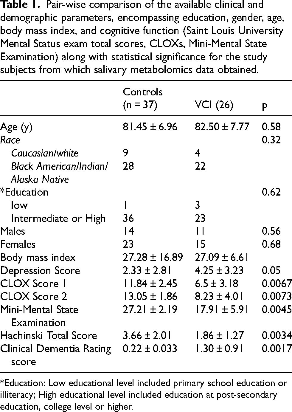
*Education: Low educational level included primary school education or illiteracy; High educational level included education at post-secondary education, college level or higher.
1H-NMR spectroscopy metabolomics
Sample preparation
Samples were thawed at room temperature. After thawing, 300 µL of each sample was filtered through washed (x7 to remove excess glycerol) 3-kDa cut-off centrifuge filter units (Amicon Micoron YM-3; Sigma-Aldrich, St Louis, MO) at 12,000 x g for 20 min to remove any proteins or macromolecules. To 200 µL of filtrate, 21 µL of 0.5 M sodium phosphate buffer (pH 7.2) containing 11.667 mmoL disodium-2, 2-dimethyl-2-silceptentane-5-sulphonate (DSS; internal standard) and 25 μL of D2O were added (final pH was 7.27 ± 0.07 for all samples). After vortexing, a total of 200 µL of sample was transferred to a 3 mm NMR tube for analysis. Samples were analyzed in a randomized order and maintained at 4°C using the state-of-the-art SampleJet™ (Bruker, Cambridge, MA) automated sample changer.
Data collection and metabolic profiling
Before 1H-NMR analysis, each sample was heated to room temperature for 3 min and then placed into the magnet. For every saliva sample, a 1H NMR spectrum was recorded using a Bruker Ascend III spectrometer operating at 600.13 MHz for proton detection, equipped with a 5 mm TCI cryoprobe, at a temperature of 298 K. The pulse sequence, developed by Ravanbakhsh et al., 12 involved an 8.10–8.97 µs 90° pulse, a 100 ms mixing time, and water suppression with a power level of 45.57 dB and an offset frequency of approximately 2350 Hz during both the mixing time and relaxation delay (RD = 4 s). The 90° pulse length and water suppression conditions were optimized for each sample. A total of 256 transients were collected over 64k data points, with a spectral width of 11,964 Hz and an inter-pulse delay of 5.4 s between transients. The free induction decay signal was zero-filled to 128k points before Fourier transformation. Chemical shifts (δ) were reported in ppm relative to the operating frequency, with all shifts internally referenced to the DSS methyl group singlet (δ 0.0). Each 1H NMR spectrum was manually phased, baseline-corrected, and profiled using professional NMR mixture analysis software Chenomx version 10.0 (Chenomx Inc., Edmonton, AB).
Liquid-chromatography mass-spectrometry (LC-MS) metabolomics
Saliva samples, standards, and quality controls were prepared and nitrogen-dried for 30 min as per biocrates’ instructions. The separation process was carried out on an MxP Quant 500 C18 column, which included a guard column and a pre-column mixer provided by biocrates Life Sciences, AG (Innsbruck, Austria). Saliva samples were derivatized with phenylisothiocyanate (PITC) at room temperature for 60 min and dried under nitrogen for 60 min. Extracts were prepared using 5 mM ammonium acetate in methanol for 30 min with orbital shaking, followed by centrifugation at 500 × g for 2 min. For LC analysis, extracts were diluted 1:1 with water, whereas for flow injection analysis, 30 µL of saliva extracts were mixed with 470 µL of kit solvent and 10 µL of quality control extract with 490 µL of kit solvent. Plates were sealed, mixed at 600 rpm for 10 min at room temperature, and loaded into a thermostatically controlled autosampler for analysis. Samples extracts were analyzed using a Waters I-class UPLC unit coupled with a Waters Xevo-TQ-S (Waters Corporation, Milford, MA, USA). The mobile phase included A: H2O with 0.2% formic acid and B: MeCN with 0.2% formic acid, delivered at 0.8 mL/min with a gradient of 0–100% B over 4.5 min. The flow rate increased to 1.0 mL/min at 100% B in 30 s, then returned to initial conditions over 70 s for column equilibration. The gradient for both positive and negative modes lasted 5.8 min, with %B composition differing between 2.0–4.5 min in the negative mode. Injection volumes were 5 µL for the positive mode and 15 µL for the negative mode. For flow injection analysis, the mobile phase (290 mL MeOH with FIA additives) was delivered at a flow rate of 0.03 mL/min, with an injection volume of 20 µL for both positive and negative modes. The area ratios and concentrations were determined using the biocrates MetIDQ software (biocrates Life Sciences, AG, Innsbruck, Austria).
Statistical analysis
All statistical analyses were performed using R programming language (v4.1.3; 2022-03-10) base package unless indicated otherwise. Before statistical analysis, preprocessing steps (data cleaning, transformation, and imputation of missing values) were undertaken to prepare the data.
In brief, any metabolites with zero intensity values were marked as missing. Then, metabolites with more than 20% missing values were removed from further analysis. Metabolites with variance in the 5th percentile were considered invariant and were removed from further analysis. Samples with more than 80% missing values were removed. The remaining missing values were imputed using the k-nearest neighbors algorithm (k = 5) using impute.knn function from the impute package (v.1.46.0) in R. 13 Principal component analysis (PCA) was carried out to identify systematic variation in the data. Outlier samples were defined as samples with variation higher than three standard deviations (SDs) of the first three principal components (PCs). To account for sample concentration differences due to dilution and minimize the technical variation, metabolite intensities were scaled sample-wise using the scale function in R. Before applying linear regression models, variance inflation analysis was carried out to detect any correlations among the independent variables using vif function from R package car (v3.1.2). Differentially abundant metabolite analysis was carried out by fitting a linear regression model on the normalized metabolite intensity data using limma (v3.50.3) package in R. 14 The model was adjusted for condition, age, and sex assigned at birth. The models were fitted using least-squares method by running lmFit function. Empirical Bayes statistics were estimated using eBayes function. The same analysis was also performed in a sex-stratified manner, although the models in these analyses did not include sex assigned at birth as a covariate. The FDR method was used to correct for multiple testing. 9 Metabolite concentration differences with q value < 0.05 were deemed significant (Supplemental Table 1).
For the metabolite-set enrichment analysis (MSEA) metabolites were ranked by the sign of fold change multiplied by the negative log-transformed p-value. Metabolites without HMDB IDs were excluded from the list, and the metabolite with the smallest p-value was kept for the analysis. Enrichment analysis was carried out using the GSEA function from the R package clusterProfiler (v4.10.1), employing the fgsea method. 15 Pathways were filtered to include at least three metabolites from the tested dataset, and p-values were adjusted using the FDR method.
Predictor analyses for biomarker identification were carried out using caret (v6.0-94) package in R. 16 For training of the predictive models, the dataset was split into 10 folds, with each fold containing 80% of the data for training and 20% for validation. The training subset from each fold was used to fit the Generalized Linear Model Network (GLMnet), Support Vector Machine with Radial Basis Function Kernel (SVM Radial) and Random Forest (RF) classifiers.17,18 Each model underwent 10-fold repeated cross-validation with 10 repeats while fine-tuning 15 different parameters for optimal performance. The best classifier in each fold was selected based on the receiver operating characteristics (ROC) metric. We then used the validation dataset from each fold to assess the performance of the best predictor on unseen data. Finally, we trained the final predictors using the entire dataset. This final training also employed 10-fold cross-validation with 10 repeats and fine-tuning 15 parameters for optimal performance, with the best predictor chosen based on the ROC metric.
Results
Metabolic profiling and quality control of the data
Comprehensive metabolomic analysis of saliva samples identified 693 metabolites using complementary analytical platforms (LC-MS and 1H-NMR). Following stringent quality control protocols including outlier removal, log-transformation, and missing value imputation, we retained 167 metabolites for subsequent analysis (Supplemental Figure 1). PCA demonstrated appropriate data clustering without spectral outliers (Supplemental Figure 2). Demographic comparisons revealed no significant differences between VCI (n = 26) and control (n = 37) groups in age or sex distribution (Table 1; p < 0.05). Variance inflation factor analysis conducted prior to linear model fitting revealed no significant multi-collinearity among the independent variables (age, sex assigned at birth, condition). Therefore, the differential abundance models were adjusted for all three covariates. A linear regression model was used with PCs as the dependent variables and all covariates as independent variables, revealing that the disease state (VCI) is positively correlated with PC1 and PC2 and no correlation between age or sex with any PCs (Supplemental Figure 3).
Differential abundance analysis using linear models
To identify metabolites with significantly altered concentration, we fitted a least-squares linear regression model that accounted for potential confounders such as sex assigned at birth and age. Among the 167 measured metabolites, 56 showed statistically significant differences (q < 0.05), with 31 elevated and 25 reduced in individuals with VCI (Figure 1(a); see Supplemental Table 1). The most affected metabolites included acetate (q = 0.00065, logFC=4.72), Cer d18:1/24:1(q = 0.0051, logFC=2.42), DG 18:1_18:1 (q = 0.00032, logFC=9.42), ethanol (q = 0.00032, logFC=11.28), methanol (q = 0.00013, logFC=8.82), and tyrosine (q = 0.00065, logFC=4.53) (Figure 1(b)). Furthermore, a sex-stratified linear model was applied to metabolomics data to identify metabolic signatures in saliva related to sex differences in VCI. Remarkably, sex-stratified analysis revealed a stronger metabolic shift in females. In the male VCI cases, only ethanol and methanol were significantly altered (q < 0.05)
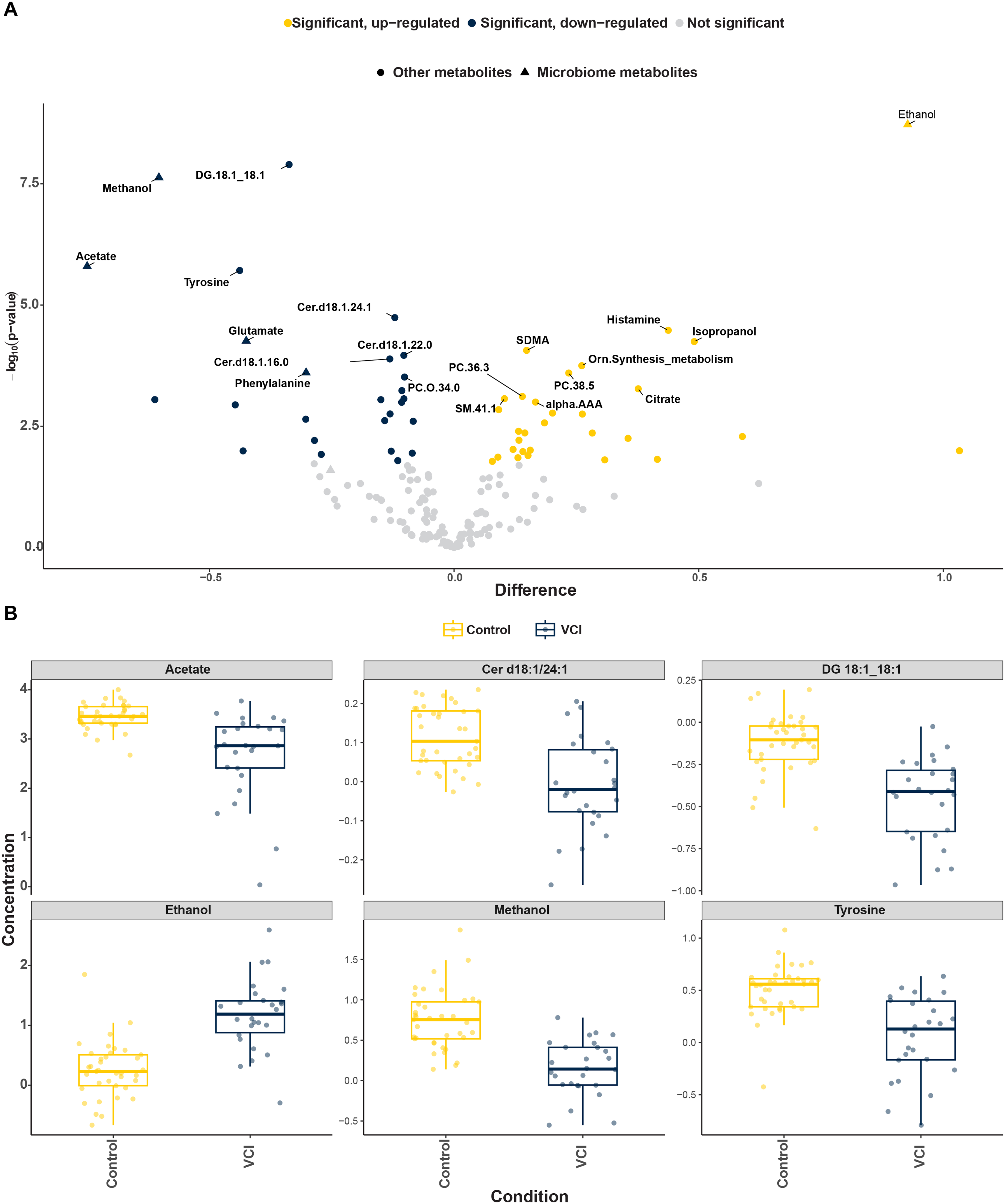
(a) The volcano plot of differential metabolite concentration. The most significant metabolites for each direction are labeled. Metabolites with q < 0.05 are colored in blue (significantly lower abundance) or yellow (significantly higher abundance). Those with q > 0.05 are colored in grey. (b) Boxplots of the six metabolites with the most significant differential concentrations when VCI case is compared to controls. (Color figure available online).
Metabolite set enrichment analysis
Metabolite set enrichment analysis (MSEA) revealed significant disturbances in several key pathways including aspartate metabolism, pyrimidine metabolism, tryptophan metabolism, mitochondrial beta-oxidation of medium-chain saturated fatty acids, and ethanol degradation (Figure 2). These pathways are represented in the diagram, with individual metabolites within them showing altered levels. To investigate how sex contributes to the metabolic differences observed in VCI, sex-stratified MSEA was conducted. In female individuals, significant disruptions were found in ethanol degradation, citric acid cycle, glycerophospholipid metabolism thyroid hormone synthesis, and transfer of acetyl groups into mitochondria, as well as in phenylalanine and tyrosine metabolism and alanine metabolism (Supplemental Table 2). Contrastingly, in male patients, MSEA analysis revealed significant disturbances in ethanol degradation, alanine metabolism, inositol metabolism, inositol phosphate metabolism, and phosphatidylinositol phosphate metabolism (Supplemental Table 3).
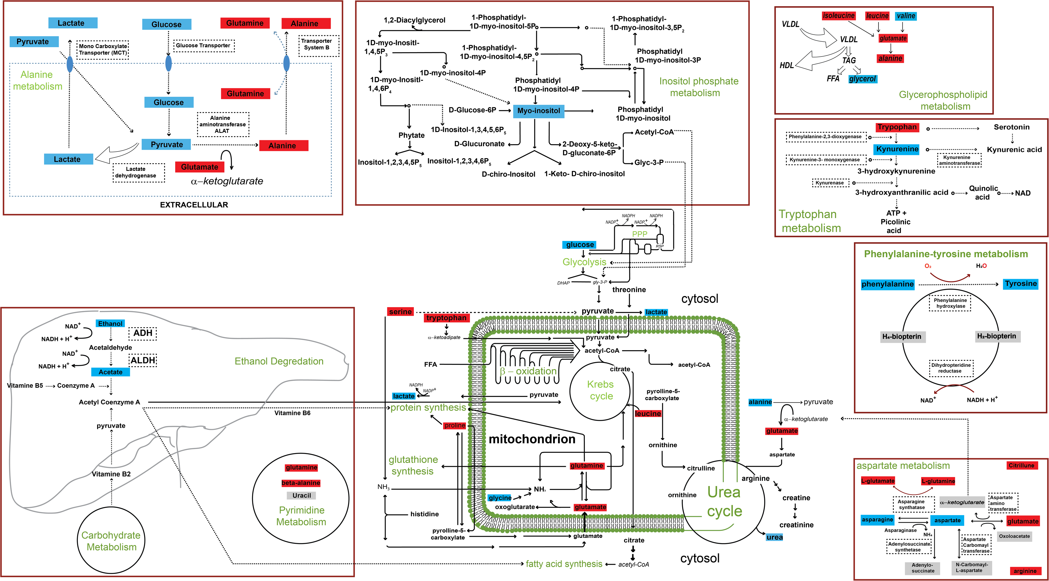
Metabolite set enrichment analysis (MSEA) of saliva metabolomics data comparing individuals with vascular contributions to cognitive impairment and dementia (VCI) to healthy controls and sex stratified metabolic pathway analysis. Metabolites significantly elevated in VCI are highlighted in red, while those reduced are shown in blue. The figure maps these altered metabolites across multiple interconnected metabolic pathways, revealing significant disruptions in inositol phosphate metabolism, alanine metabolism, ethanol degradation, phenylalanine-tyrosine metabolism glycerophospholipid metabolism, tryptophan metabolism, and mitochondrial dysfunction. (Color figure available online).
Diagnostic model building through a machine learning approach
Finally, to assess the potential of salivary metabolomics for discriminating against VCI from cognitively control case, we developed several diagnostic models using ensemble methods, namely, random forest (rf), generalized linear and similar models via penalized maximum likelihood (glmnet), and Support Vector Machine with Radial Basis Function Kernel (SVM Radial). The average ROC in the validation datasets was 0.988 for RF, 0.962 for the glmnet and 0.994 for svmRadial classifiers. While the final predictor showed the best training ROC of 0.944 for svmRadial classifiers, 0.921 for Glmnet and 0.910 for the random forest, respectively (Figure 3(a), (b)). The top 10 salivary metabolites utilized in SVM Radial, Random Forest and glmnet that were selected by feature selection algorithms are listed in Table 2.
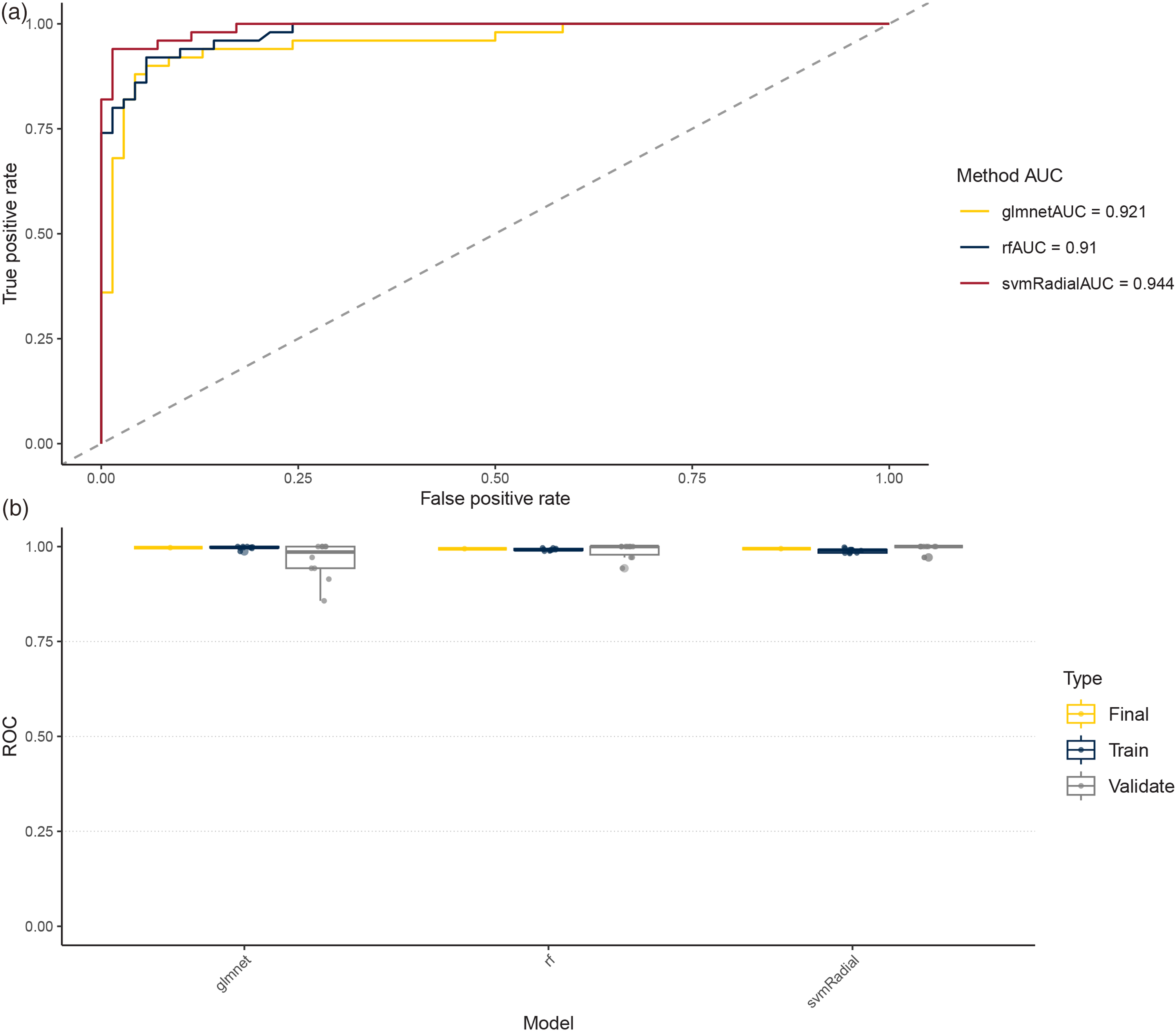
Performance evaluation using AUC for Random Forest, Glmnet, and SVM radial machine learning models. (Color figure available online).
The top 10 salivary metabolites utilized in Random Forest, Glmnet. and SVM Radial machine learning models that were preselected by feature selection algorithms.

Discussion
VCI represents a spectrum of cognitive disorders caused by cerebrovascular damage from ischemic, hemorrhagic, or hypoperfusion events. 19 VCI includes any form of cognitive decline due to a vascular origin, from mild vascular cognitive impairment to various forms of vascular dementia, but its role is unclear. Therefore, the identification of reliable biomarkers that can stratify individuals at elevated risk for cognitive decline and inform the selection of targeted therapeutic and lifestyle interventions is of critical importance. Saliva, rich in cells, microbiota, and biomolecules, is a valuable source for chronic diseases biomarkers. 20 Saliva contains proteins, RNA, metabolites, and lipids that mirror systemic and neurodegenerative changes, as many salivary compounds pass from plasma through diffusion or transport. Its proximity to the CNS highlights its value for diagnosis and prognosis. 21 Saliva is an ideal, noninvasive, and cost-effective sample for collection and storage. Using 1H NMR spectroscopy, we identified distinct salivary metabolomic profiles that accurately distinguished MCI and AD patients from controls through regression models. 9
To our knowledge, this is the first targeted metabolomics study using both 1H-NMR spectroscopy and LC-MS to comprehensively profile the salivary metabolome in VCI patients comparing matched controls, to identify diagnostic biomarkers and uncover novel pathophysiological mechanisms. Using our approach, 9 we measured 167 salivary metabolites and identified 56 significantly altered in VCI, with acetate, Cer(d18:1 24:1), DG(18:1 18:1), ethanol, methanol, and tyrosine showing the most pronounced changes. Notably, several significantly dysregulated metabolites including methanol, acetate, ornithine, phenylalanine, glutamate, and ethanol are known microbial-derived compounds, suggesting the potential involvement of oral microbiome-brain communication in VCI pathogenesis. The role of the oral microbiome in cognitive decline and dementia is an emerging research area.21–23 Alterations in the composition of the oral microbiome, along with the reduced diversity, have been observed in individuals experiencing cognitive decline compared to those with normal cognitive function. 21 Evidence suggests a potential link between disruptions in the oral microbiome (dysbiosis) and dementia. 23 Furthermore, periodontal disease, which is associated with oral microbial dysbiosis, is connected with greater cognitive impairment and dementia. 24 While nervous, endocrine, and immune pathways shape the gut–brain axis, mechanisms linking oral bacteria to VCI remain unclear. 25 Notably, Rivière et al. hypothesized that oral bacteria might access the brain via trigeminal nerve branches. 26 The potential for oral bacteria to enter the bloodstream presents a risk of sepsis.27–29 These bacteria may impair the blood-brain barrier (BBB), allowing microbial metabolites like short-chain fatty acids to enter the brain and affect its function. 30 Although BBB disruption has been noted in AD patients and animal models, its role in VCI defies. 31 Moreover, detection of lipopolysaccharides from T. denticola, T. forsythia, and P. gingivalis in the brain correlates with increased proinflammatory cytokines (IL-1β, IL-6, TNFα, IFNγ) and decreased IL-10, suggesting oral microbiota may contribute to VCI via neuroinflammation. 32 Taking these into account, further research is needed to clarify exact role of oral microbiome in VCI.
MSEA identified disruptions in aspartate, arginine–proline, ornithine, pyrimidine metabolism, catecholamine biosynthesis, mitochondrial β-oxidation of medium-chain fatty acids, and mitochondrial tRNA pathways (q < 0.05). Mitochondria are critical for brain energy metabolism and neuronal survival, with dysfunction increasingly implicated as an early driver of neurodegeneration.33,34 MSEA results reinforce this, revealing enrichment in metabolites associated with mitochondrial β-oxidation and tRNA aminoacylation. Paglia et al. consistently found disrupted mitochondrial aspartate metabolism in demented brains using unbiased mass spectrometry imaging. 35 It is important to note that N-methyl-D-aspartate receptors (NMDARs) play a crucial role in excitatory synaptic transmission cardiovascular regulation and plasticity and are linked to neurodegenerative and psychiatric disorders. 36 NMDAR activation depends on the binding of glutamate or aspartate and glycine.37,38 We are purposing altered aspartate metabolism may impair NMDAR function, contributing to VCI. Multi-omics analysis of postmortem VCI brain tissue revealed both gain- and loss-of-function alterations in aspartate-related pathways, linking metabolic changes to transcriptomic and epigenomic profiles (unpublished), highlighting aspartate metabolism as a potential therapeutic target. Pyrimidine metabolism is essential for vascular function, supporting DNA/RNA, lipid, and carbohydrate synthesis, cell proliferation, and energy metabolism. Its dysregulation is associated with neurodegeneration through impaired repair, inflammation, mitochondrial dysfunction, and disrupted protein synthesis. 39 Supporting our findings, altered de novo pyrimidine biosynthesis is linked to OxPhos dysfunction in post-mitotic AD brain cells. 40 Additionally, studies report age-related accumulation of pyrimidine intermediates in human CSF 41 and aging mouse brains 42 whereas aged C. elegans show reduced levels, indicating age-dependent alterations in pyrimidine metabolism. 43
Ornithine metabolism, critical for neuronal function, 44 is disrupted in VCI, causing toxic metabolite accumulation (e.g., ammonia), reduced acetyl-CoA synthesis, energy deficits, and consequent neurodegeneration. 45 Dementia patients show elevated arginine and reduced ornithine, reflecting disrupted energy metabolism. Ornithine decarboxylase 1 (ODC1), which converts ornithine to putrescine in polyamine synthesis, is expressed in astroglia. This activity promotes GABA production, contributing to memory deficits in AD and linking ornithine metabolism dysfunction to cognitive decline in VCI.46–48 Arginine and proline metabolism support cognition, and their disruption is associated with cognitive decline and neurological diseases. L-arginine, a precursor to nitric oxide, supports CNS regulation and neurotransmitter synthesis, including glutamate and GABA. 49 Using untargeted metabolomics and 16S rRNA sequencing He et al. reported L-arginine metabolism helps maintain intestinal homeostasis, influencing cognition. 50
Additionally, L-arginine was found to exert anti-stress effects that may prevent cognitive decline. 51 Notably, arginine metabolism also contributes to neurotransmitter synthesis, converting into glutamate and GABA essential for brain function. 52 Agmatine, derived from L-arginine, may modulate neurotransmitter systems, 53 while proline supports protein synthesis, signaling, stress response, and collagen metabolism for vascular integrity.54–56 Disruptions in these pathways may impair vascular health and contribute to VCI. Delwing et al. showed proline inhibits acetylcholinesterase in the cortex, linking it to cognitive impairment in vascular dementia. 57 Proline metabolism also supports nitric oxide synthesis for vascular health 58 ; its disruption reduces NO, impairing blood flow and increasing VCI risk.59–61 Catecholamines (dopamine, epinephrine, norepinephrine) regulate cognition, memory, attention, and autonomic function. Their dysfunction contributes to neurodegeneration, making them potential therapeutic targets for cognitive impairment.62,63
A study by Hong et al. using 3×Tg-AD mice and 2VO rats found distinct central catecholamine and metabolite expression patterns in AD and VaD, suggesting a unique catecholamine-related pathogenic mechanism in VaD. 64 Recent genetic studies in mice by Kazuto et al. demonstrate that catecholamines are crucial for brain function, with deficiencies impairing latent learning and long-term memory. 62 Both schizophrenia and dementia involve disruptions in peripheral and central catecholamine systems. Cognitive impairment is associated with increased platelet monoamine oxidase activity, while demented schizophrenic individuals show elevated plasma dopamine β-hydroxylase activity. Additionally, exposure to polyhalogenated aromatic hydrocarbons may impair cognitive function by disrupting brain glucose metabolism and catecholamine synthesis.
Interestingly, our study found that angiotensin receptor blockers treatment elevated salivary acetone and isopropyl alcohol across AD, MCI, and cognitively normal groups, without obscuring AD-specific biomarkers. Given hypertension's impact on vascular integrity, extending salivary metabolomic analysis to VCI is key for validating clinical biomarker panels.
Importantly, growing evidence indicates that sex plays a significant role in dementia development. 65 Subtypes of dementia, including AD, VaD, Lewy body dementia, and Parkinson's disease, show sex-specific differences, yet the role of sex in brain aging and its impact on VCI remains poorly understood. This gap highlights the need for sex-specific research to inform personalized treatment strategies66–68 Exalto et al. found that in VCI, females had more white matter hyperintensities, while males showed more lacunar infarcts and smaller brain volumes; cognitive decline was similar, but mortality was higher in males. 69 Herein, we investigated sex-specific salivary metabolites in VCI and strikingly found significant male–female differences. Both differential abundance and pathway enrichment analyses suggest distinct biological mechanisms by sex. In females, ethanol degradation, the citric acid cycle, and thyroid hormone synthesis were most disrupted, while in males, ethanol degradation, alanine metabolism, and inositol metabolism were predominantly affected.
It is important to recognize that there is a pressing need for easily accessible biomarkers with high diagnostic accuracy to aid in the widespread screening of VCI. This study identifies five salivary metabolites (ethanol, methanol, acetone, tyrosine, propionate) that predict VCI with high accuracy (AUCs 0.944, 0.921, and 0.910 for svmRadial, Glmnet, and RF, respectively). These results outperform cross-validated training, demonstrating strong generalizability and supporting saliva as a non-invasive alternative to CSF and serum. Validation on independent cohorts and optimal model selection remain essential.
Our findings have two clinical implications. First, they may provide novel insights into pathogenic mechanisms underlying VCI; it is possible that modifying levels of specific metabolites through diet or supplementation could help reduce the risk of cognitive decline and dementia in individuals with VCI. Second, metabolomics panels could predict progression, guide treatment, reveal additional disease markers and deepen understanding of VCI-related mechanisms.
The strengths of this study are threefold, enhancing the reliability and significance of its findings. First, it employs two metabolomics platforms and, for the first time, analyzes salivary metabolites in VCI with validated, high-precision methods and strong quality control. Second, it uniquely examines sex-specific metabolic pathways via saliva, considering the oral–microbiome–brain axis. Third, it adjusts for demographics to identify sex-specific signatures, providing a more precise understanding of VCI. This study suggests salivary metabolites may serve as VCI biomarkers, with pathways consistent with prior research. Given saliva's accessibility, larger multicenter studies are needed to validate their use for risk assessment and subtype differentiation.
Our study also has some limitations. First, it includes a small patient sample, which may have reduced our ability to identify statistically significant associations and draw definitive conclusions about causality; however, even with a small sample size, we have successfully developed highly accurate predictive models and uncovered specific details regarding the pathophysiology of VCI. Second, the metabolites we identified could be on the causal pathway for cognitive decline or dementia but secondary to tissue damage caused by demyelination. 69 We were unable to confirm this because not all the patients were evaluated for multiple sclerosis, and myelin loss was not measured, so further mechanistic and longitudinal studies should address this point.
Moreover, given the cross-sectional nature of this study, the observed metabolic differences likely reflect alterations that have already occurred in individuals with VCI. Nonetheless, several metabolites identified in this panel participate in biological pathways that are known to shift early in disease development, suggesting potential utility for detecting metabolic disruption before major clinical changes emerge. While the present data provide an initial foundation for salivary metabolomics–based stratification, longitudinal studies will be essential to determine whether these metabolites can prospectively identify individuals at risk for progression to VCI and to characterize temporal trajectories of metabolic.
Taking all limitations into consideration, to obtain a more reliable and comprehensive understanding of VCI, future studies using larger sample cohorts should incorporate a broader range of clinical demographics, imaging data, and longitudinal observations, providing a more dynamic picture of disease course, including data on pharmacological interventions.
Conclusion
To the best of our knowledge, this is the first multiplatform targeted metabolomics study combining 1H NMR spectroscopy and LC-MS that uses saliva as a potential biomatrix for predicting those at greatest risk of developing VCI. This study offers initial evidence suggesting that salivary metabolites hold promise as biomarkers for VCI. MSEA was used to provide insights into disease mechanisms. Further, the metabolic pathways that were found to be significantly affected in VCI appear to be consistent with previously published results. Metabolomic analysis of this fluid may help provide insights into the mechanism of VCI. Given the convenience and frequency with which saliva can be obtained, for the generalizability of our findings to be validated, conducting larger, multicenter studies representing broader and more diverse populations is warranted. Such studies could further validate these findings and explore the potential of salivary metabolites as reliable biomarkers for VCI risk assessment or even differentiating subtypes of the disease.
Supplemental Material
sj-docx-1-alz-10.1177_13872877261423158 - Supplemental material for Salivary metabolomics for early detection of vascular contributions to cognitive impairment and dementia: Exploring microbiome dysbiosis and sex differences
Supplemental material, sj-docx-1-alz-10.1177_13872877261423158 for Salivary metabolomics for early detection of vascular contributions to cognitive impairment and dementia: Exploring microbiome dysbiosis and sex differences by Ali Yilmaz, Nadia Ashrafi, Zoe Guerra, Delaine Goniwiecha, Nazia Saiyed, Juozas Gordevičius, Karolis Krinickis, Migle Gabrielaite, Tammy Osentoski, Nicole Schumacher, Suriah Khan, Amita Pai, Stacey Ruff, Michael E. Maddens, Khaled Imam, Roberto Monastero and Stewart F. Graham in Journal of Alzheimer's Disease
Footnotes
Acknowledgements
We express our gratitude to the John and Marilyn Bishop Charitable Foundation, the Fred A. & Barbara M. Erb Foundation, and the Maibach family for their generous contributions. Their support has been invaluable in making this work possible.

Ethical considerations
All procedures involving human participants complied with the ethical standards of the institutional and/or national research committees, as well as the 1964 Declaration of Helsinki and its subsequent revisions or comparable ethical guidelines. The study was approved by the institutional review board.
Consent to participate
Written informed consent was obtained from all participants or their authorized representatives at the time of enrollment.
Consent for publication
Not applicable.
Author contribution(s)
Funding
The authors disclosed receipt of the following financial support for the research, authorship, and/or publication of this article: This work was supported by the John and Marilyn Bishop Charitable Foundation, the Fred A. & Barbara M. Erb Foundation, and the Maibach family. Their generous financial contributions were instrumental in enabling the completion of this study.
Declaration of conflicting interests
The authors declared no potential conflicts of interest with respect to the research, authorship, and/or publication of this article.
Data availability statement
The data supporting the findings of this study are available within the article and metabolomics data will be available upon request.
Supplemental material
Supplemental material for this article is available online.
References
Supplementary Material
Please find the following supplemental material available below.
For Open Access articles published under a Creative Commons License, all supplemental material carries the same license as the article it is associated with.
For non-Open Access articles published, all supplemental material carries a non-exclusive license, and permission requests for re-use of supplemental material or any part of supplemental material shall be sent directly to the copyright owner as specified in the copyright notice associated with the article.
