
Abstract
For individuals undergoing mastectomy, reconstruction of the nipple–areola complex (NAC) is a critical step in emotional and psychological recovery. However, current clinical approaches—including flap suturing, tattooing, or grafting—are limited by loss of projection, poor mechanical stability, and absence of sensation. Additive manufacturing and tissue engineering offer promising alternatives by enabling the development of hybrid scaffolds that maintain long-term projection and support the potential return of sensation. This review summarizes the state-of-the-art in NAC reconstruction and highlights how advances in additive manufacturing can address existing limitations. Emerging scaffold design strategies allow precise fabrication of biomimetic architectures that replicate the anatomical form and function of the NAC, while supporting tissue integration and mechanical durability. The use of biocompatible polymers such as poly-ε-caprolactone, combined with bioactive coatings and plasma surface modification, enhances cell attachment and vascularization. Additionally, the incorporation of stem cells, multicellular constructs, and conducting polymers is explored to enable multifunctional tissue regeneration and restore sensation through electrical stimulation. By integrating innovations in biomaterials science, regenerative medicine, and advanced fabrication technologies, the field is moving toward nipple reconstructions that are not only more life-like in appearance but also biologically responsive and sensate.
Impact Statement
Reconstruction of a neurovascularised nipple–areola complex remains a major challenge in postmastectomy breast reconstruction. This review highlights advances in plasma-modified 3D-printed poly-ε-caprolactone scaffolds that improve surface hydrophilicity, cell adhesion, and neurovascular integration. Combining plasma surface engineering with additive manufacturing and regenerative strategies offers a pathway to anatomically accurate, mechanically durable, and functionally innervated constructs for next-generation nipple reconstruction.
Introduction
Breast cancer is one of the most common cancers worldwide and despite advancements in early detection and treatment, it remains the second leading cause of cancer death among women. 1 The most widely performed surgical treatment for breast cancer is a mastectomy which is a procedure to remove all or part of the breast. In recent years, there has been an increase in the use of breast-conservation surgery and nipple-sparring mastectomy. Typically, for nipple–areola complex (NAC) preservation in patients, a tumor to nipple distance needs to measure 2.5 cm or more using a digital mammogram image. 2 When these options are not available, an NAC reconstruction is performed. This is an important part of the breast reconstruction process as the psychological impact this has on the women who receive it is significant. NAC reconstruction provides a sense of completeness at the end of the treatment and helps to improve body image and self-esteem as well as help those to feel more like themselves again. 3
Numerous techniques have been explored to reconstruct the NAC, with popular options being tattooing or nipple reconstruction surgery (NRS) (Fig. 1). 4 Nipple reconstruction is the final step in breast reconstruction, performed 4–6 months after a mastectomy. NRS uses local skin flaps to create a raised nipple-like projection; however, there are numerous limitations, such as the loss of nipple projection long-term, with repeat procedures required. 5 Most surgical nipple reconstructions lose 40%–70% of their projection within 1–3 years, even when grafts or matrices are used.6–8 Furthermore, the reconstructed nipple lacks sensation, and the skin used may not replicate the pigmentation or texture of the original tissue. Areola tattooing, which is often used to enhance appearance, can fade over time and may not achieve a satisfactory match for all skin tones. Additionally, surgical complications such as infection, asymmetry, or scarring can affect cosmetic outcomes and patient satisfaction.9,10 Recently, the integration of tissue engineering and 3D printing technologies has emerged as a promising solution, providing a more durable, functional, and aesthetically pleasing results compared with conventional methods. 11 Tissue engineering integrates cells, biomaterial scaffolds, and bioactive cues to create functional constructs for tissue repair and regeneration. 12 Cells from autologous, allogeneic, or xenogeneic sources are seeded onto 3 D-printed scaffolds that provide structural support and guide attachment, proliferation, and differentiation. Scaffold composition and architecture, derived from natural or synthetic biomaterials, are tailored to the mechanical and biological requirements of the target tissue. 13
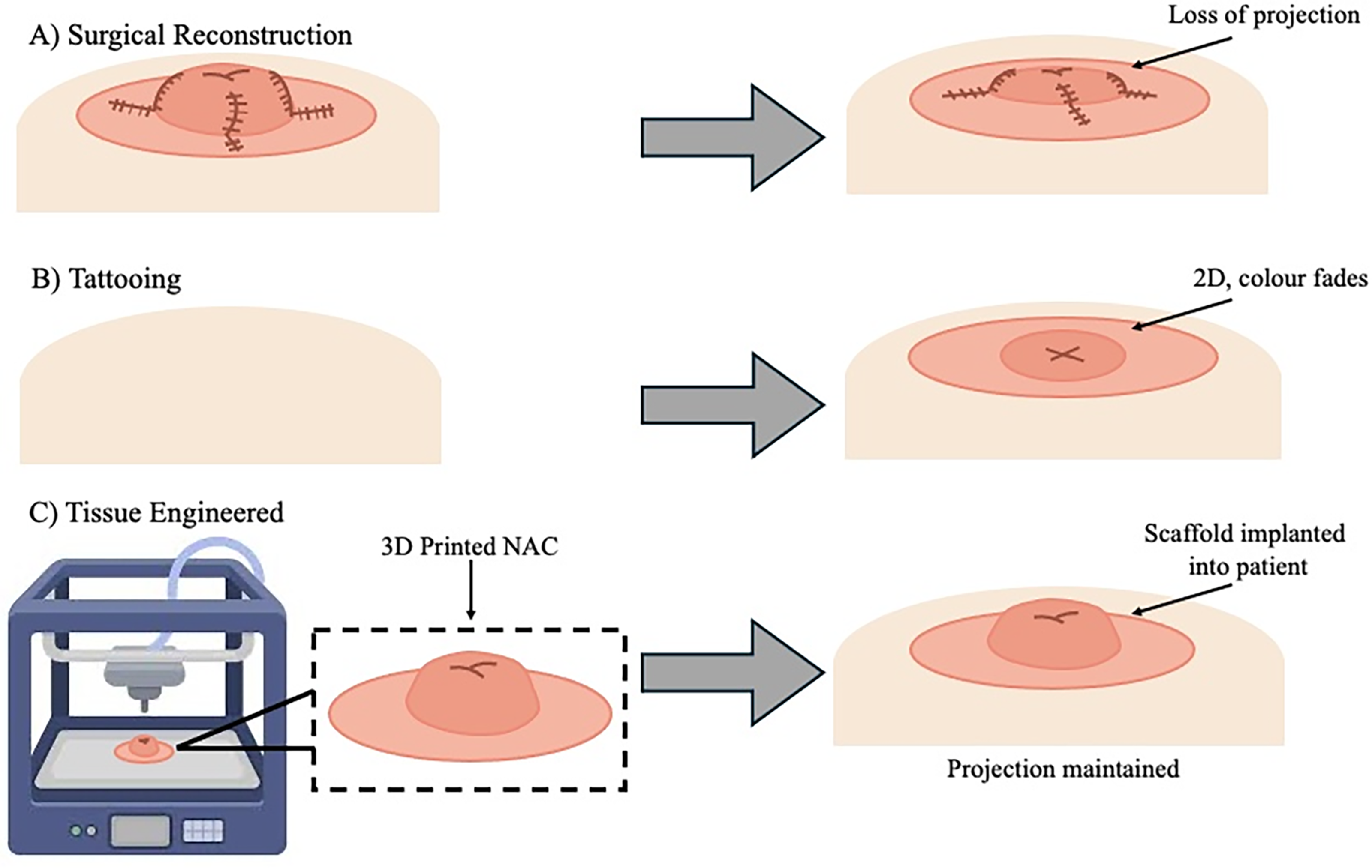
Current options for nipple reconstruction schematic.
To improve scaffold integration and performance, surface modification techniques such as nonthermal plasma treatment have been explored to enhance biocompatibility and cellular adhesion. Nonthermal plasma treatment alters the surface chemistry and morphology of a biomaterial without affecting its bulk characteristics.14,15 In the context of NAC reconstruction, plasma-treated scaffolds could significantly improve biocompatibility and cellular integration, supporting the long-term stability and functionality of the reconstructed tissue. Additionally, hybrid scaffold designs—combining conductive polymers, biologically active coatings, and multilayered architectures—offer new opportunities to mimic native tissue properties and promote both vascularization and innervation. 16 These strategies are essential to overcoming challenges such as limited nutrient diffusion in thicker constructs, ensuring long-term viability and functionality of NAC reconstructions.
Anatomy of NAC
The anatomy of the NAC is important to understand to accurately reconstruct the structure using 3D printing methods. There are two basic structures that make up the NAC which include the areola and the nipple. The areola can range in sizes, with an average range of 30 to 60 mm, and has sebaceous glands which enlarge during pregnancy to give rise to the tubercles of Montgomery to keep the nipples moisturized and protected. The nipple emerges from the center of the areola and also ranges in size (from 10 to 12 mm wide by 9 to 10 mm in height). 17 The cellular morphology is similar to that of the areola, however, there are no sebaceous glands present. A prominent anatomical feature of the nipple is the 10 to 20 milk duct orifices. These ducts are arranged in a bundle in the center with the ducts narrowing as they near the tip of the nipple. 18
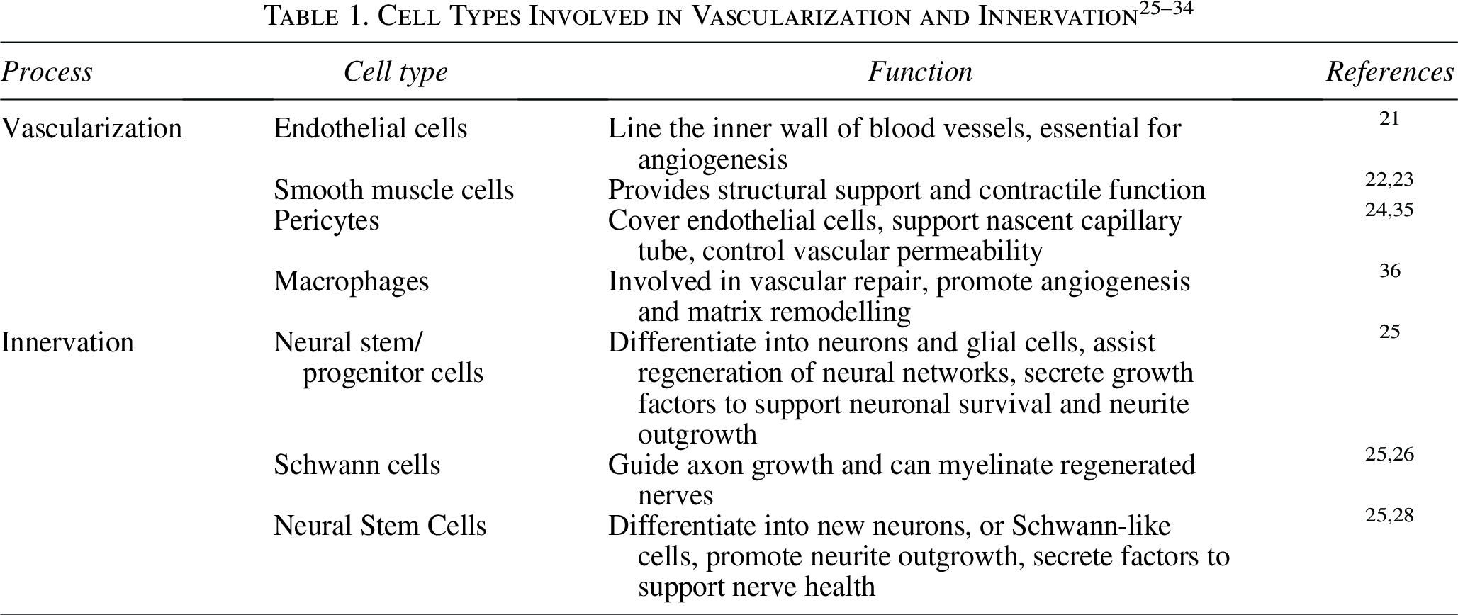
Vascularization and innervation of tissue constructs
When designing tissue engineered constructs, vasculature is important to consider as cells must be within 200 µm of a blood vessel to remain viable. 19 Neoangiogenesis, the growth of new blood vessels from existing vasculature (Fig. 2), is highly regulated through growth factors, chemokines, and angiogenic factors. Achieving functional vasculature is important for meeting the nutritional requirements of tissues, allowing for regenerative constructs to be produced. The establishment of functional vasculature remains one of the primary challenges in tissue engineering. Adequate perfusion is essential for nutrient delivery, waste removal, and overall tissue maturation, yet most engineered constructs lack the hierarchical vascular networks found in native tissues. 20 To address this limitation, various strategies have been developed to enhance perfusion and promote neovascularization. Recent efforts have focused on the integration of perfusion bioreactor systems that provide controlled flow dynamics, enabling more physiologically relevant conditions that support endothelial cell organization and vessel formation. These dynamic culture systems have shown promise in improving vascular density, tissue viability, and functional integration following implantation.21,22

Simplified schematic of neovascularization under physiological conditions.
In addition to vascularization, innervation plays a critical role in regulating tissue and organ development as well as physiological functions such as sensation, movement and homeostasis. Notably, angiogenesis and innervation and closely interconnected processes, with blood vessels providing the nutrients for the growth of neural networks and nerve fibers simulating the formation of new blood vessels which have a synergistic influence on tissue regeneration. 23 Additionally, both endothelial and neural cells respond to similar guidance cues that regulate cell migration, differentiation, and growth. For example, vascular endothelial growth factor (VEGF) mediates cross-talk between these two systems, with neural cell-derived VEGF guiding endothelial cells and influencing vascular differentiation, while endothelial VEGF enhances neuronal proliferation, differentiation, and migration. 24 Various cell types are commonly used to support these processes (Table 1). It has been demonstrated that electrical signals can improve tissue regeneration by promoting innervation, therefore, incorporating conducting polymers or electroactive materials into scaffolds is a promising strategy to improve the vascularization and integration of tissue engineered constructs, an area which has been overlooked in the past.35,36
Design and development
In the context of nipple reconstruction, tissue engineering offers the potential to overcome current limitations associated with traditional surgical techniques, specifically the loss of nipple projection and long-term deformation. 37 The reconstruction of the NAC requires careful consideration of various aesthetic factors including color, projection, size, and texture. These features play an important role that influences patient satisfaction and the overall outcome of breast reconstruction. Current methods, such as using local flaps, often fail to achieve long-term projection of the nipple construct. The use of additive manufacturing techniques, namely 3D printing, allows for greater control of the NAC’s architecture and allows for tailoring of the structural integrity, preventing the collapse of the nipple construct. Compared with traditional methods for producing tissue engineered scaffolds (e.g., solvent casting, electrospinning, or freeze drying), 3D-printed scaffolds allow for high reproducibility, accuracy, and customization, making it an attractive option for reconstructing the nipple.38,39
A successful tissue-engineered nipple scaffold has certain criteria that must be met; these criteria include biodegradability, low immunogenicity, structural integrity, and porosity. Biodegradability ensures that the scaffold gradually degrades as native tissue regenerates, eliminating the need for surgical removal. Low immunogenicity minimizes the risk of adverse immune responses, promoting tissue integration and healing. The native human nipple exhibits a stiffness in the range of 0.1–0.2 MPa, which serves as a critical benchmark for scaffold design. Replicating this mechanical profile is essential to ensure that the engineered construct maintains comfort, structural integrity, and long-term shape fidelity following implantation. 40 Incorporating this physiological reference helps guide biomaterial selection and fabrication strategies towards scaffolds that better mimic the native tissue environment. Finally, an appropriately engineered porosity promotes cell infiltration, vascularization, and nutrient diffusion, all of which are vital for effective tissue regeneration and long-term scaffold viability. 37
Additive manufacturing approaches
To achieve these design and biological requirements, various 3D printing methods can be used, each with their own advantages and limitations. The use of 3D printing in tissue engineering and regenerative medicine has demonstrated great promise due to its ability to create geometrically complex constructs and have precise control over the macro- and microarchitecture with the production of interconnected pore structures and pore size. This allows for the generation of constructs that can accurately recreate the native in vivo environment. 40 Common 3D printing methods that are utilized for tissue engineering include fused deposition modeling (FDM), inkjet bioprinting, and melt electrowriting (MEW), which have been summarized in Table 2 49 (Fig. 3).
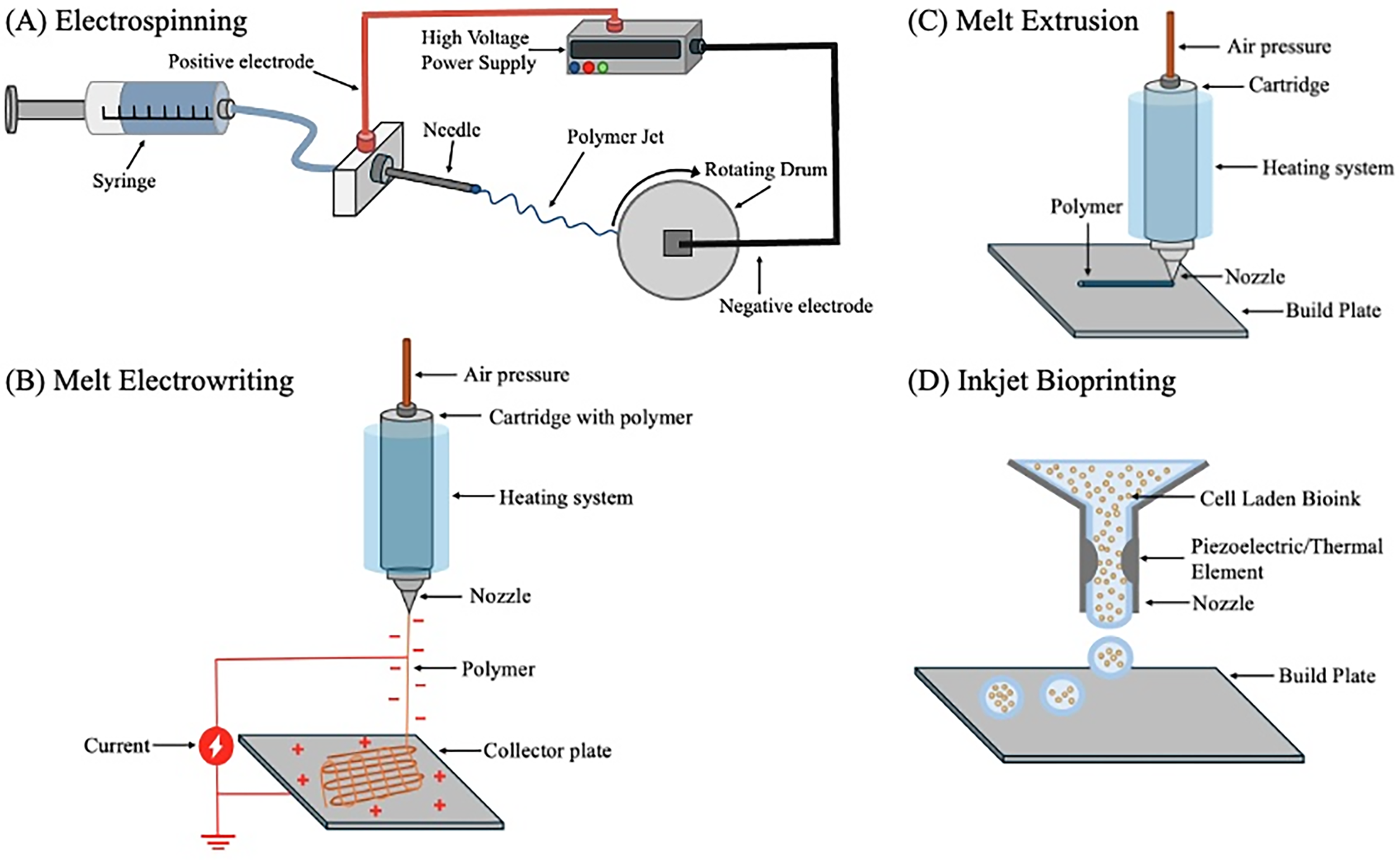
Schematic overview of different additive manufacturing techniques commonly used in tissue engineering.
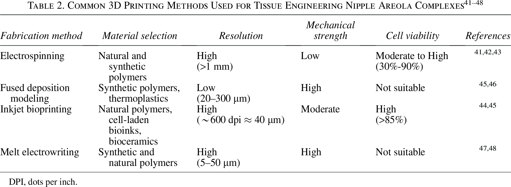
DPI, dots per inch.
In addition to 3D printing, electrospinning is a well-established fabrication method widely used for creating nanofibrous scaffolds that mimic the extracellular matrix (ECM) (Fig. 3). It is a highly effective fabrication technique, which is simple, versatile, and cost-effective. 50 Electrospinning can manufacture these nanofibers (<1 µm) by applying a high voltage to a polymer solution, which produces a jet that elongates and solidifies into fibers once it reaches a collector plate.41,51 The resulting scaffold can replicate key physical and biochemical cues found in the ECM such as fiber orientation, porosity, and interconnecting pores, all of which are important for facilitating the development of functional tissues. 52 Common materials used in electrospinning include synthetic polymers such as poly-ε-caprolactone (PCL) and polylactic acid (PLA) as well as natural polymers like collagen and fibrinogen. These materials can also be coelectrospun to combine and tailor their physical and chemical properties, resulting in scaffolds with enhanced functionality and biocompatibility. 42 Despite its advantages, electrospinning can have limitations in creating large, 3D structures and produces scaffolds with limited mechanical strength. This can be overcome by combining electrospinning with 3D printing methods. 53
In addition to 3D printing, electrospinning is a widely used method for creating nanofibrous scaffolds that mimic the ECM. It is simple, versatile and cost-effective. 50 By applying a high voltage to a polymer solution, nanofibers (<1 µm) are formed as the jet elongates and solidifies on a collector plate.41,51 The resulting scaffold can replicate key ECM features such as fiber orientation, porosity and interconnecting pores, supporting functional tissues development. 52 Commonly used materials include PCL, PLA, collagen, and fibrinogen which can be coelectrospun to tailor scaffold properties and enhance biocompatibility. 42 Despite its advantages, electrospinning can have limitations in creating large, 3 D structures, often producing scaffolds with limited mechanical strength. This can be overcome by combining electrospinning with other 3 D printing methods. 53
FDM is a common 3D printing method where a thermoplastic, synthetic polymer filament is heated and deposited layer-by-layer. It produces clean, durable, and detailed constructs at a low cost without organic solvents, making it popular in bone tissue engineering and drug delivery.54,55 However, high processing temperatures limit material choice and prevent direct cell incorporation. 56 Among additive manufacturing techniques, the choice of fabrication method strongly influences both plasma compatibility and collagen deposition potential. An additive manufacturing method that is compatible with cells, inkjet bioprinting dispenses droplets of cell-laden bioink using thermal, piezoelectric, or microvalve processes, enabling the fabrication of high-resolution tissue constructs. 57 It offers low cost, high cell viability and high spatial resolution. However, bioink aggregation and viscosity can reduce print quality and clog the printer heads, limiting printable materials to alginate, poly (ethylene glycol), dimethyl methacrylate, and collagen. 58 Inkjet bioprinting commonly employs hydrogel-based bioinks, which provide a hydrated, ECM-like environment that supports cell attachment and facilitates collagen synthesis and matrix remodeling. 59 Because hydrogel inherently contain hydrophilic functional groups, they naturally promote cell adhesion and wettability; thus, plasma surface medication is rarely applied to hydrogels. 60 Instead, plasma treatment is more often used to functionalize synthetic polymer substrates, like PCL or PLA, 61 prior to hydrogel coating, enhancing interfacial bonding between layers. 62 MEW combines melt extrusion with electrospinning to fabricate highly organized, porous scaffolds with precise microarchitecture 63 (Fig. 4). The voltage differential between the printer nozzle and the build plate induces the formation of a Taylor cone at the nozzle tip, from which a fine fiber is drawn.64,65 Specifically, MEW, like electrospinning, is able to produce ultra-thin fibers with high surface area-to-volume rations, increasing the number of available sites for collagen deposition and improving the efficiency of plasma activation. 66 This makes MEW- or electrospinning-based scaffolds particularly compatible with plasma surface modification, offering a synergistic strategy for enhancing collagen integration and bioactivity compared to techniques that generate smoother or less porous surfaces, such as traditional FDM printing.
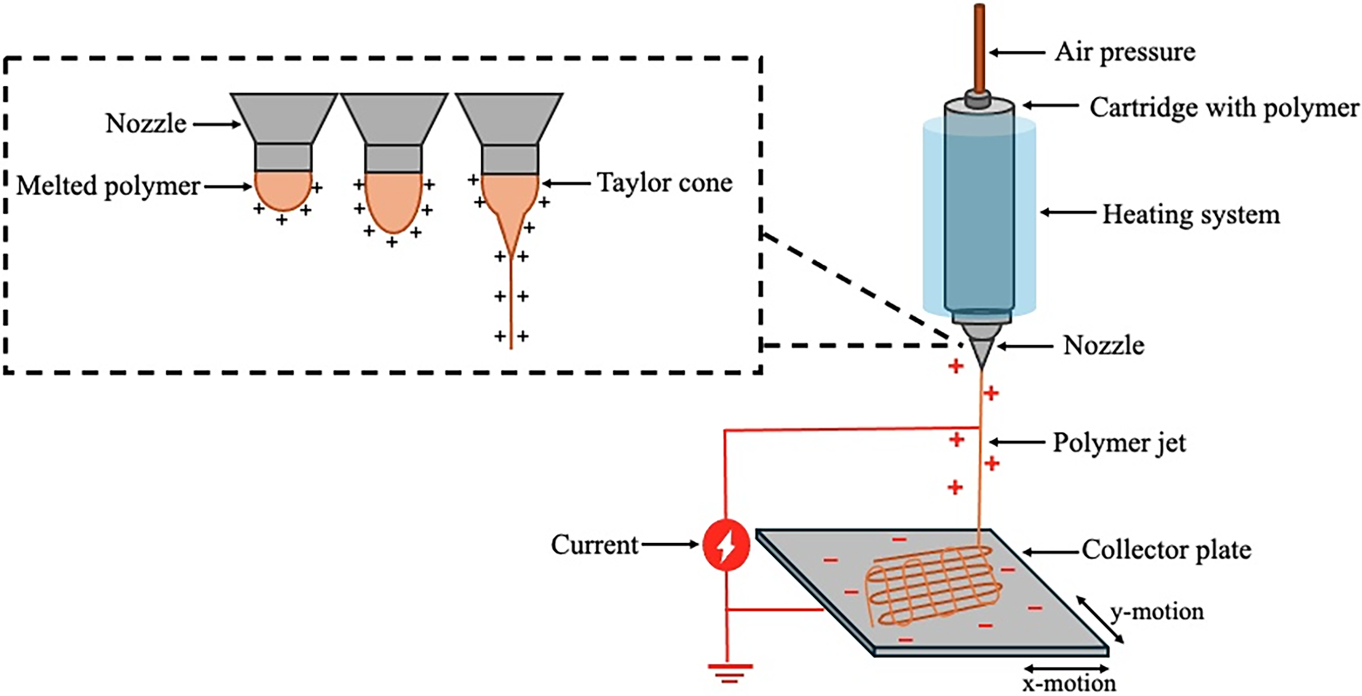
Schematic of a melt electrowriting (MEW) system, showing the formation of a Taylor cone. The Taylor cone is formed because of a high electric field acting on the extruded polymer.
Biomaterial selection
Selecting suitable biomaterials is crucial for developing scaffolds that replicate the ECM and offer adequate structural support. This involves combining natural and synthetic materials to mimic the ECM environment and promote cell growth and tissue regeneration. 67 Among FDA-approved synthetic polymers, PCL is frequently used in bioengineering applications such as heart valve replacement, nerve tissue engineering and soft tissue repair. PCL gradually degrades through hydrolysis, eliminating the need for surgical removal once it has fulfilled its purpose. Comparing PCL to other commonly used polymers, its lower degradation rate helps contribute to its reduced cytotoxicity in vivo, making it an attractive option for its use in tissue engineering and regenerative medicine. 68 Additionally, PCL exhibits excellent mechanical properties, including flexibility and strength, allowing it to mimic the natural structure of tissues. Its mechanical strength can be tailored by adjusting the molecular weight or combining it with other materials. In addition to PCL, other FDA-approved polymers are widely applied in tissue engineering and demonstrate potential for NAC scaffold development. Poly(lactic-coglycolic acid) (PLGA) offers tuneable degradation kinetics through the adjustment of the lactic-to-glycolic acid ratio. This tunability comes from differences in crystalline and hydrophobic structures between the two monomers. PLGA also demonstrates excellent biocompatibility and processability, with its degradation products being natural metabolized by the Krebs cycle, ensuring minimal cytotoxicity. PLGA tends to degrade faster than PCL, producing lactic acid and glycolic acid byproducts that are readily metabolized by the body.39,69 PLA is another FDA-approved, biodegradable polymer with favorable mechanical strength and chemical modifiability, making it suitable for a range of soft tissue applications. 70 Polyurethane (PU) and polyethylene terephthalate (PET) have also been employed in biomedical scaffolds and implantable devices due to their mechanical robustness, elasticity, and long-term stability. PU, in particular, exhibits excellent fatigue resistance and flexibility, 71 properties advantageous for dynamic soft tissue environments, 72 while PET provides high tensile strength and durability, although its nondegradable nature may limit its use in resorbable tissue scaffolds. Overall, while these polymers offer distinct advantages, PCL remains a preferred choice for soft tissue engineering applications due to its slower degradation rate, elasticity, and mechanical resemblance to native tissue. 73 Despite this, PCL and other synthetic polymers share a limitation, which is the absence of bioactive surface functional groups, reducing its cell-surface interaction. 74 Bioactive sites are required to bind with cell receptors which aids in cell adhesion, proliferation and activation. 68 One method of overcoming this is using nonthermal plasma treatment to modify the surface properties of the PCL scaffold.
Collagen
Collagen is a key component in the ECM which forms the primary building blocks for skin, muscle, bone and other connective tissues. 75 Its use in tissue engineering and regenerative medicine has been extensive due to its excellent properties such as its biocompatibility, biodegradable and its promotion of cell adhesion, proliferation, and differentiation. These properties make it a suitable biomaterial which can be used to create biomimetic scaffolds. 76 Type 1 collagen is widely used in tissue engineering because of its abundance in skin and bone and supports the mechanical and structural integrity of connective tissues. Additionally, it promotes cell adhesion, migration, and proliferation, making it valuable for regenerative applications.77,78 Collagen can be sourced from various origins (Table 3), traditionally bovine and porcine, though these have limitations due to cultural, religious, and disease transmission concerns.

Alternative sources of collagen have been investigated with the purpose of overcoming the limitations surrounding mammalian-sourced collagen. Jellyfish-derived collagen, or type 0 collagen, is of interest due to its high collagen content and seasonal abundance. Marine-based collagen, such as fish, sea urchins and jellyfish, are emerging as a popular biomaterial because they are environmentally friendly and more inclusive for groups avoiding porcine or bovine collagen. 84 Type 0 collagen resembles types I, II, III, and V and offers benefits such as long-term anti-inflammatory effects, maintenance of cell phenotypes without dedifferentiation, nontoxicity, and supports higher fibroblasts viability compared with bovine collagen. 82
Despite the advantages that marine-based collagen provides, there are still limitations for the use of collagen in tissue engineering. Modifying pure collagen is challenging due to collagen’s poor mechanical properties and rapid degradation. Various strategies have been developed to overcome these limitations, with crosslinking being the most common approach.85,86 Crosslinking can modulate the degradation rate of collagen and enhance both its biological and mechanical properties of the scaffold by reinforcing interactions within the matrix. Physical crosslinking methods, such as UV irradiation or dehydrothermal treatment, modify the collagen structure without chemical reagents, while chemical crosslinkers, such as glutaraldehyde or genipin, form covalent bonds that stabilize the matrix. Enzymatic crosslinkers, such as transglutaminase or lysyl oxidase, provide a biocompatible and controllable means to strengthen collagen scaffolds while maintaining bioactivity.87,88 However, crosslinking may not sufficiently improve these properties under physiological conditions, with some crosslinking methods reducing biocompatibility. 88 A hybrid design approach, which combines collagen with other biomaterials or incorporates advanced fabrication and postprocessing techniques is often needed to achieve the optimal structural, mechanical, and biological properties for effective tissue engineering applications. 89
Conducting polymers
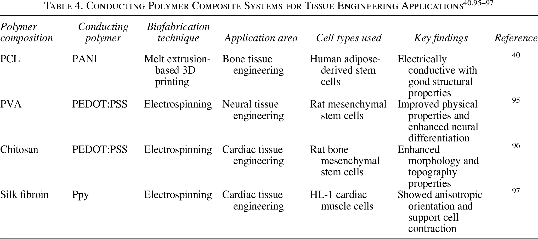
In addition to hybrid synthetic-natural polymer systems, the incorporation of conducting polymers offers a further dimension to scaffold functionality in tissue engineering applications. Being a class of ionic electroactive organic polymers, conducting polymers are able to integrate the optical and electrical characteristics of metallic and inorganic semiconducting materials with the flexibility and ease of processing found in synthetic polymers. 90 Conducting polymers are characterized based on their conjugated backbones, which consist of alternating single and double bonds. These bonds allow electrons to move more freely along the chain. The π-bonds involved are weaker than the strong σ-bonds holding the chain together, but they enable a high degree of electron overlap, which enables the electrical conductivity. 91 Polyaniline (PANI), polypyrrole (PPy), and poly(3,2-ethylenedioxythiophene) polystyrene sulfonate (PEDOT:PSS) are examples of conducting polymers which have generated interest in the tissue engineering field for its ability to enhance nerve regeneration, among other benefits such as improving cell proliferation, differentiation, and migration.92,93 Additionally, the incorporation of conducting polymers into scaffolds aid in modulating cellular specialization and differentiation, the success of which has been demonstrated in various tissue engineering applications, including skin, bone, cardiac, and neural. 94 Because of these attributes, conducting polymers have been used in regard to wound healing and tissue engineering vasculature constructs (Table 4).
PANI is a popular conducting polymer used to induce electroactivity in a tissue engineered scaffold. This is due to its biocompatibility, conductivity, ease of processing and its antimicrobial effect. There are three different oxidation states that PANI can exist; the fully reduced leucoemeraldine, the fully oxidized pernigraniline, and the partially oxidized emeraldine base. 98 The emeraldine base form is particularly significant in tissue engineering applications, as it exhibits both electrical conductivity and relative stability under physiological conditions. When doped with acids, such as camphorsulfonic or hydrochloric acid, PANI can become even more conductive, which is essential for electrical signal propagation in applications like neural tissue engineering or electrically responsive drug delivery systems. 99 One example of conductive polymer integration in tissue engineering is the incorporation of PANI with PCL scaffolds through extrusion-based 3D printing. Wibowo et al., demonstrated that using low concentrations of PANI could enhance the conductivity of the scaffold while maintaining cytocompatibility over 21 days. Although this work was primarily looked at for bone tissue engineering, it highlights the potential of conducting polymers to promote cellular responses relevant for innervation in soft tissue constructs, such as a NAC. 40 Another study showed that the incorporation of PLA and PANI was able to greatly improve the extension of neurite outgrowths after being treated with an atmospheric pressure plasma jet. The neurite density increase was significant on the PLA and doped PANI compared with the PLA scaffolds with 6.7 × 10³/cm2 and 4.6 × 10³/cm2, respectively. This demonstrates the potential of incorporating a conducting polymer into the scaffold, in the absence of external electrical stimulation, can encourage cell behaviors that are electrically sensitive. 92
PEDOT:PSS is a poly(thiophene) derivative which has been widely used in tissue engineering scaffolds due to its biocompatibility, chemical stability. Additionally, it has a higher electrical conductivity compared with other conducting polymers such as PANI, which makes it an attractive option for innervated tissue engineered scaffolds. 96 PEDOT:PSS can be incorporated into a variety of scaffold fabrication methods, including electrospinning, 100 3D printing, 101 and surface coating, 102 enabling tuneable electrical properties while maintaining structural integrity. Its conductive properties has been shown to promote cell adhesion, proliferation, and neurite outgrowth with studies showing that there is neurogenic differentiation in as early as 7 days.95,103 These characteristics make PEDOT:PSS a promising material for the engineering of electrically responsive tissues such as skin, nerve, cardiac, and skeletal muscle. The use of PEDOT:PSS in fabricating an electrically conductive scaffold has been explored in the literature, with one example being its incorporation with electrospun silk scaffolds. Previous studies have shown improved biological results in neuronal differentiation when PEDOT:PSS is incorporated into the scaffold. Specifically, Magaz et al., demonstrated that although all their samples supported neurite outgrowths, coating the scaffold with PEDOT:PSS at 3 mg/mL and dimethyl sulfoxide significantly improved the average neurite extension. It was concluded that electroactive scaffolds were a promising platform for peripheral nerve regeneration, however, further research is needed. 104 Another study demonstrated the benefit of coating a freeze dried chitosan/gelatin scaffold with PEDOT:PSS through the promotion of cell adhesion and proliferation as well as the enhancement of neural stem cell differentiation. The results showed an upregulation of various proteins, such as βIII-tubulin, that are prominent in neuronal cell differentiation in the presence of PEDOT:PSS scaffolds compared with the control. 105 Overall, the incorporation of conductive polymers into tissue engineered scaffolds holds significant potential for fabricating innervated constructs that can function and integrate with the host environment. 106
Hybrid design approach
When considering tissue engineering for the creation of 3D vascularized and innervated NACs, the combination of synthetic and natural polymers presents a promising hybrid design approach. Specifically, this approach can address the challenges that are involved in creating functional, biologically integrated tissues such as NACs, which require both structural integrity and biological function. Extrusion-based 3D printing is an effective tool which allows for accurate fabrication of scaffolds with tuneable properties; however, there are limitations to the materials used when this printing technique is utilized in soft tissue engineering. 107 The combination of PCL and collagen is a one solution to over these limitations, as it uses the mechanical integrity of synthetic polymers like PCL with the inherent bioactivity of natural polymers such as collagen. 108 This not only improves the scaffold’s performance, but it is also involved in promoting vascularization in tissue engineered constructs. Additionally, a hybrid design approach allows for greater spatial and compositional control within the scaffold. 109 Therefore, extrusion-based 3D printing of hybrid materials is one pathway for developing functional, vascularized, and anatomically accurate NAC constructs.
While PCL-collagen scaffolds support vascularization and structural integration, they offer limited capacity to promote functional innervation, highlighting the needs for incorporating electrically conductive polymers into the scaffold design. The integration of PEDOT:PSS into the hybrid scaffold enables the creation of a multifunctional scaffold which not only supports vascularization and mechanical integrity but also contributes to the restoration of neural functionality. 110 There are various methods that can be used to integrate PEDOT:PSS into the scaffold, such as blending, 111 dip coating, 102 or electrospinning; 96 however, each approach presents specific challenges. Blending PEDOT:PSS with PCL for melt extrusion would allow for uniform conductivity as well as being a one-step fabrication method; however, it is challenging due to the polymers’ thermal incompatibles and chemical properties. 111 Although PEDOT:PSS does not fully degrade until temperatures above 200°C, its electrical conductivity begins to decline at much lower temperatures. Stepien et al., demonstrated that PEDOT:PSS films undergo chemical decomposition at temperatures above 160°C, with their conductivity degrading exponentially at temperatures as low as 55°C.112,113 Melt extruding PCL occurs within this range, therefore, blending PEDOT:PSS by this method would likely compromise its conductivity. 114 Direct reports on melt blending PEDOT:PSS with PCL are limited in the literature, which likely reflects these structural and chemical incompatibilities. Dip coating the PCL scaffold with PEDOT:PSS is the simplest method for introducing conductivity. This method involves immersing the scaffold in a solution of PEDOT:PSS, then allowing the solvent to evaporate. A potential problem with this method is that the PEDOT:PSS coating detaches from the scaffold when it is exposed to a physiological environment. This occurs due to both the weak physical interaction between the hydrophilic PEDOT:PSS layer and the hydrophobic PCL scaffold, and the inherent water dispersibility of PEDOT:PSS, which makes it prone to swelling and leaching in aqueous environments. Without additional surface treatments or crosslinking, the PEDOT:PSS layer remains vulnerable to physical degradation.115,116 Therefore, electrospinning a PEDOT:PSS layer provides a low-temperature alternative that maintains electrical functionality and minimizes the risk of leaching in physiological conditions. This can also be minimized by incorporating plasma treatments into the scaffold design. The plasma treatment forms functional groups which allows for the interaction between the scaffold and PEDOT:PSS, thereby improving the coating stability and durability. 117 Overall, combining synthetic, natural, and conductive materials via multiple fabrication methods offers a modular and synergistic approach for engineering innervated, vascularized NAC constructs.
Plasma: a surface modification technique
The interaction between biomaterials and native tissue is influenced by their surface properties. Surface modification techniques can be used to enhance the biocompatibility and integration of biomaterials that would otherwise not interact effectively with the surrounding tissue. By changing characteristics such as surface roughness, chemical composition, or hydrophilicity, these techniques can promote cell adhesion, proliferation, and differentiation, which ultimately leads to better functional integration and reduced risk of adverse reactions. 118 Plasma, one of the four states of matter, has been shown to be a promising candidate for modifying the surface properties of 3D scaffolds. Briefly, plasmas are fully or partially ionized gases, which have had enough energy added to remove electrons from atoms. This creates a blend of free electrons, free radicals, neutrals, and positively charged species which induces chemical and physical changes on a materials’ surface. 119 Plasma can be categorized as thermal or nonthermal with the category of nonthermal plasma (NTP) gaining interest for its use in material science. The benefits of NTP are that it is environmentally friendly, inexpensive, fast, and it is effective for modifying the surface various materials. The use of NTP for surface modification and activation is preferred over thermal plasma as it does not cause thermal damage to the temperature-sensitive materials, such as PCL, nor compromise the bulk properties. 120 Researchers have investigated different types of gases which could be used for surface modification, common ones being argon (Ar), nitrogen (N2), and oxygen (O2). There has been a particular focus on nitrogen-containing plasma surface modifications as it has been demonstrated that the incorporation of amine groups promotes the immobilization of biomolecules (e.g., collagen, polysaccharides and enzymes). Additionally, nitrogen plasma introduces a localized positive charge onto the surface of the polymer to electrostatically interact with cells and proteins which are negatively charged (Fig. 5). This enhances cell adhesion and proliferation, thereby improving the integration and function of the 3D scaffold. 121
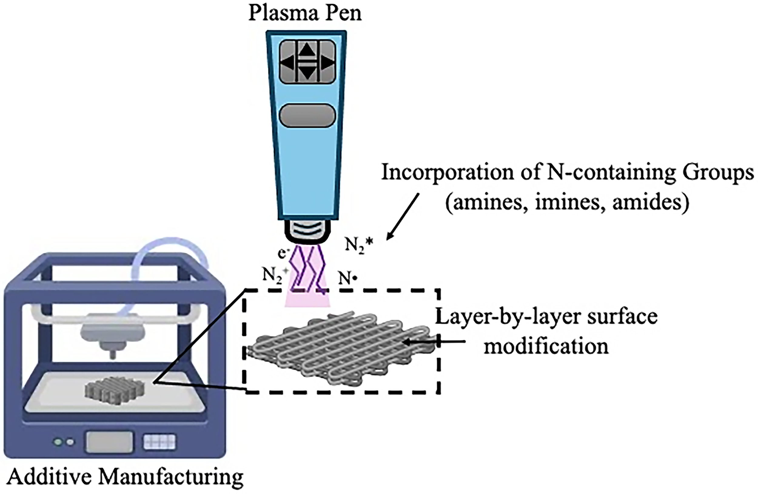
Scaffold fabrication schematic. Sequential melt extrusion of polymer layers, with plasma treatment with nitrogen gas applied after each layer.
Previously, plasma treatments were completed as a postprocessing technique, after 3D printing the scaffold construct. This presented a challenge to produce a homogenous surface throughout the scaffold, particularly in the inner strut architecture, that are particularly problematic for tissue engineering applications where uniformity is critical. 122 Because plasma treatments are performed after the scaffold has been fully printed, the functional groups that the plasma adds to the surface of the scaffold can fail to deeply penetrate the internal architecture of the scaffold. As a result, only the outer surfaces receive effective modification, while the inner struts and pores may remain untreated or inconsistently modified. This uneven treatment can lead to gradients in surface properties such as wettability, chemical functionality, and roughness, which in turn may cause nonuniform cellular responses, inconsistent mechanical properties, and unpredictable degradation behavior. 121 To address these challenges, Kim et al. (2023) proposed and demonstrated that a layer-by-layer method for plasma treatment can produce a homogenous surface modification. This method involves applying plasma treatment to each individual layer immediately after it is deposited, allowing the plasma to uniformly treat every surface.123,124
Recent work has demonstrated how plasma surface modification can play a significant role in improving the performance of scaffolds for soft tissue engineering applications. Meghdad et al. (2019) used NTP treatment to surface modify a PCL nanofibrous scaffold to enable gelatin immobilisation. This enhances the scaffold’s hydrophilicity and cell attachment, demonstrating the efficacy of NTP in improving scaffold biofunctionality. Plasma treatment of PCL scaffolds enables effective bioactive molecule immobilization, enhancing cell adhesion, infiltration, and differentiation. These are key factors for successful NAC scaffold integration and regeneration. 125 Additionally, the use of plasma-treated PCL scaffolds with conducting polymers incorporated has been demonstrated to be beneficial by Licciardello et al. (2021). They fabricated electrospun nanofibers composed of a PCL-PANI blend, which created a porous, ECM-like scaffold that maintained structural stability. However, the scaffold was highly hydrophobic, with having a water contact angle of 133.5 ± 2.2°. By using atmospheric plasma treatment, the surface of the PCL-PANI scaffold was able to become hydrophilic, with the water contact angle decreasing to 67.1 ± 2°. Additionally, biological assessments demonstrated that cells cultured on the plasma treated PCL-PANI scaffolds showed enhanced viability and improved biocompatibility through its cell-material interactions. This work demonstrates that combining conducting polymers with plasma-treated PCL scaffolds can reduce hydrophobicity, leasing to improved cell attachment and viability. For NAC reconstruction, this is promising because scaffold hydrophilicity and cell compatibility are essential for dermal integration, while conductivity provides a pathway for promoting innervation and potentially restoring sensation. Overall, these properties support long-term scaffold durability and functional tissue regeneration. 90
Conclusion
The loss of the NAC following mastectomy profoundly impacts body image and psychological well-being. Recent advancements in tissue engineering now enable the fabrication of patient-specific scaffolds that mimic the structural and elastic properties of the native NAC. Hybrid additive manufacturing approaches that combine melt extrusion with electrospinning offer tunable control over mechanical stiffness, porosity, and surface topography, enhancing cell adhesion and long-term projection stability. Moreover, nonthermal plasma surface activation—particularly under nitrogen-rich gas chemistries—combined with collagen coating improves scaffold hydrophilicity and bioactivity, facilitating superior cell attachment and tissue integration. Future studies should systematically evaluate the long-term mechanical fatigue behavior of hybrid NAC scaffolds under cyclic loading to ensure structural stability postimplantation. The incorporation of conductive polymers and bioelectrical stimulation presents a promising avenue for promoting neural differentiation and functional sensory recovery; however, optimization of polymer conductivity, degradation kinetics, and biocompatibility remains essential. Importantly, the translation of conductive and hybrid biomaterials into clinical applications must also address regulatory challenges related to safety, degradation by-products, and long-term host response. In vivo validation of vascularization, innervation, and sensory restoration are critical next steps toward developing clinically viable, multifunctional NAC regeneration.
Authors’ Contributions
C.R.O.D.: Conceptualization, investigation, methodology, formal analysis, investigation, writing—original draft preparation, visualization. M.A.A.: Resources, supervision, writing—review and editing. J.D.C.: Resources, conceptualization, supervision, writing—review and editing.
Footnotes
Disclosure Statement
J.D.C. serves as Guest Editor for the special issue who had no role in the editorial review or decision-making process for this article, which was handled independently by the Editor-in-Chief. The authors declare no other competing interests.