



Abstract
Three-dimensional (3D) culture systems have emerged as powerful tools to model tumor biology and bridge the gap between conventional two-dimensional (2D) assays and in vivo studies. Here, we evaluate a poly(ethylene glycol)-based hydrogel as a simplified yet functionally relevant platform for modeling premalignant lung adenocarcinoma using the A549 cell line. Cells cultured in hydrogels exhibited transcriptional profiles that more closely resembled xenograft tumors than conventional 2D monolayers. Pathway-level analysis and regression-based benchmarking revealed restoration of critical hallmark programs, including proliferation, immune signaling, developmental pathways, and stress response cascades. While the 3D model does not fully recapitulate the complexity of the tumor microenvironment, its chemically defined, tunable, and reproducible design offers an accessible, physiologically informative model of lung adenocarcinoma that restores key transcriptional and functional features lost in conventional culture systems.
Impact Statement
This research establishes a poly(ethylene glycol)-based 3D culture as a more translationally relevant lung cancer model than 2D systems and provides a general framework for benchmarking engineered tumor models against in vivo biology.
Introduction
Lung cancer imposes a major global health burden, accounting for more deaths each year than any other malignancy. 1 Although advances in surgery, radiation therapy, targeted therapies, and immunotherapies have improved outcomes for many patients, overall long-term survival remains limited.2–5 Therapeutic progress in part depends on the quality of preclinical models used to study disease mechanisms and evaluate therapies. Historically, two-dimensional (2D) culture and subcutaneous xenografts have been considered the gold standard experimental systems in lung cancer research. 6 In 2D culture, cells are grown on flat surfaces with uniform exposure to nutrients and mechanical cues, and it is widely used because it is simple, rapid, and inexpensive.7–10 However, these systems lack the structural and microenvironmental context of tumors in vivo, leading to altered morphology, signaling, and treatment responses.11–13 Alternatively, xenograft models preserve tumor heterogeneity and architecture more faithfully,14,15 but their high cost, lengthy establishment times, and low-throughput nature restrict their widespread use. 16 As a result, neither system alone fully captures the complexity of lung tumors nor meets the practical needs of translational research.
To address the limitations of conventional lung cancer models, three-dimensional (3D) culture systems have been widely adopted to introduce matrix-dependent signaling and spatial constraints that regulate tumor cell state and behavior.13,17,18 A range of 3D approaches exists, including spheroid cultures, hydrogels, organoids, microfluidic devices, and tissue slices. Much of this work has relied on naturally derived matrices such as Matrigel and collagen due to their ease of use and inherent bioactivity. 18 However, these materials suffer from important limitations that complicate mechanistic interpretation. In particular, Matrigel exhibits substantial batch-to-batch variability and contains hundreds to thousands of poorly defined growth factors, introducing experimental confounders that are difficult to control or reproduce. 19 In addition, both Matrigel and collagen display relatively low mechanical stiffness (G′ < 1 kPa), limiting their relevance for modeling the broader stiffness ranges observed in tumors and common metastatic sites. 20 Furthermore, in these matrices, changes in mechanical properties are often intrinsically coupled to changes in biochemical composition, making it difficult to decouple the independent effects of matrix stiffness and extracellular matrix (ECM) ligands.
Synthetic hydrogels provide a controlled alternative to biologically derived matrices by enabling independent specification of biophysical and biochemical cues. Poly(ethylene glycol) (PEG)-based hydrogels are particularly well suited for this purpose because PEG is biologically inert, biocompatible, and chemically defined, with cross-linking strategies that allow precise tuning of stiffness, degradability, and ligand presentation.21–25 Owing to their hydrophilicity and resistance to nonspecific protein adsorption, unmodified PEG hydrogels function as a noninteractive baseline in which cellular responses arise only from experimentally introduced signals. 24 Importantly, the modular design of PEG systems permits independent control of mechanics and bioactivity, enabling systematic interrogation of matrix-dependent effects that cannot be readily isolated in natural matrices.
Our laboratory and others have demonstrated that PEG-based hydrogels can support the growth and heterogeneity of nonsmall cell lung cancer (NSCLC) cells while enabling the systematic incorporation of ECM ligands and mechanical cues to study processes such as epithelial–mesenchymal transition and cell invasion.26,27 Hybrid PEG hydrogels have been applied to breast and lung cancer models, supporting cell viability, morphology, and matrix-dependent behavior while recapitulating aspects of in vivo tumor responses, including therapeutic sensitivity.28,29 PEG hydrogels have also been used to model vascular interactions by coculturing endothelial and stromal cells with tumor cells, enabling the study of angiogenic signaling and vascular morphogenesis in a controlled 3D environment. 30 In addition, our work has shown that PEG systems provide a tractable platform for studying immune–tumor interactions, including natural killer cell migration and cytotoxicity under physiologically relevant conditions.26,31 Despite these advances, PEG-based tumor models are most often validated using phenotypic, functional, or pathway-specific readouts rather than through direct molecular comparison to tumors grown in vivo, leaving a critical gap in understanding how well these engineered systems recapitulate the global transcriptional programs of native tumors.
As a result, it remains unclear which transcriptional programs characteristic of lung tumors are preserved in PEG-based 3D culture and which are lost or altered relative to tumors grown in vivo. Although 3D culture platforms offer clear advantages over conventional systems, most transcriptomic studies benchmark these models only against 2D culture, revealing how cells adapt to a 3D environment but providing limited insight into their fidelity to native tumors.32–36 Direct comparisons to in vivo tumors are typically restricted to phenotypic or functional readouts rather than global molecular programs.18,37–39 This lack of systematic molecular benchmarking limits the interpretability and translational relevance of synthetic hydrogel models and underscores the need to define which aspects of tumor biology are faithfully captured in 3D culture.
Here, we compare a PEG-based 3D lung cancer model against both 2D culture and xenograft tumors to assess its translational relevance (Fig. 1). Using bulk RNA sequencing and pathway-level analyses, we compare the global transcriptional programs across systems to identify (i) processes that are preserved in vitro relative to tumors in vivo, (ii) features selectively captured in 3D culture but absent in 2D, and (iii) contexts where 3D models provide unique advantages for preclinical research. Finally, this work develops a framework for evaluating the fidelity of engineered tumor models and establishes PEG hydrogels as a facile platform for modeling lung tumor biology.
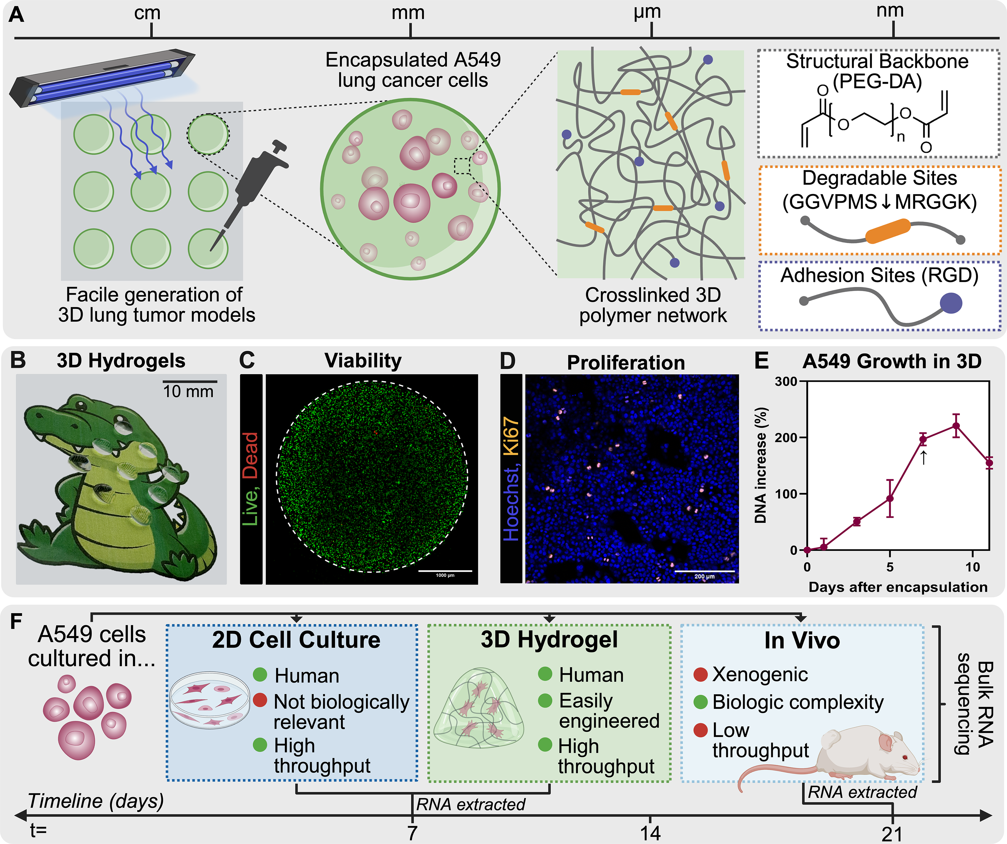
Experimental design.
Materials and Methods
Cell culture
A549 luciferase-expressing human lung adenocarcinoma cells (ATCC, CCL-185-LUC2) were maintained in RPMI 1640 supplemented with 10% (v/v) fetal bovine serum (Corning), 1% (v/v)
Peptide functionalization and polymer synthesis
To enable cell-mediated degradation, hydrogels were functionalized with matrix metalloproteinases (MMP)-sensitive peptide sequence, VPMS↓MRGG, which can be cleaved by multiple MMPs, including collagenase-1 (MMP-1), gelatinase A (MMP-2), and gelatinase B (MMP-9). 40 The sequence was modified with glycine (G) spacers and a C-terminal lysine (K), yielding the final sequence GGVPMS↓MRGGK (Biomatik) and subsequently conjugated to PEG-acrylate (Laysan Bio Inc.) groups as described previously.26,40–43 The resulting degradable block copolymer, acryl-PEG-dMMP-PEG-acryl (PEG-dMMP-PEG, MW: 7,900 Da), was synthesized by reacting the terminal and lysine amine groups of the peptide with succinimidyl valerate ester (SVA; Laysan Bio Inc.) of acryl-PEG-SVA in 50 mM sodium bicarbonate buffer (pH 8) at a 1:2 molar ratio for 4 h. The reaction mixture was dialyzed against deionized water for 24 h, lyophilized for 48 h, and stored at −20°C until use.
Cell adhesion was promoted by the incorporation of an RGD-functionalized PEG (BioSynth). Acryl-PEG-RGD (MW: 4,000 Da) was synthesized as described previously.26,41–43 Briefly, the terminal amine of cyclo(Arg-Gly-Asp-D-Phe-Lys) (cRGD) was reacted with acryl-PEG-SVA in a 50 mM sodium bicarbonate buffer (pH 8) at a 1.05:1 molar ratio for 4 h. The reaction mixture was dialyzed against deionized water for 24 h, lyophilized for 48 h, and stored at −20°C until use. The cRGD motif incorporated into the hydrogel mimics the vitronectin adhesion site, enabling interactions with ανβ3 and ανβ5 integrins.44–46
3D hydrogel preparation
Hydrogels were synthesized from a precursor solution comprising 5% (w/v) PEGDA (3.4 kDa), 5% (w/v) PEG-dMMP-PEG (7.9 kDa), and 5 mM acryl-PEG-RGD (4 kDa), all dissolved in phosphate-buffered saline (PBS), as described previously. 26 The photoinitiator Irgacure 2959 (Millipore Sigma) was added at 0.05% (w/v) prior to polymerization. These hydrogel parameters were chosen based on our prior findings demonstrating that this PEG-based hydrogel not only supports A549 lung cancer cell growth but also facilitates natural killer cell infiltration and migration. 26 This feature presents a promising avenue for future studies modeling immune cell–lung cancer cell interactions in a preclinical setting, which is beyond the scope of the current work.
For mechanical testing, hydrogels were polymerized in cylindrical silicone molds (8 mm diameter × 1.7 mm depth; Grace Biolabs). Precursor solution (108 µL) was exposed to long-wave ultraviolet A light (6.0 mW/cm2, OmniCure S2000; Excelitas Technologies) for 4 min. Following photopolymerization, hydrogels were rinsed with PBS and incubated at 37°C, 5% CO2 for 24 h before testing. Mechanical properties were measured using an Anton Paar MCR 302 rheometer with parallel plate geometry, following established protocols.47,48 An 8-mm sandblasted top plate was used to minimize slip. To approximate physiological conditions, the Peltier stage was maintained at 37°C, and samples were enclosed in a humidity chamber. Amplitude sweeps (0.01–100% strain at 1 Hz) were first performed to define the linear viscoelastic range (LVE). Frequency sweeps (0.1–100 Hz) were then conducted at 0.1% strain, which fell within the LVE. Storage (G′) and loss (G″) moduli were determined, and the complex shear modulus (G*) was calculated using equation 1. Young’s modulus (E) was then determined from equation 2, assuming a Poisson’s ratio (υ) of 0.549–52:


For swelling analysis, hydrogels were polymerized from 10-µL precursor solution, rinsed in PBS, and incubated for 24 h at 37°C and 5% CO2. Hydrogels were blotted to remove excess liquid, and the swollen weight (Ws) was recorded. Samples were lyophilized for 24 h and weighed again to obtain the dry weight (Wd). The swelling ratio was calculated using equation 3
53
:

Cell encapsulation and culture in 3D hydrogels
A549 cells were suspended in the hydrogel precursor solution at a density of 5 × 10⁶ cells/mL, and 10 µL droplets of cell-laden precursor solution were pipetted. The droplets were photopolymerized using the OmniCure S2000 for 4 min at 6.0 mW/cm2 to encapsulate cells in 3D. The crosslinked tumor models were then transferred to a 96-well, washed with 200 µL of PBS, and cultured in 300 µL of complete medium at 37°C and 5% CO2, with media exchanged daily. Cell viability was assessed postencapsulation using Calcein-acetoxymethyl (AM) (Thermo Fisher) and Ethidium Homodimer-1 (Thermo Fisher) according to manufacturer protocols. Cell proliferation was monitored by Ki67 immunofluorescence (abcam) and Quant-IT PicoGreen dsDNA assay on homogenized samples (Invitrogen) according to manufacturer protocols over 11 days.
Confocal microscopy
For all confocal imaging, a Zeiss LSM 880 confocal microscope in scanning mode was used. The microscope had diode (405 nm), argon (458, 488, and 518 nm), and HeNe (543, 594, and 633 nm) laser modules. Images of the entire hydrogel were acquired using z-stacks and tiling features in the ZEN imaging software (Carl Zeiss), and max intensity projections were used to generate 2D images (Fiji ImageJ).
In vivo xenograft model
All animal experiments were approved by the Institutional Animal Care and Use Committee at the University of Florida (protocol 202009835). Male NOD SCID mice (The Jackson Laboratory, 6–8 weeks old) were injected with a 28-gauge needle subcutaneously with 1 × 10⁶ A549-luciferase-expressing cells suspended in Matrigel (Corning) in the flank region. Tumor growth was monitored every other day via calipers (Supplementary Fig. S4). Tumors were harvested at 21 days postinjection, and RNA was immediately extracted.
RNA isolation and quality assessment
Total RNA from 2D cell culture (n = 5), 3D hydrogel (n = 5), and xenograft samples (n = 4) was extracted using the RNeasy Mini Kit (Qiagen) following manufacturer instructions. Briefly, cells from 2D culture were lysed in TRIzol buffer (Invitrogen), and RNA from tumor tissue and 3D hydrogel samples were first loaded in tubes prefilled with 3 mm zirconium beads (Benchmark Scientific) and mechanically dissociated using a BeadBug homogenizer (Benchmark Scientific) prior to column purification. To collect a large enough sample for RNA sequencing, four hydrogel samples were pooled together.
RNA concentration and purity were assessed using a NanoDrop spectrophotometer (Thermo Fisher Scientific), and integrity was evaluated with an Agilent TapeStation to obtain RNA Integrity Numbers (RINs). Samples with RIN ≥ 9 were used for library preparation (Supplementary Fig. S5).
RNA sequencing
For library construction, poly(A)-selected, directional/stranded RNA libraries were prepared using Illumina RNA-Seq Library Prep Kit, following the manufacturer’s protocol. Paired-end sequencing was performed on an Illumina NovaSeq platform by the University of Florida ICBR NextGen DNA Sequencing Core Facility (RRID: SCR_09152), generating 50 million reads per sample.
RNA sequencing data processing
Sequencing quality was evaluated using FastQC to confirm base quality, adapter content, and overread metrics. Reads passing quality control were used for downstream alignment and differential expression analyses. Data filtering and trimming were performed before analysis (Supplementary Fig. S6). Raw and normalized RNA sequencing data and corresponding metadata were imported to R (v4.3.2). To address duplicated gene symbols, expression values were averaged across duplicates before downstream analyses. Processed data matrices were stored in R as numeric matrices with gene names as row identifiers and samples as columns. Gene-level count matrices were imported into edgeR 54 and normalized using the trimmed mean of M-values method. A design matrix encoding experimental groups was constructed, and mean-variance modeling was applied using the limma-voom workflow. 55 Log-transformed counts per million values adjusted by voom were used for principal component analysis (PCA), correlation analysis, and visualization of gene expression patterns.
Differential expression analysis
Differentially expressed genes (DEGs) were identified using limma. 56 Contrasts were defined for (i) 3D versus 2D culture, (ii) 2D culture versus in vivo, (iii) 3D hydrogel versus in vivo, and (iv) in vivo versus 2D culture. Empirical Bayes shrinkage was applied to moderated t-statistics, and results were extracted as ranked gene lists (Supplementary Fig. S6). Genes were considered significantly differentially expressed if they met a false discovery rate (FDR) threshold of adj. p < 0.05 and an absolute log fold change (logFC) > 0.58 (corresponding to approx. 1.5-FC). The number of significantly upregulated, downregulated, and nondifferentially expressed (non-DEG) genes was generated for each comparison (Supplementary Fig. S2 and S3).
Data visualization
Gene set overlap was assessed using Venn diagrams (VennDiagram package) and Upset plots (UpSetR package). DEG numbers were summarized by condition and visualized as bar plots. Heatmaps of the top 50 variable DEGs per contrast or the top 1,000 variable expressed genes were generated using pheatmap. 57 PCA plots were created using ggplot2 with ellipses indicating group dispersion. Pairwise sample correlations were calculated using Spearman’s rank correlation, visualized with corrplot.
Pathway and gene set analyses
Hallmark gene sets were obtained from MSigDB v7.5.1 via msigdbr. 58 Gene set variation analysis (GSVA) was applied to determine pathway-level expression scores and visualized by a heatmap. Genes were defined as restored if they were significantly differentially expressed in the 2D culture versus in vivo contrast (adjusted p < 0.05, logFC > 0.58) but showed minimal or nonsignificant changes in 3D hydrogel versus in vivo (adjusted p > 0.1, logFC < 0.1). Restored genes were mapped to Entrez identifiers using the org.Hs.eg.db database. Functional enrichment was performed using Gene Ontology (GO) biological processes, kyoto ecyclopedia of genes and genomes (KEGG) pathways, and Hallmark pathways via clusterProfiler. 59 For enrichment analysis, multiple hypothesis testing was corrected using the Benjamini–Hochberg method, and pathways with a q-value < 0.2 were considered significant. For visualization, the top 10 pathways per database were selected based on the lowest adjusted p-values and plotted as −log(adjusted p), with bar colors representing the source database.
To assess concordance between 3D culture and in vivo tumors at the pathway level, normalized enrichment scores (NES) values from fgsea
60
were computed for two contrasts: 3D hydrogel versus 2D culture and in vivo versus 2D culture. Hallmark pathways were manually assigned to functional categories (Supplementary Table S1). Each pathway’s residual was calculated as the difference between in vivo NES and 3D NES (Equation 4), representing the deviation from perfect concordance (y = x):

Weighted residuals were defined by normalizing the absolute residuals by −log(minimum adjusted p value across contrasts) to account for statistical confidence (Equation 5). A NES scatterplot was generated with pathways colored by functional category and point-size proportional to −log(FDR). Selected pathways of interest were labeled, and a dashed diagonal line (y = x) indicates perfect agreement. Category-level residual distributions were visualized as horizontal dot plots, ordered by median weighted residual to highlight categories with the closest approximation of in vivo pathway activity. The median was used to summarize category-level residuals to reduce the influence of extreme outliers:

Software
All analyses were performed in R version 4.3.2. 61 The following packages were used: edgeR, limma, fgsea, msigdbr, org.Hs.eg.db, clusterProfiler, ReactomePA, ComplexHeatmap, pheatmap, ggplot2, biomaRt, AnnotationDbi, corrplot, and supporting tidyverse libraries. Figures were generated in R or exported for postprocessing in GraphPad Prism.
Results
To evaluate the fidelity of PEG-based 3D hydrogels as a lung adenocarcinoma model, A549 cells were cultured under three conditions: conventional 2D culture, 3D PEG hydrogel encapsulation, and in vivo subcutaneous xenografts (Fig. 1). The hydrogels were functionalized with RGD adhesion motifs and MMP-degradable crosslinks (Fig. 1A) and tuned to a stiffness of 13.2 ± 1 kPa (Supplementary Fig. S1) to approximate the mechanical properties of solid lung tumors.62–64 Encapsulated A549 cells remained viable throughout the 7-day culture period, proliferating within the hydrogel, as demonstrated by Ki67 staining and DNA assays (Fig. 1C–E). Proliferation plateaued by day 7, at which point samples were collected for RNA sequencing, while xenograft tumors were grown for 21 days to reach sufficient size for RNA extraction (Fig. 1F). We hypothesized that the transcriptomic profiles of A549 cells cultured in 3D hydrogels would more closely resemble in vivo tumors than those of cells grown in 2D culture.
Bulk RNA sequencing revealed distinct transcriptional landscapes across the three systems. PCA showed clear segregation, with 3D hydrogel samples clustering closer to the in vivo tumors along principal component 1 (PC1), which explained 54% of the total variance (Fig. 2A). Pairwise correlation analyses confirmed stronger similarity of 3D cultures to xenografts than to 2D cultures, with an average Spearman’s rank correlation of 0.957 (Fig. 2B). Hierarchical clustering of the top variable genes further showed that 3D samples collected at 7 days clustered with in vivo tumors harvested at 21 days (Fig. 2C), highlighting the rapid convergence of transcriptomic profiles.

Comparative analysis of transcriptional profiles in two-dimensional (2D) cell culture, three-dimensional (3D) hydrogel, and in vivo xenograft models.
To investigate functional similarities, we applied GSVA using the 50 MSigDB hallmark gene sets. Each condition exhibited distinct pathway enrichment patterns, with 3D culture clustering with in vivo tumors before merging with 2D cultures (Fig. 2D). Core A549 processes, including DNA repair,32,65 KRAS signaling, 66 and inflammatory responses, 32 consistently mimicked in vivo profiles, indicating that 3D hydrogels capture in vivo-like features absent in monolayer culture.
Differential expression analyses highlighted the divergence of 2D cultures from in vivo xenografts. The number of DEGs was greater in the 2D versus in vivo comparison than in the 3D versus in vivo comparison, consistent with stronger transcriptomic alignment of 3D cultures (Fig. 3A and B). In both contrasts, downregulated genes outnumbered upregulated genes (Supplementary Fig. S2). Heatmaps of the top 50 DEGs further illustrated these differences, where in 2D cultures, many genes that are highly expressed in tumors are lost or reduced in 2D but were maintained or partially recovered in 3D culture (Fig. 3C). Conversely, Figure 3D highlights the genes driving distinctions between 3D cultures and tumors, emphasizing residual transcriptional differences that persist in the hydrogel model. Of note, A549 cells cultured in 3D retained their expression of genes such as ERBB3, CLDN4, TSPAN8, and CEACAM5, but showed aberrant expression of genes like TGFβR2, RSPO2, and TPPP, suggesting that 3D culture preserved key epithelial identity and tumor-associated signaling while altering genes involved in growth factor responsiveness, Wnt-mediated regulation, and cytoskeletal organization.

Transcriptomic differences relative to in vivo xenografts.
To define shared biology, we examined genes that were not differentially expressed (non-DEGs) between in vivo tumors and either 2D or 3D culture conditions. Notably, a greater fraction of non-DEGs was observed for 3D versus in vivo than for 2D versus in vivo contrasts (Supplementary Fig. S3), highlighting the superior transcriptomic fidelity of the 3D hydrogel model. To identify biologically meaningful convergence, we focused on genes that were dysregulated in 2D culture relative to in vivo but restored in 3D hydrogels, yielding 2,783 “restored” genes (Fig. 4A). Pathway enrichment analysis of the restored genes revealed recovery of genome maintenance, DNA repair, metabolic programs, ribosome biogenesis, telomere and RNA processing, and cell proliferation across GO, KEGG, and Hallmark databases (Fig. 4B). These restored pathways indicate that 3D hydrogels reinstate essential cellular processes, including cell cycle control, protein synthesis, mitochondrial translation, and RNA trafficking, that are compromised in 2D culture. Together, these results demonstrate that PEG hydrogels not only approximate in vivo transcriptional states but also actively restore critical aspects of tumor cell biology lost under conventional 2D conditions.

Characterization of genes restored in three-dimensional (3D) culture.
Finally, we identified how 3D PEG-based hydrogels aligned with in vivo tumor biology at the pathway level using NES across seven biologically defined hallmark categories: proliferation, growth signaling, metabolism, stress response, immune signaling, angiogenesis, and other cellular programs. A scatterplot of NES values for 3D versus 2D and in vivo versus 2D contrasts revealed that many pathways clustered along with the diagonal, indicating strong concordance of 3D culture with in vivo enrichment patterns (Fig. 5). Notably, E2F targets, Myc targets, TNFα signaling via NF-κB, KRAS signaling, hypoxia, and angiogenesis were among the most concordant, highlighting cellular programs effectively recapitulated in 3D but disrupted in 2D. Weighted residuals calculated from the diagonal provide a quantitative measure of category-level fidelity, with smaller residuals indicating greater similarity to in vivo tumors. Boxplots of these residuals demonstrated that immune signaling, proliferation, angiogenesis, and stress response pathways, as well as other cellular programs, showed high concordance, emphasizing domains in which the hydrogel model most faithfully recapitulates in vivo tumor biology.

Pathway-level concordance between three-dimensional (3D) culture and in vivo tumors. Scatterplot of normalized enrichment scores (NES) comparing 3D versus two-dimensional (2D) (x-axis) and in vivo versus 2D (y-axis). Each point represents a Hallmark pathway, colored by functional category, with circle size proportional to −log10(FDR). The dashed diagonal line (y = x) indicates perfect agreement between contrasts. Selected pathways of interest are labeled. Inset: box plots of weighted residuals
Discussion
Recognizing the translational limitations of conventional approaches, national initiatives such as the NCI Cancer Moonshot have prioritized the development of advanced preclinical tumor models. These programs emphasize physiologically relevant platforms that mimic human tissue complexity to accelerate therapeutic discovery and reduce failure rates in clinical trials.67–70 The paradigm shift away from the reliance on animal-only preclinical pipelines has accelerated the adoption of 3D hydrogels, spheroids, organoids, and engineered microtissues as preferred in vitro platforms for modeling tumor biology and for drug development workflows. 18 Numerous studies have shown that 3D systems better replicate tissue architecture, cell-ECM interaction, and physiochemical gradients than 2D culture and are increasingly championed as complementary, or in some contexts, alternative models to in vivo studies. 11 While established 3D lung adenocarcinoma models have demonstrated strong predictive power in capturing tumor responses to therapy, their broader application in high-throughput drug screening remains limited by structural complexity, reliance on specialized instrumentation, and prolonged culture timelines that reduce scalability and throughput.33,71–74 This motivates the development of simpler, chemically defined, and modular 3D systems that aim to retain essential microenvironmental cues while increasing throughput and reproducibility.
Mishra et al. and Gamerith et al. were among the first to evaluate transcriptional changes in A549 cells grown in 3D relative to traditional 2D monolayers, using ex vivo lung scaffolds and hanging-drop spheroids, respectively.32,37 In both cases, cells grown in 3D exhibited slower growth kinetics and altered expression relative to traditional 2D monolayer cultures, supporting the premise that dimensional context reshapes tumor behavior. More recently, Zou et al. developed a bioprinted model in which A549 cells were patterned within a sodium alginate/gelatin/fibrinogen ink, enabling long-term culture and transcriptomic profiling. 34 Similarly, Dong et al. engineered multicellular lung spheroids within gelatin hydrogels where ECM stiffness and biochemical composition could be tuned to probe cellular confinement effects. 35 Collectively, these systems demonstrate the potential of simple, 3D formats to restore important features of the tumor microenvironment for mechanistic studies. However, most studies emphasize differences between 2D and 3D conditions without anchoring these profiles to in vivo tumors. Moreover, the design of many of these platforms is fixed, preventing systematic modification of microenvironmental features such as matrix stiffness, composition, or architecture, which are critical for modeling diverse tumor phenotypes and optimizing drug response studies. To our knowledge, our study is among the first to quantitatively benchmark a lung adenocarcinoma hydrogel model against xenograft transcriptomes, thereby providing a direct measure of convergence toward in vivo biology.
In this study, we demonstrate that PEG-based hydrogels offer a simple yet effective platform for modeling NSCLC, striking a balance between biological relevance and experimental accessibility. Within only 7 days of encapsulation, A549 cells encapsulated in PEG hydrogels acquired transcriptomic and pathway profiles that more closely resemble aspects of 21-day tumors than conventional 2D culture. The relatively rapid convergence highlights the importance of dimensionality and matrix cues in reprogramming cancer cell states. While our hydrogel system was not designed to fully replicate the complexity of the tumor microenvironment, it restores several tumor hallmark pathways absent in 2D culture and does so in a chemically defined, reproducible, and tunable format. These features position PEG hydrogels as a practical bridge between simple monolayer assays and highly complex in vivo or tissue-engineered models, enabling mechanistic and drug testing within a tractable time.
Pathway-level analysis identified hallmark categories in which 3D hydrogel culture resembled in vivo tumor biology, including sustained proliferation, immune signaling, developmental programs, and stress response pathways. These categories represent fundamental drivers of tumor growth and therapeutic response, many of which are poorly captured in conventional monolayer systems. 75 Proliferative control, mediated through E2F targets, G2/M checkpoint activity, and Myc targets, is a defining feature of uncontrolled tumor growth in NSCLC.34,76 In hydrogels, these pathways were markedly attenuated relative to 2D, aligning more closely with xenograft profiles. Immune programs, including NF-κB and interferon signaling, and stress responses were also recapitulated in 3D cultures, likely reflecting the generation of diffusion gradients in the hydrogel. These stress and immune-modulatory networks, often silenced in 2D, represent critical axes of tumor–host interaction.32,77,78 Likewise, angiogenic and developmental pathways, such as angiogenesis and epithelial to mesenchymal transition, were enriched in the hydrogel system, suggesting that even in the absence of vasculature, ECM cues can prime cells toward angiogenic programs and stem-like phenotypes.32,79,80 Core oncogenic signaling cascades, such as KRAS and PI3K-AKT, were more faithfully modeled in hydrogels, highlighting pathways that are highly druggable yet notoriously context-dependent in lung cancer.81,82 Conversely, other pathways, such as NOTCH and TGFβ signaling, were not well captured in the hydrogel, underscoring opportunities to integrate additional hydrogel design components for expanded model utility. Together, these results highlight functional domains where synthetic 3D hydrogel restores tumor-relevant biology absent in 2D culture, opening translational opportunities for mechanistic interrogation and preclinical drug testing.
Despite these advantages, several limitations must be acknowledged. Our analysis was restricted to a single NSCLC cell line (A549) and time-point, which may not capture the molecular and phenotypic heterogeneity observed across patient tumors at different stages of disease. 83 The hydrogel model also lacks stromal, vascular, and immune components that shape tumor progression, as well as key physiological features of the lung, including the air–liquid interface and mechanical cues associated with breathing.33,84,85 Consequently, certain signaling axes, like TGFβ, were not fully reproduced in the hydrogel, and other categories, including DNA damage response, appeared exaggerated in 3D relative to in vivo tumors, potentially due to the effects of photocrosslinking or other hydrogel-associated factors.
We benchmarked PEG-based 3D culture against murine xenografts rather than patient samples to provide a standardized and interpretable in vivo reference. Xenografts enable direct comparison across experimental systems while holding cellular genotype constant, thereby isolating the influence of microenvironmental context on tumor transcriptional programs. In contrast, comparisons to patient tumors would introduce substantial confounding from interpatient genetic variability, differences in treatment history, and tumor stage, which would obscure model-specific effects and complicate interpretation of concordance or divergence. Thus, xenografts serve as a necessary intermediate benchmark for assessing how well-defined in vitro systems recapitulate tumor intrinsic programs in an in vivo setting.
Future work will be required to determine whether the transcriptional concordance observed here extends to additional NSCLC cell lines, patient-derived tumor cells, and more complex coculture systems incorporating stromal or immune components. Nevertheless, by establishing a genotype-controlled in vivo reference, this study provides a framework for evaluating the molecular fidelity of engineered tumor models. In doing so, it aligns with growing efforts to leverage PEG-based hydrogels as reproducible and scalable platforms for dissecting tumor-microenvironment interactions while maintaining experimental control.
In conclusion, PEG-based 3D hydrogels provide a rapid, tunable, and reproducible platform that captures key aspects of lung adenocarcinoma biology lost in 2D culture. Within 7 days, A549 cells in hydrogels modulated proliferation, immune signaling, developmental, and stress response pathways to states closely resembling in vivo tumors. Benchmarking against xenografts establishes a framework for assessing functional fidelity, positioning PEG hydrogels as a practical bridge between monolayer assays and complex models. This platform enables reproducible modeling of tumor-microenvironment interactions and accelerates preclinical testing, offering a versatile tool for mechanistic and translational cancer research.
Authors’ Contributions
S.L. and B.S. conceived the experiments. S.L. conducted the experiments. K.S. processed and filtered the RNA sequencing data. S.L. and H.R.K. performed the RNA sequencing data analysis. J.O.B. provided oversight on RNA sequencing experimental design and analysis. S.L. wrote the article, and B.S. contributed to revisions. All authors reviewed and approved the final article.
Footnotes
Acknowledgments
The authors would like to thank Dr. Folly Patterson for assistance on RNA extraction and purification protocols. The authors would also like to acknowledge the University of Florida Health Cancer Center Division of Quantitative Sciences and Biostatistics Shared Resource and the Interdisciplinary Center for Biotechnology Research, particularly Dr. Yanping Zhang and Dr. Alberto Riva, for assistance with experimental planning.
Data Availability
All code used for data processing and figure generation is available on GitHub at:  . Processed data and analysis outputs are included in the repository in the data directory. Raw sequencing data generated during the study are available through Gene Expression Omnibus (GEO) under accession number GSE312045.
Disclosure Statement
No competing financial interests exist.
Funding Information
This work was supported by the University of Florida Health Cancer Center Pilot Center (Award
Supplemental Material
References
Supplementary Material
Please find the following supplemental material available below.
For Open Access articles published under a Creative Commons License, all supplemental material carries the same license as the article it is associated with.
For non-Open Access articles published, all supplemental material carries a non-exclusive license, and permission requests for re-use of supplemental material or any part of supplemental material shall be sent directly to the copyright owner as specified in the copyright notice associated with the article.
