



Abstract
Background:
Familial exudative vitreoretinopathy (FEVR) is a rare inherited ocular disorder characterized by abnormal peripheral retinal vascular development. KIF11 variants are known to cause autosomal dominant FEVR, but novel pathogenic variants remain to be identified.
Objective:
This study aimed to identify a novel causative variant of FEVR and provide evidence for genetic counseling.
Methods:
Whole-exome sequencing was performed on members of a Chinese family with FEVR. Rare variants with a gnomAD allele frequency <0.1% in East Asian and general populations were prioritized. Sanger sequencing was used for validation, and bioinformatic analyses (including Combined Annotation Dependent Depletion [CADD] score and cross-species conservation analysis) were conducted to assess pathogenicity.
Results:
A novel heterozygous frameshift variant KIF11 c.1239_1240del (p.V414Sfs*9) was identified in the proband, his brother, and his mother. This variant, absent from public databases (1000 Genomes, ExAC, gnomAD), had a CADD score of 26.9 and affected a highly conserved residue, suggesting disruptive effects on protein structure. The proband’s mother showed macular involvement.
Conclusions:
The novel KIF11 frameshift variant (c.1239_1240del p.V414Sfs*9) identified in a Chinese family expands the mutation spectrum of autosomal dominant FEVR, with macular involvement aiding clinical prognostic assessment.
Introduction
Familial exudative vitreoretinopathy (FEVR, OMIM 133780) is a rare genetic disorder characterized by abnormal development of peripheral retinal vasculature, with clinical manifestations of retinal vascular anomalies, and is most commonly inherited in an autosomal dominant manner (Criswick and Schepens, 1969; Li et al., 2016).
To date, several FEVR-associated genes have been identified in approximately 50% of cases, including canonical Wnt/Norrin signaling pathway genes, such as LRP5, NDP, TSPAN12, FZD4, ATOH7, and ZNF408 (Mao et al., 2022; Soetikno et al., 2025). Kinesin family member 11 (KIF11, OMIM 148760), localized to chromosome 10, band 10q23.33, encodes KIF11 (also known as EG5), which is a crucial ciliary regulator localized at the basal body of the primary cilium that is primarily involved in normal mitotic cell division. Studies have confirmed that KIF11 plays a critical role in centrosome separation and spindle assembly, and its dysfunction induces vascular defects in zebrafish and chick embryos (Zhou et al., 2024). Previous studies have indicated that KIF11 regulates retinal vascular development independently of the Wnt/Norrin pathway. The major impact of the pathogenic variants of KIF11 is mostly reflected in their interference with the normal proliferation and migration of retinal endothelial cells in the developing neural retinal vasculature (Li et al., 2023; M. Yang et al., 2022). Previous KIF11-inhibited mouse models demonstrated that chemical inhibition of KIF11 leads to retinal developmental defects, rosette formation, photoreceptor ciliary abnormalities, and vision loss (Xu et al., 2025). KIF11 variants cause severe retinal detachment with ischemic vitreoretinal proliferation, showing marked phenotypic variability among family members; in eyes without retinal detachment, peripheral vascular anomalies are often subtle (Kondo et al., 2021).
Materials and Methods
Study participants and clinical evaluations
A 6-month-old male child who had been diagnosed with FEVR from a Chinese family was recruited at the hospital. Relevant ophthalmological examinations were conducted on this proband and his family members. The current medical history, past medical history, personal history, family history, and marital history of the family were inquired about in detail and recorded, and a pedigree chart was drawn.
Whole-exome sequencing
Genomic DNA from peripheral blood samples was extracted using a QIAamp DNA Blood Mini Kit (Qiagen, Germany), according to the manufacturer’s protocol. Using the SureSelect Human All Exon V6 Enrichment kit (Agilent, CA, United States, USA), exome capture was performed for DNA samples. Sequencing was performed on the NovaSeq platform (Illumina, CA, US, USA) with a depth of 100×, and the percentage of target regions with coverage ≥20× was 99.2%. All reads were aligned to the human genome assembly (UCSC hg38) using Burrows-Wheeler Aligner software v0.7.9. Candidate genes were filtered according to the following criteria: missense, nonsense, frameshift, non-frameshift, or splicing site variants; variant frequency <0.1% from East Asian and the total population in the gnomAD v4.1.0 database (http://gnomad.broadinstitute.org).
Identification of allelic variants
The Sanger validation method was used to exclude false positive results of candidate pathogenic variants. Sanger sequencing was employed to further verify the co-segregation of genotypes and phenotypes in the proband and his family members. Primers for the KIF11 were designed. Forward primer- GAGTGGTGTTTGTGCGTGTT and reverse primer- GGCAGGAGAATCGCTTGA.
Rare and potentially deleterious variants were prioritized through data filtering and bioinformatics analysis. The allele frequencies of these variants in different populations were retrieved from the public database GnomAD (https://gnomad.broadinstitute.org). The effects of variant sites on protein function and the pathogenicity of the candidate variants were predicted by publicly available servers such as SIFT (https://grch37.ensembl.org/Tools/VEP), Polyphen-2 (http://genetics.bwh.harvard.edu/pph2/), Mutation Taster (http://mutationtaster.org/) and Combined Annotation Dependent Depletion (CADD) (https://cadd.gs.washington.edu/snv). The three-dimensional structures of the wild-type and mutated proteins were predicted and assessed for potential structural damage using online SWISS-MODEL (https://swissmodel.expasy.org/). Variants of interest were scanned against the 1000 Genomes (https://www.internationalgenome.org/), ExAC (http://exac.broadinstitute.org), and GnomAD databases to determine if they had been previously identified and reported.
Results
Ocular irregularities of proband and family members revealed by fundus photography
Fundus photography and fluorescence angiographies of the proband and his immediate family members were performed to determine the nature and extent of ocular irregularities. The main fundus changes in patients with FEVR include retinal pigment changes, peripheral retinal ischemia, temporal dragging of the optic disc, falciform retinal folds, and chorioretinal atrophy. The proband presented with total retinal detachment, dilated vessels, and fluorescent leakage in the right eye, while vascular development was confined to Zone II in the left eye, with arteriovenous anastomoses at inferotemporal vessel terminals. The proband’s older brother had bilateral 360° avascular zones and temporal V-shaped ridge-like retinopathy, with temporal fluorescent leakage and vascular proliferation in the right eye, and straight vessels with arteriovenous anastomoses at the terminals in the left eye. The proband’s mother exhibited extensive peripheral crystalline deposits in the right eye and temporal retinal detachment involving the macula in the left eye (Fig. 1A). The father displayed no ocular anomalies consistent with FEVR.
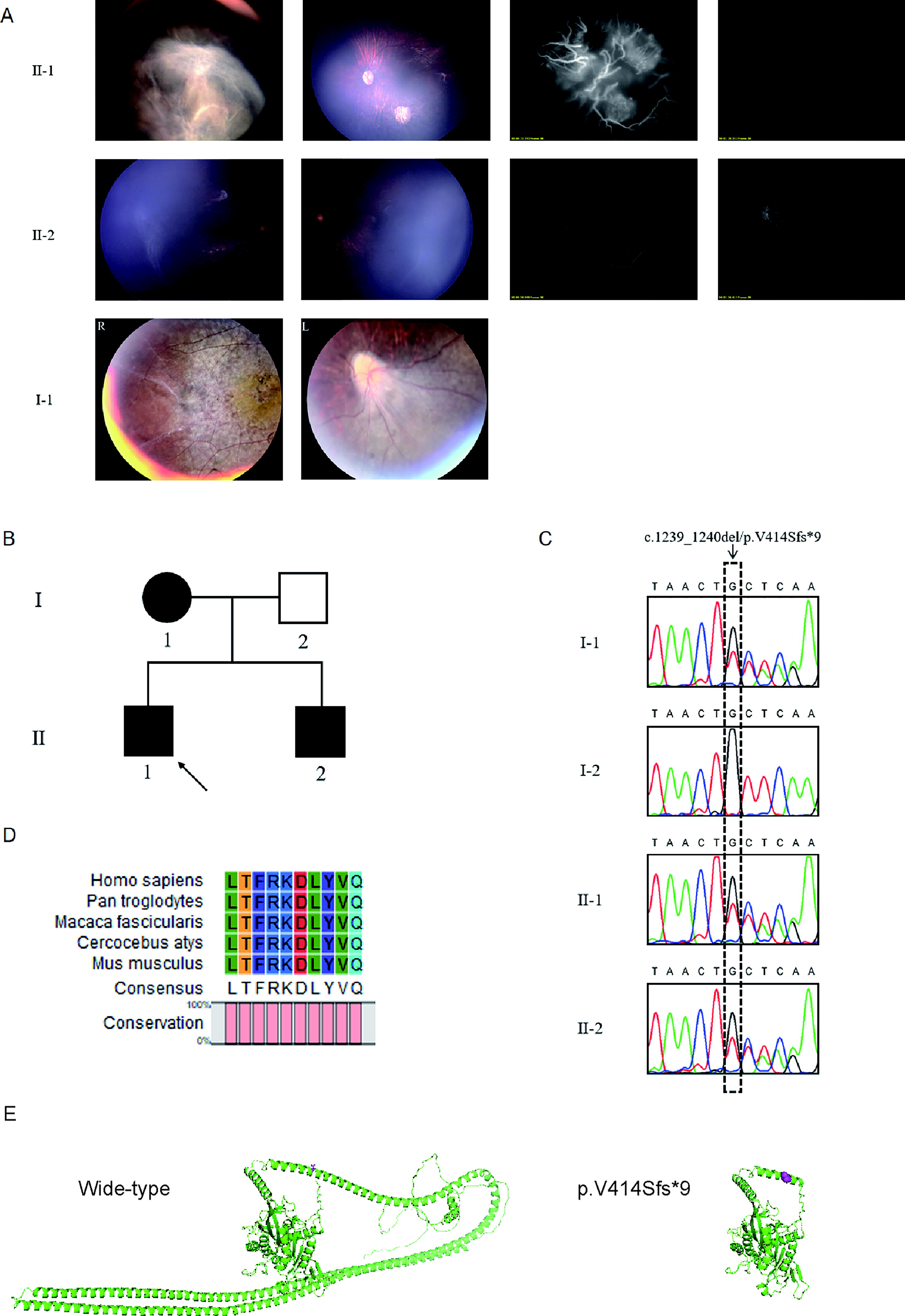
Identification of the novel KIF11 variant in a patient with FEVR from a Chinese family.
Mutation analysis
Our analysis revealed that 42 rare and likely deleterious variants among 38 genes were prioritized by data filtering and bioinformatic analyses (Supplementary Table S1). One particular frameshift deletion variant in exon 11 of KIF11 (designated c.1239_1240del: p.V414Sfs*9) was identified. This heterozygous variant was shared by the proband, his older brother, and his mother, but not the father (Fig. 1B), which was verified by Sanger sequencing (Fig. 1C). This variant had not been reported previously and was not detected in 1000 Genomes, ExAC, or gnomAD. The mutation was predicted by CADD to have a disruptive impact on the structure and function of the KIF11 protein, with a CADD score of 26.9 (Table 1), resulting in a truncated protein. The implicated valine residue at position 414 (V414) in the wild-type protein is highly conserved across multiple species (Fig. 1D). SWISS-MODEL-generated three-dimensional structure of KIF11 with p.V414Sfs*9 variant shows marked truncation and structural alterations relative to the wild-type (Fig. 1E). On the basis of these results, we speculate that this novel KIF11 variant is the cause of the proband’s FEVR and the ocular anomalies observed in his immediate family members. The proband’s mother and brother both carry the KIF11 variant and exhibit FEVR-related symptoms. This co-segregation of genotype and symptoms provides clinical support for the pathogenicity of the variant.
In Silico Prediction Findings Related to the V414Sfs*9 Variation in KIF11

The accession number of human KIF11 is GenBank: NM_004523.
gnomAD, The Genome Aggregation Database, http://gnomad.broadinstitute.org; SIFT, sorting intolerant from tolerant, http://sift-dna.org; PolyPhen-2, PP2, Polymorphism Phenotyping, version 2, http://genetics.bwh.harvard.edu/pph2/; MT, mutation taster, http://mutationtaster.org; CADD, Combined Annotation Dependent Depletion, https://cadd.gs.washington.edu, higher scores are more deleterious.
Discussion
In this study, we identified a novel potentially pathological variant of KIF11 in a Chinese infant who presented with severe ocular pathologies typical of FEVR. Since FEVR was first reported in 2014, multiple KIF11 variants have been identified as causative for autosomal dominant FEVR, with significant intra- and interfamilial phenotypic variability (Robitaille et al., 2014). KIF11 variants follow an autosomal dominant pattern, though most pathogenic variants are de novo (Chen et al., 2020; Wang et al., 2022). Subsequent cohort studies have shown that KIF11 variants account for 5% to 9% of FEVR cases (Hu et al., 2016; J. Yang et al., 2022). Our findings are consistent with previous studies, where most KIF11 variants causing FEVR are protein-truncating variants (Karjosukarso et al., 2018). The currently reported heterozygous truncating variants of KIF11 include c.131_132dupAT (p.P45Ifs*92), c.2230C>T (p.Q744*), c.2717del (p.L906*), c.2863C>T (p.Q955*), and c.2952_2955delGCAG (p.G985Ifs*6) (Hu et al., 2016; Karjosukarso et al., 2018). The variant identified in this study (c.1239_1240del, p.V414Sfs*9) is located in exon 7, suggesting that exon 7 may be a novel variant hotspot region of the KIF11. Meanwhile, this finding also supplements the differences in the distribution of KIF11 variants among different populations.
Previous studies have indicated that KIF11 regulates retinal vascular development independently of the Wnt/Norrin pathway (Le V et al., 2023). FEVR patients with KIF11 variants are more susceptible to foveal hypoplasia, and sectoral or panretinal chorioretinopathy is a common specific manifestation of KIF11 variants (J. Yang et al., 2022). In the present study, the macular involvement observed in the proband’s mother is consistent with previous findings that KIF11 variants may correlate with more severe retinal phenotypes, suggesting potential value of KIF11 as a prognostic marker (Ju et al., 2024; Tao et al., 2023). Hence, our results further expand and clarify the understanding of KIF11-related ocular anomalies.
However, because of limited family genetic data and small sample size, the variant’s frequency, phenotypic correlation, pathogenic mechanism, and genetic pattern in FEVR require further validation with larger cohorts. Despite these limitations, this study provides new insights into the genetic etiology of FEVR and references for clinical genetic counseling.
Conclusions
In conclusion, we identified a novel heterozygous frameshift variant (c.1239_1240del p.V414Sfs*9) of KIF11 in a Chinese family affected by FEVR, expanding the known mutation spectrum of KIF11. Combined with recent findings, the present case suggests that KIF11 variants may be commonly associated with FEVR, although further studies are needed to determine their frequency in FEVR patients. The present findings deepen our understanding of the molecular pathogenesis of FEVR and may facilitate the development of diagnostic and preventive strategies.
Authors’ Contributions
B.W. conceived this study. J.Z. and Y.Z. drafted the article and analyzed data. P.Z., W.L., and Y.L. performed experiments and validation.
Footnotes
Acknowledgments
The authors thank the patient and his family members for participating in our study.
Author Disclosure Statement
No competing financial interests exist.
Funding Information
This work was supported by the Central Government to Guide Local Scientific and Technological Development (2022YRZ0102).
Ethical Compliance
The authors adhered to the Declaration of Helsinki (World Medical Association, revised in 2013), and the ethical committee of the National Research Institute for Family Planning approved this study. All family members participating in this study signed informed consent.
Supplemental Material
References
Supplementary Material
Please find the following supplemental material available below.
For Open Access articles published under a Creative Commons License, all supplemental material carries the same license as the article it is associated with.
For non-Open Access articles published, all supplemental material carries a non-exclusive license, and permission requests for re-use of supplemental material or any part of supplemental material shall be sent directly to the copyright owner as specified in the copyright notice associated with the article.
