
Abstract
Microbial fuel cells (MFCs) are bioelectric devices that use bacterial metabolic activity to convert chemical energy to electricity, which offers a potential way to green energy production. Exoelectrogenic bacteria that can transfer their metabolic electrons outside their cells are the primary contributing organisms in this process. This study aims to screen and identify exoelectrogenic bacteria for electricity generation in MFCs. Seawater samples were collected from Kuakata, Bangladesh, and a dual-chambered MFC was used to detect the presence of exoelectrogenic bacteria in the sample. The exoelectrogenic bacterium was screened and isolated from the anode biofilm of the MFC using an MnO2-supplemented agar medium. The ability of electricity generation by the isolated bacteria was also determined in a dual-chambered MFC. Then, the isolated exoelectrogen was identified via biochemical assay and 16S rRNA gene sequencing. The isolated exoelectrogenic bacteria generated a voltage of up to 0.341 volts, a current density of 0.044 mA/cm2, and a maximum power density of 0.015 mW/cm2 in the MFC. Biochemical assay and sequencing analysis reported the bacteria as Pseudomonas aeruginosa (similarity index: 99.45%), which demonstrates potential for bioelectricity and sustainable energy production, contributing to novel, eco-friendly resources for biofuel production in Bangladesh.
Introduction
Society is dealing with the continuous decline of fossil fuels and chemical feedstocks and the proliferation of pollutants produced during fuel production. The increasing global energy demand and the decline of fossil fuels stimulated the exploration of alternative approaches for sustainable and renewable energy production. 1 Industries are solely dependent on conventional nonrenewable energy sources such as natural gas, coal, petroleum, and others for power generation. 2 However, its constant reliance on fossil fuels has significant disadvantages, such as declining resources, greenhouse gas emissions, and air and water pollution. 3 These elements urge the transition to more sustainable, eco-friendly energy sources to accommodate the world’s increasing energy demands and prevent environmental harm. 4 In this context, microbial fuel cells (MFCs) are an innovative bioelectrochemical technology that utilizes the metabolic processes of microorganisms, particularly exoelectrogenic bacteria, to directly transform the chemical energy in organic matter into electricity. 5 Exoelectrogenic bacteria are a group of microorganisms able to pump out electrons outside the cell instead of delivering them to intracellular electron acceptors during respiration. These electrons are accepted by insoluble electron acceptors, including metal oxides and solid electrodes in MFCs, enabling electricity generation. 6 This phenomenon of transferring electrons outside the cell is known as extracellular electron transfer (EET).7,8 Unlike the nonexoelectrogens, which deliver electrons to intracellular acceptors like oxygen during respiration, exoelectrogenic bacteria have developed complex mechanisms to bind with extracellular electron acceptors. 9 These mechanisms vary within different species of the exoelectrogenic bacteria. The mechanisms include direct contact via proteins in the outer membrane, using either endogenous or exogenous compounds; electron shuttles; and conductive nanostructures via nanowires or pili.10,11 Exoelectrogenic bacteria develop a biofilm on the anode, consume organic substrates, and generate and transfer metabolic electrons, which is vital to MFC operation. 12 Researchers are investigating these bacteria for enhanced efficiency and ergonomics in MFCs. The diversity of exoelectrogenic bacteria and their metabolic adaptability enable MFCs to harness a wide range of organic substrates as their nutrients. These substrates encompass carbohydrates, organic acids, alcohols, and complex biological matter found in organic waste and wastewater. This feature sets MFCs as a potentially revolutionary technology in power generation as well as waste management.13,14
While previous studies solely focused on well-known model organisms, including Geobacter sulfurreducens, Geobacter metallireducens, Shewanella oneidensis, etc. These bacteria are found in complex microbial communities in their native habitats, making it challenging to isolate and characterize. 15 Though previous studies involved the utilization of mixed microbial populations, such as lake sediments, marine sediments, wastewater, anaerobic and activated sludges, etc., in MFCs, the isolation and characterization of the underlying exoelectrogenic bacteria are limited.16,17 The isolation of exoelectrogenic bacteria involves the use of electrodes as the terminal electron acceptor, allowing selective enrichment of the microorganism in electrochemical conditions. Only microorganisms that are able to transfer their cellular electrons to the electrodes are able to proliferate and dominate in the medium under such conditions. 7 The microorganisms are then subsequently isolated using culture-based techniques. The culture techniques involve supplementing reducible electron acceptors (e.g., Fe(III) and Mn(IV)) in the nutrient medium, which facilitates the selection and differentiation in the growth of microorganisms able to undergo EET under oxygen-deficient conditions.18–20
Researchers are investigating approaches to expanding the technology and incorporating it into existing wastewater treatment plants and other facilities in order to advance the MFC technology from laboratory-scale experiments to practical applications. 21 Therefore, isolation of exoelectrogenic bacteria is vital, as a pure and well-characterized strain can improve the performance, efficiency, and scalability of MFC technologies. However, the screening, isolation, and characterization of these exoelectrogenic bacteria from natural environments are poorly accomplished, especially from the marine ecosystem of Bangladesh. The coastal and marine systems of Bangladesh are rich in biodiversity due to a warm tropical climate and abundant annual rainfall, which support diverse microbial and marine life. 22 Sediments in these regions are organic-rich and largely anaerobic, creating a favorable habitat that supports microbial communities capable of utilizing alternative electron acceptors and adopting EET processes, making them suitable sources for isolating electroactive microorganisms, such as exoelectrogenic bacteria. 23 In this study, we aimed to isolate and identify exoelectrogenic bacteria from sea sediment–water suspension collected from Kuakata Sea Beach, Bangladesh. MnO2-supplemented agar medium was used to screen the exoelectrogenic bacteria. We have also evaluated the potential of electricity generation by the isolated exoelectrogenic bacteria in MFCs (Fig. 1). The isolated bacteria can be used in the development of a cost-effective and efficient MFC for bioenergy production and waste management in the coastal region of Bangladesh.
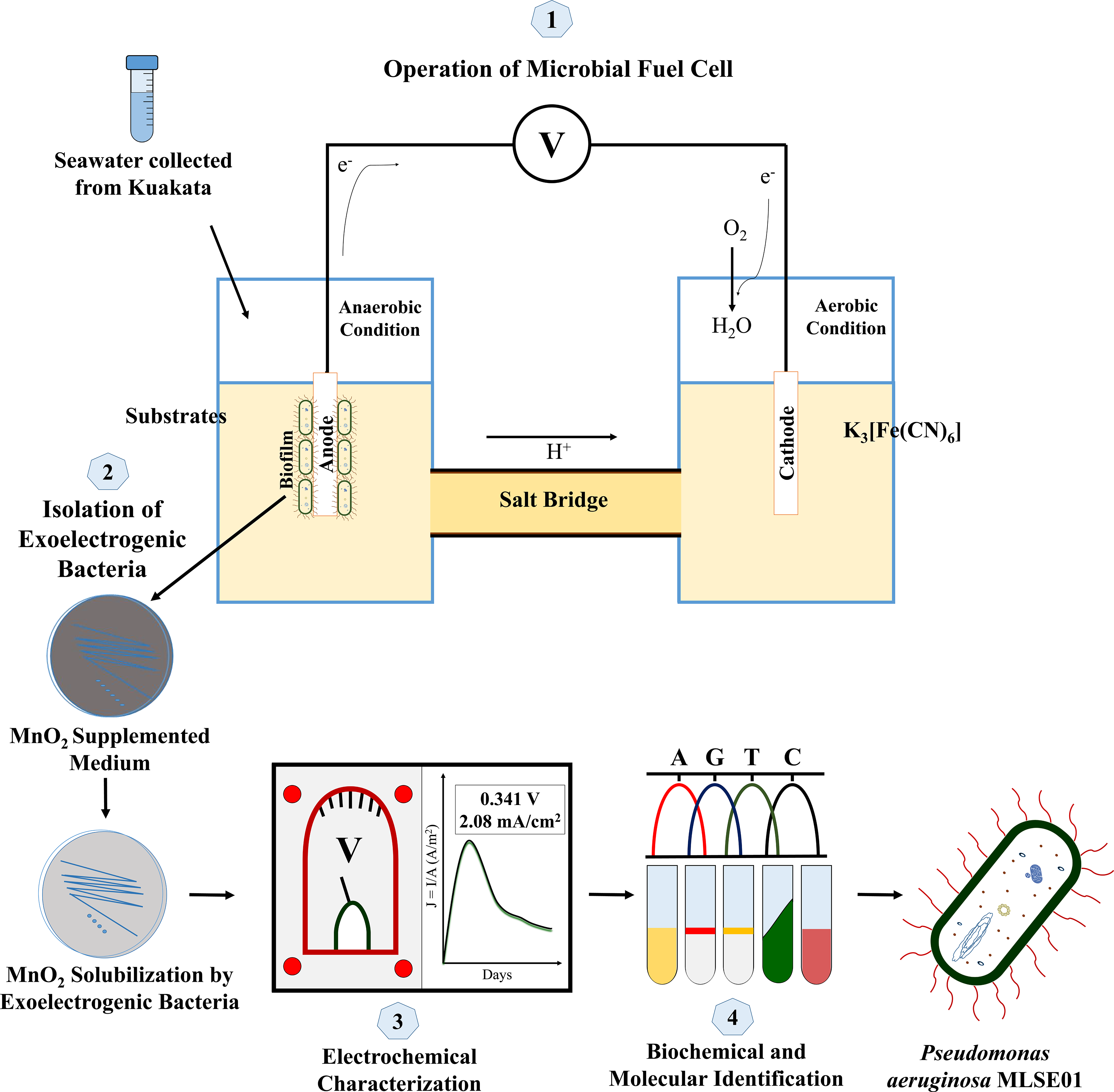
Graphical representation of the present study.
Materials and Methods
Sample collection
The seawater-containing bacterial consortium was collected from Kuakata Sea Beach, Patuakhali, Bangladesh (located at 21.81372°N, 90.12240°E). Approximately 15 mL of sandy water from about 10 cm below the sea sediment surface was collected in a sterile cap-secured sample collecting tube and transported immediately to the laboratory at 4°C. The samples were kept airtight and opened only when inoculating MFCs.
Inoculation and operation of MFCs
A dual-chamber MFC was constructed using plastic containers (1 L each) with graphite rod electrodes (6 cm in length and 0.5 cm in diameter), and no variable external resistor was used. The electrodes had a projected lateral surface area of 9.42 sq. cm (both anode and cathode). An agar-salt bridge (containing KCl, NaCl, and NaNO3) was placed between the two chambers to enable ion transfer. 24 A salt bridge was used instead of other cutting-edge techniques due to its simplicity, low cost, and ease of operation, which makes it suitable for preliminary isolation of exoelectrogenic bacteria. 25 Tryptone-based minimal media was provided in the anode chamber for the growth of exoelectrogenic bacteria, while the cathode contained 100 mM K3Fe(CN)6 in KH2PO4 buffer (pH 7.0) with air exposure. Both media were autoclaved and cooled down to room temperature before inoculation with the seawater sample. The sample was inoculated in the anode compartments of the MFC, which was operated in anaerobic conditions for 6 days for the feasible growth of exoelectrogenic bacteria present in the sample. The MFC anode was inoculated with 1 cm³ of seawater. The anode compartment was kept in an anaerobic condition, which is facilitated by AnaeroPack™. The cathode chamber remained uninoculated and exposed to air for the continuous supply of oxygen. The operation of MFC was carried out in batch mode under closed conditions, with no continuous inflow or outflow of substrate during the experimental period.
Isolation of exoelectrogenic bacteria
Exoelectrogenic bacteria from a fully functioning MFC were introduced on the MnO2-supplemented proposed medium “E-Z exoelectrogen growth and isolation medium” with some modification. 26 Biofilm sample collected from the MFC anode was streaked onto solid minimal medium plates using the quadrant streak method, with flame-sterilized inoculating loops between each quadrant to ensure colony isolation. The bacterial colony decolorizing the MnO2-supplemented medium was taken and isolated on a fresh nutrient medium. Another MFC was prepared and inoculated with the isolate for further confirmation of the bacteria having exoelectrogenic properties. Plates were incubated anaerobically at 37°C for 16–24 h in sealed jars containing AnaeroPack sachets to maintain an oxygen-free environment.
Electrochemical characterization of the isolated exoelectrogenic bacteria
Again, two MFCs were constructed and operated for 10 days. The closed circuit voltage and the electricity generated in the MFCs were measured using a multimeter.
Current density
The ammeter current was recorded daily at a particular time with a fixed resistance. Then, the current density (A/m2) was calculated by the following equation:
Current density,

Here, I = current and A = projected lateral surface area of anode.
Power density
The voltage difference between the anode and cathode (V) was recorded, and the power density (mW/m2) was calculated according to the following equation:
Power density,

Biochemical identification of the isolated exoelectrogenic bacteria
The biochemical identification of isolated exoelectrogenic bacteria was conducted following “Bergey’s Manual of Determinative Bacteriology.” 27 The bacterial colony was identified via Gram staining, catalase test, oxidase test, indole test, MR-VP test, and citrate utilization test.
Molecular identification of the isolated exoelectrogenic bacteria
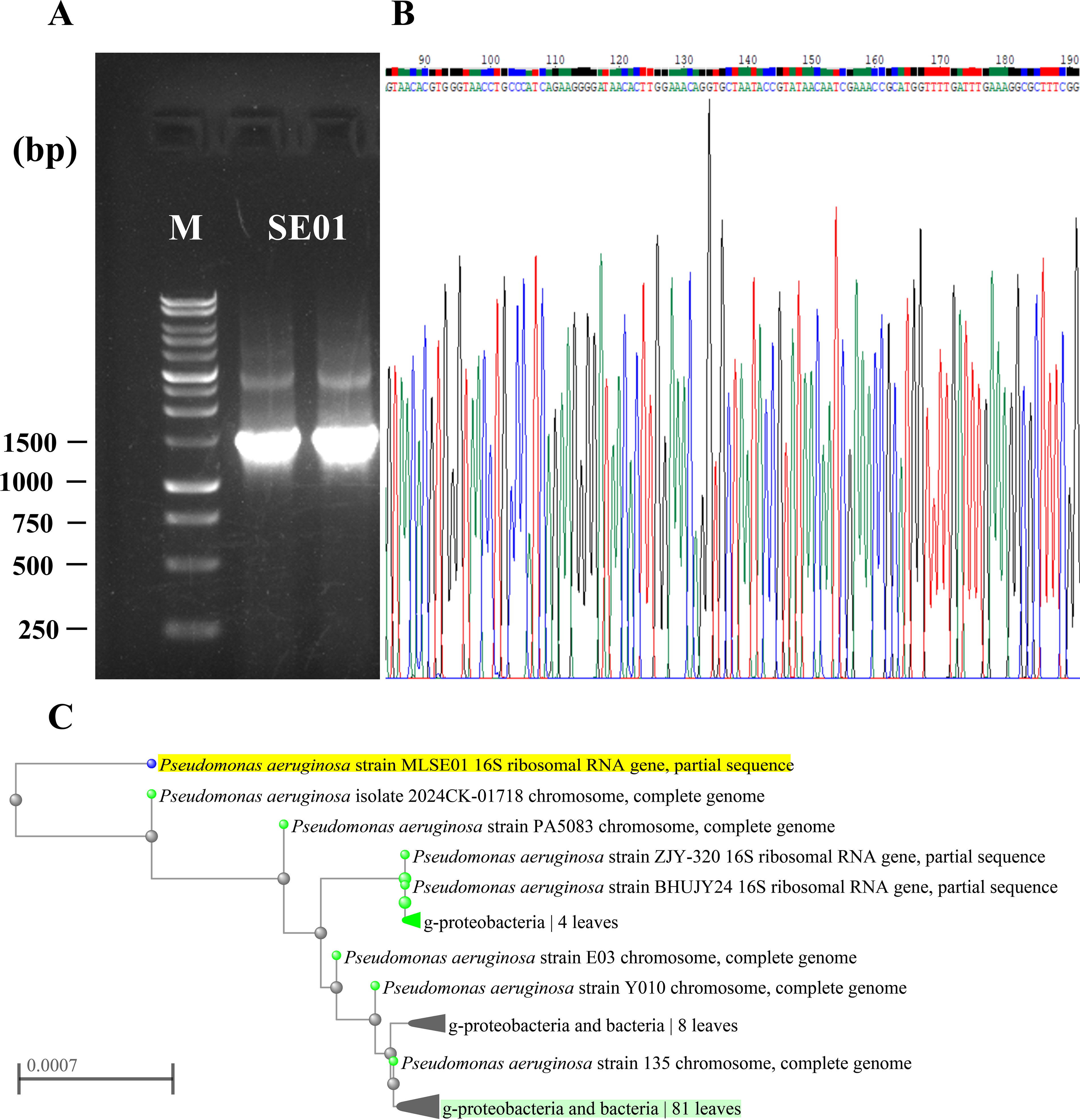
The isolated bacteria were then further identified via 16S rRNA gene sequencing. The DNA was extracted using the DNAzol DNA extraction kit. Followed by DNA extraction and PCR of the 16S rRNA gene, the amplified products were sequenced. The 16S rRNA gene was amplified using 27 F-1492R primers, and the amplicon was sequenced. The sequence reads were checked and assembled using the software BioEdit, version 7.2.5. The assembled and checked sequence was compared with the 16S rRNA gene sequences submitted in the GenBank database by using the neighbor-joining method. Nucleotide BLAST was used to identify the species. The sequences were subjected to a homology search using the National Centre for Biotechnology Information (NCBI) BLAST program (https://blast.ncbi.nlm.nih.gov). Based on the homology index, the bacteria were identified, and the phylogenetic tree was constructed using NCBI: https://www.ncbi.nlm.nih.gov/blast/treeview/treeView.cgi?request=page&blastRID=022SZ7B7016&queryID=gb. Max seq difference was set as 0.5.
Results
Screening of exoelectrogens on minimal growth medium
A dual-chamber MFC was used to detect the presence of exoelectrogenic bacteria in the sample and to generate electricity from the exoelectrogenic bacteria. The anode chamber was inoculated with the Kuakata sample and operated for several days to ensure substantial biofilm growth of exoelectrogenic bacteria present in the sample. The readings were taken regularly with a multimeter that measured the flow of electricity and the voltage generated between the anode and cathode chambers. A slight rise was observed in the flow of electricity and the generation of voltage between the anode and cathode chambers.
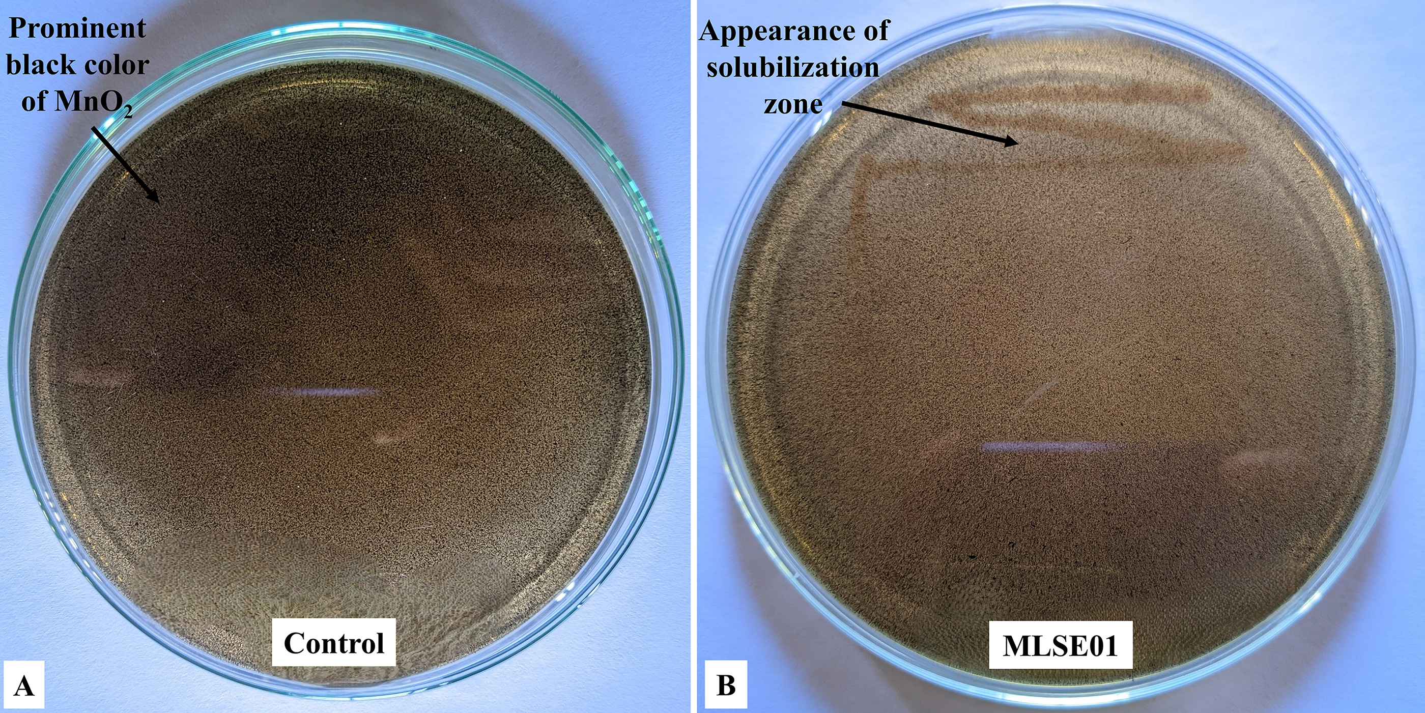
The biofilm bacteria from the anode electrode surface were cultured on the minimal media supplemented with MnO2 for 16–24 h in anaerobic conditions. The growth of bacteria was observed after the incubation period. The exoelectrogenic bacteria were screened on the plate based on their color formation. White-colored colonies indicate the solubilization of MnO2 in the media. The uninoculated area remained prominently black due to the presence of the MnO2 particles in the media (Fig. 2A). A colony was taken and underwent pure culture to further evaluate the individual performance of electricity generation in a dual-chambered MFC. The isolated colony was marked as MLSE01 (Fig. 2B).
Electrochemical characterization of exoelectrogenic bacteria in MFCs
The initial acclimation and selection of exoelectrogenic bacteria from seawater was done in two-chambered MFCs operating in closed operation, inoculated with isolate MLSE01. The MFCs exhibited a very repeatable and stable operation for 10 days. The voltage difference between the anode and cathode (V) was measured using a multimeter. The average voltage and current value of two fully functional MFCs inoculated with MLSE01 were taken for analysis. The values of electricity generation in the MFCs indicate a dynamic system.
The MFCs were initiated with low output in both current and voltage, which indicates a potential lag phase during microbial adaptation. After that, a significant increase in performance was observed on day 5, with 0.41 mA current and 0.341 V voltage reaching peak values, suggesting enhanced bioelectrochemical activity (Table 1).
Electricity Generation of MLSE01 in the Microbial Fuel Cell
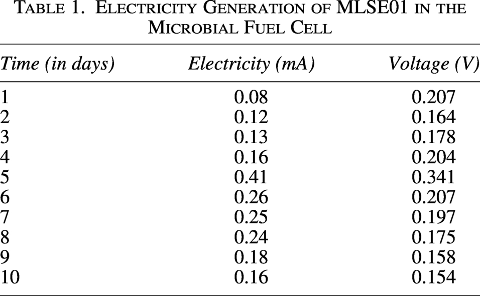
Eventually, both the current and the voltage gradually declined and became more stable. This time-dependent fluctuation of both the current and the voltage refers to the dynamic nature of electricity generation in MFCs, which is likely to be influenced by the growing microbial community and substrate availability in the anode compartment. In addition, a similar drift was observed in both current density and power density analysis. The MFC was initiated with a gradual increase in current density from approximately 0.008 mA/cm2 on day 1 to 0.017 mA/cm2 on day 4, indicating the adaptation and biofilm formation of the exoelectrogenic bacteria in the anode. On day 5, a rise to 0.044 mA/cm2 was detected, which indicates the exponential growth phase of the exoelectrogenic bacteria in the anode, allowing the electron release at the highest peak. Subsequently, current density decreased to 0.017 mA/cm2 by day 10, which is anticipated due to the depletion of nutrients in the anode (Fig. 3A). Power density also increased gradually from 0.002 mW/cm2 on day 1 to 0.004 mW/cm2 on day 4, demonstrating the development of electrochemical activity of the bacteria. A sharp peak of 0.015 mW/cm2 on day 5 suggests the optimal bacterial activity in the anode. At the end of the MFC operation period, the power density declined to 0.003 mW/cm2 on day 10, likewise the current density (Fig. 3B). This fluctuation in both the current density and the power density over time signifies that the electricity generation in MFCs was not consistent. It likely occurred due to the growth of the MLSE01 isolate and the amount of nutrients available in the anode for the isolate.
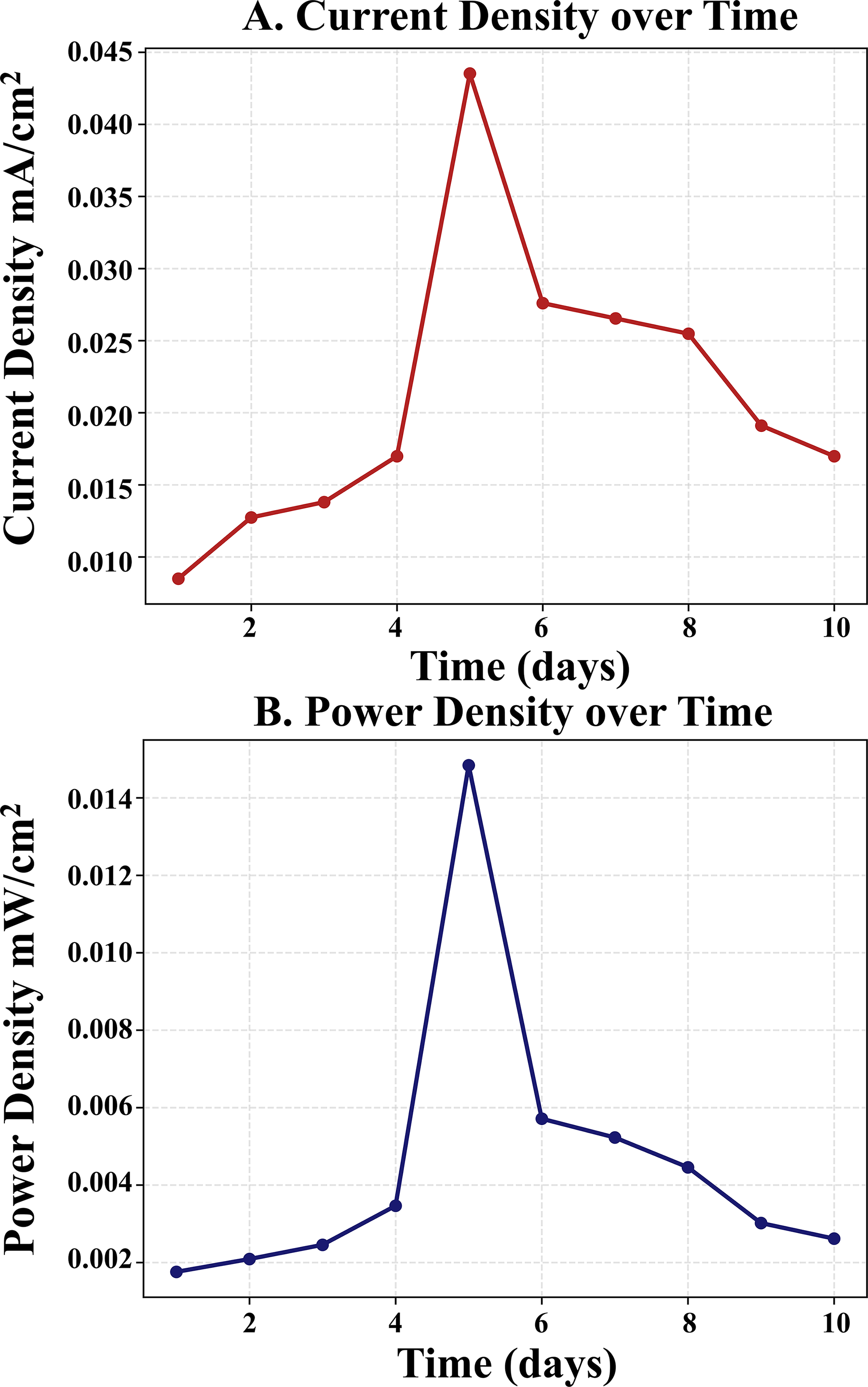
Power generation of MLSE01 isolate in a dual-chambered microbial fuel cell.
Identification of the isolated bacteria
Biochemical tests identified the bacterium as a Pseudomonas species. Gram staining confirmed it as a Gram-negative (−ve) rod-shaped bacterium under a light microscope (Fig. 4A). The KOH string test was also positive (+ve), supporting its Gram-negative nature (Fig. 4B). The result of the oxidase test was positive (Fig. 4C). In addition, the Methyl Red, Voges–Proskauer, and indole tests were negative (−ve) (Fig. 4D–F), while the catalase test and citrate test were positive (+ve), indicating catalase enzyme activity and citrate utilization (Fig. 4G–H). These results indicate that the bacterium’s classification is a Pseudomonas species. Following DNA extraction, the 16S rRNA gene was amplified, and the PCR product was visualized at around 1400 base pairs. After sequencing via BioEdit software, the result confirmed the isolate as P. aeruginosa. The sequenced 16S rRNA gene was submitted to and is openly available in the NCBI database (https://www.ncbi.nlm.nih.gov/). The Accession ID of the gene is PV484567. The 16S rRNA gene sequence of isolated P. aeruginosa was considered to have a significant degree of similarity (99.45%) to the sequences of existing P. aeruginosa, and the isolate was designated as MLSE01 for laboratory identification purposes. The use of the 16S rRNA gene as a “gold standard” for bacterial identification and phylogenetic study has gained popularity. Phylogenetic tree analysis demonstrating the evolutionary relationships of the isolated bacteria, revealing the relationship of P, aeruginosa MLSE01 and P. aeruginosa isolate 2024CK-01718 (Fig. 5A–C). The MLSE01 isolate formed a distinct phyletic group like the other P. aeruginosa strains in the tree, signifying greater similarity to each strain than the gamma-proteobacteria group.
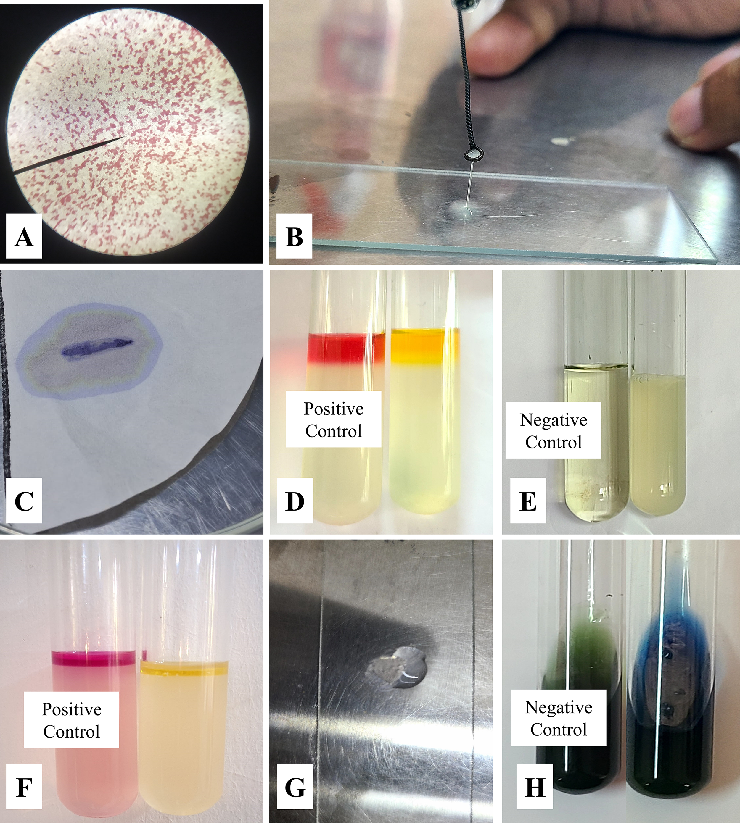
Biochemical Identification of MLSE 01 isolate:
Discussion
MFCs have gained notable attention because of their multitude of applications in power generation and bioremediation. 28 The adaptability of MFCs to diverse organic substrates, including agricultural, municipal, and industrial wastes and their derivatives, makes them attractive. 16 The MFCs act as bioreactors and use the metabolic activity of exoelectrogenic bacteria to transform chemical energy stored in organic matter into electrical energy under anaerobic conditions. 29 Seawater comprises a diverse number of underexplored microorganisms with unique metabolic features. 30 Marine environments are abounding with electroactive microorganisms that play a pivotal role in natural biogeochemical cycles, including redox reactions of nitrate, sulfur, and iron.31–33 These microorganisms are adapted to extreme environments like high salinity and exhibit metabolic diversity, allowing them to survive depending on a wide range of substrates. 34 In this study, an exoelectrogenic bacterium was isolated from the seawater-driven MFC using an MnO2-supplemented agar medium. The supplemented MnO2 acts as the terminal electron acceptor for the exoelectrogenic bacteria in the absence of other acceptors like oxygen, sulfate, or nitrate ions. MnO2 (Mn (IV) dioxide) particles are insoluble and form a prominent black color in the media. These particles accept metabolic electrons produced by the exoelectrogenic bacteria and convert Mn4+ ions into Mn2+ ions, decolorizing the medium. This change indicates the production of electrons by the bacteria. When the MLSE01 isolate was grown on the MnO2-supplemented agar medium, a MnO2 solubilization zone was observed around the streak line of the isolate, confirming the potential of using MnO2 as the terminal electron acceptor. 26
The MLSE01 isolate demonstrated a dynamic pattern in power generation in the MFCs over a period of 10 days. As no variable external resistance was applied in the operation, the reported voltage and current reflect the performance of the MFC under fixed load conditions. 35 The first 4 days indicate the initial lag phase of the MLSE01 isolate is characterized by low current and power density output, reflecting the adaptation and the development of biofilm in the MFC anode. Eventually, a significant rise in both current and power density was observed, which indicates the exponential growth phase and optimal utilization of the substrates in the anode, resulting in the highest rate of electron transfer. The peak output of the current and the voltage were recorded as 0.41 mA and 0.341 V, respectively. Following the highest peak, both the current and voltage gradually decreased.36,37 This decline primarily results from the depletion of nutrients in the anode. Besides several factors like changes in biofilm structure, shifts in pH due to the production of metabolic byproducts may also contribute to the decline.38–40 Similar performances of exoelectrogenic bacteria have been documented in the existing literature, confirming the reliability of these observations. 41 The generation of electricity by exoelectrogenic bacteria is strongly attributed to the formation of a biofilm on the anode. Biofilm formation is vital as it facilitates the efficient electron transfer to the electrode by the exoelectrogenic bacteria. 42 Biofilm formation with stable communities enhances electricity generation, whereas weak biofilms can limit the electron transfer efficiency.43,44 The electricity generation also depends on different factors such as the pH of the medium, operating temperature, presence of oxygen, substrate concentration, etc. 45 Most exoelectrogenic bacteria generate optimum electricity under pH 7.0–8.0. Fluctuation towards acidification or alkalinity can inhibit microbial growth, disrupt biofilm stability, and decrease electron transfer rates. 46 Temperature influences bacterial growth kinetics and metabolic activity. The optimal current generation has been reported in the mesophilic range (25–35°C), where the metabolic activity and electron transfer activity are maximal. The overall electricity generation can be lowered outside the temperature range. 47 The presence of oxygen in the anode can affect the overall power output in a MFC, as it can compete as the final electron acceptor. It can reduce the flow of electrons to the electrodes, thereby decreasing the current output.48,49 Higher ionic strength in the medium generally promotes the electron flow. 50 Additionally, the availability of nutrients acts as a mediator of microbial growth. Limiting the amount of substrates reduces electron production in terms of reducing the bacterial growth, whereas excessive concentrations can cause metabolic imbalances and inhibit growth. 51
Gram staining, biochemical tests, and 16S rRNA gene sequencing identified the MLSE01 isolate and named it P. aeruginosa MLSE01. The negative result in Gram staining and rod-shaped morphology under microscopic observation aligns with the well-documented characteristics of Pseudomonas species. 52 The positive result for oxidase, citrate utilization, and catalase test is consistent with the metabolic profile of Pseudomonas species with MLSE01. 53 Sequencing and phylogenetic tree analysis also revealed the relatedness of the isolate to the other P. aeruginosa strains. 54 The isolated P. aeruginosa MLSE01 demonstrated a maximum power density of 0.015 mW/cm2 and a voltage of 0.341 V, which is higher than other Pseudomonas strains like the model P. aeruginosa PA01 strain that produces 1.0 µW/cm2 power density in textile-based biofuel cells. 55 Other strains like MFC-EW603 and MFC-EW819 generate power density around 40.1 and 21.3 mW/m2, respectively, whereas Pseudomonas species like Pseudomonas sp. BJa5 and Pseudomonas putida B6-2 generate power densities of 39 mW/m2 and 411 mW/m2, justifying the superiority in exoelectrogenic properties of the P. aeruginosa MLSE01 over them.56–58
The findings of the proposed work evidence the potential role of the isolated exoelectrogenic bacteria in bioelectrical devices, particularly MFCs. The electricity generation ability of P. aeruginosa MLSE01 demonstrates its applicability in bioenergy production using low-cost and renewable substrates like industrial wastewater and organic wastes. This bacterium can be used in an effluent treatment plant or wastewater treatment systems to simultaneously degrade pollutants and generate electricity. 14 Furthermore, the isolate can be employed in bioremediation, including the removal of environmental contaminants like heavy metals, xenobiotic compounds, etc., from aquatic and sediment environments via bacterial metabolic redox processes.59,60 MFC-based systems can also act as biosensor devices to monitor water quality by correlating the electric output with different pollutant levels. 61 Overall, the present study and the findings contribute to a basis for the development of cost-effective, eco-friendly, and sustainable technology. Therefore, the isolation of the exoelectrogenic P. aeruginosa MLSE01 from the marine ecosystem of Bangladesh is significant, as it is largely underexplored in the context of MFC technology.
Conclusion
This study involves the isolation and identification of exoelectrogenic bacteria from the sea sediments of Kuakata, Bangladesh. The isolate demonstrated a stable performance in generating electricity in the MFCs and was identified as P. aeruginosa MLSE01. The findings of the study may contribute to insights into using local microorganisms for green energy production and waste treatment. Future studies may emphasize optimizing MFC conditions, discovering cheap substrates, exploring continuous nutrient supply strategies, and investigating the combined effect with other microbes to enhance the performance of MFCs using P. aeruginosa MLSE01. This study would contribute to the practical application of MFC technology in coastal areas and beyond.
Data Availability Statement
All data will be made available upon reasonable request to the corresponding authors.
Authors’ Contributions
S.S.: Conceptualization, methodology, formal analysis, data curation, visualization, writing—original draft; T.A.: Methodology, formal analysis, writing—original draft; R A.H.: Formal analysis, writing—original draft, writing—review & editing; B.K.P.: Writing—original draft; S.K.: Writing—original draft; N.S.H.: Writing—original draft; M.A.H.M.J.: Conceptualization, methodology, data curation, writing—review & editing, supervision, validation.
Author Disclosure Statement
No competing financial interests exist.
Footnotes
Funding Information
This research work has been conducted with the financial support of a special allocation research fund from the BANBEIS, Ministry of Education, Bangladesh. Ref- 37.20.0000.004.33.020.23-5-27, LS20232358, Year 2023-26.