




Abstract
Introduction
Foam sclerotherapy has gained a great popularity among phlebologists worldwide, although a major lack of homogeneity in the material used to produce sclerosant foam (SF) and to inject SF has been reported.
Aims
To highlight the literature data and a few personal clinical and experimental outcomes concerning the main variables in SF production and injection.
Methods
A review of the published literature and of our own 12 year clinical and experimental experience has been undertaken in order to focus on a few variables of the material and methods used to produce SF with Tessari method and to inject SF.
Results
In SF production, differences in gas components, liquid to gas ratio, as well in disposable material can variably influence the resulting SF. Similarly SF injection through ultrasound guidance, with needle, or with short/long catheter may exhibit different foam behaviours according to the variable material and techniques which are employed. More recently the introduction of long catheters, possibly together with hook phlebectomy, seems to potentiate the short-mid term outcomes of foam sclerotherapy.
Conclusion
SF formation is greatly influenced by the choice of the gas component, the liquid-to-gas ratio, the type of syringes; also larger needles are to be preferred for injection of SF, while long catheters seem to represent a valid alternative especially when combined with tumescence to minimise saphenous diameter.
Introduction
In the last decades, the use of sclerosant foam (SF) has progressively spread among sclerotherapy practitioners. The Tessari method, 1 as an easy, inexpensive and repeatable extemporary method to create a durable and dense SF, contributed to this diffusion. Ultrasound-guided foam sclerotherapy (UGFS) has significantly increased in popularity and acceptance in the international vascular community, although a lack of standardization in the process of SF formation and SF injection has been highlighted by different authors 2–4 and several variables objectively interfere with SF formation/injection. In one study, Tessari and colleagues 5 showed, through a reproducibility test, no significant differences in the resulting SF produced by means of his method by 20 non-medical volunteers as to density, half-life and bubble size parameters. In fact, recently, first Wollmann and colleagues proposed the DSS method, 6 which has been basically derived from the Tessari method, but it is based on the use of a two-way connector that connects two syringes in a straight fashion; later C Hamel Desnos and collaborators 7 also introduced a mechanical device that aims to improve the standardization of SF formation in the Tessari/DSS method.
Material and methods
With reference to the variability of the methods of SF preparation, the material to be used has to be selectively chosen in order to standardize and optimize the method and the resulting SF as much as possible. The material that can be used in SF formation through the Tessari method comprises syringes, a three-way valve, drugs and air/gas (Figure 1). Additional material, such as filters, 8 has been tested to possibly improve SF quality.
Among the variables that significantly affect SF formation and foam sclerotherapy safety (and possibly efficacy), the gas component of the SF seems to be of paramount importance.
For many years, room air has been used in common practice to form SF together with sodium tetradecylsulphate (STS) or with polidocanol (POL). However, following Juan Cabrera's 9 experience about his patented microfoam and Antonio Cabrera's 10 report, a significant use of biocompatible gases (namely CO2 and O2) has diffused in the scientific community in recent years.
Eckmann et al.
11
published his results from experimental studies concerning differences in the vascular distribution of air-based and CO2 + O2-based SF microbubbles, showing improved safety for the latter. Similarly, Morrison et al.
4,12
reported the statistically significant difference between the lower incidence of side-effects resulting from CO2 or CO2 + O2-based SF and the higher incidence of side-effects from air usage, especially when injecting high volumes.
Material to form and to inject sclerosant foam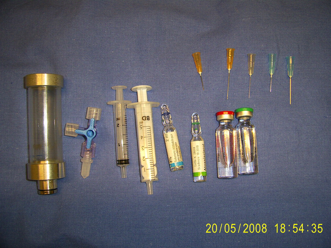
The formula TP = r 2 d/2D Sf (where TP is the time of persistence of the bubbles, r the radius of the microbubble, d the density of the gas inside the microbubble, D the diffusibility of the gas through the microbubble membrane and Sf the saturation factor of the gas in the blood) 13 summarizes the most important gas-related factors that may interfere with the time of persistence of foam bubbles, as in SF or in ultrasound contrast agents. In fact, CO2 is 50 times more diffusible than nitrogen through the capillary–vein wall and through the bubble membrane; hence the use of CO2 alone leads to a very short-lasting SF, which can be overcome by the mix of this gas with another physiologically acceptable gas (such as oxygen) that has a lower diffusibility. This gas mixture, in variable percentages, can also reduce microbubble size when compared with air SF microbubbles, both under experimental conditions 5 and in the blood stream where presumably the diffusion of biocompatible gases from microbubbles into the blood fluid favours progressive destruction of the SF microbubbles and their reduction in size, although a minimal part of nitrogen in the SF microbubbles is virtually always present. 14 It is equally possible to speculate that on the one hand physiological CO2 clearance in the lungs will likely enhance bubble destruction at this level, and on the other hand arterial capillaries of the lung (and of the brain in case of A-V shunts or of patent foramen ovale) should undergo a better resorption of foam bubbles due to the higher diffusibility of CO2 through vessel walls. With reference to the use of CO2 in medicine, alone or in combination with other substances, since 1957 this gas component has been used as a contrast agent in radiology, 15 echocardiography, angiography, 16–18 magnetic resonance, 19 or ultrasound diagnostics in general with good efficacy and a low rate of side-effects. More recently, CO2 has been employed to create the microbubbles that potentially may deliver (mainly anti-cancer) drugs and genes. 20
Through an experimental and clinical study, 5 a clear reduction of microbubble size when using CO2 was highlighted at a 10–60 seconds interval (30–50% of the air-SF bubble size), but the half-life of the resulting CO2-based SF was significantly decreased for STS and POL. When CO2 and O2 were combined, the half-life of SF increased and bubble size was still significantly smaller than with air SF.
The liquid-to-gas ratio may play a remarkable role in foam formation; in fact, a dry foam (e.g. 1:6 ratio) has completely different chemical–physical properties from a wet foam (e.g. 1:3). Significantly, the maximum displacement distance and the duration of this displacement are greatly increased in dry foams, but the duration of dry foam is shorter; similarly, the bubble size can be smaller in dry foam. 2,3 All these theoretical and practical considerations led Tessari et al. 2 1 to propose from the very beginning a 1:4 liquid-to-air/gas ratio as the best compromise to form a dense, durable SF that exhibits a valid displacement time and displacement distance. In fact, several published literature data 22,23 include a different ratio in SF formation, which may jeopardize data about the efficacy and safety of foam sclerotherapy.
The syringes that are used in current practice greatly vary among practitioners and this variable may strongly influence the resulting SF, because of the major role that silicon content plays in SF duration and bubble size. A few brands significantly perform better than others, 5,24 with syringes containing no rubber in the plunger producing a longer-lasting SF with smaller bubble size. Similarly, 2–3 mL syringes may expose the formed SF to a lower content of silicon, as compared with 5–10 mL syringes. On the other hand the turbulence effect, which is basic for SF formation, is objectively greater when syringes of different volumes are used. The 2.5 + 5 mL syringe combination was in our own experience the best compromise to obtain a viscous SF, although larger syringes of different volumes (i.e. 5 + 10 mL) are to be used if higher volumes have to be delivered in one single injection.
Our own experience is in agreement with other authors' experiments 24 with reference to the limited influence of the kind and brand of the three-way valve to be used in SF formation. On the contrary, the narrowing (30–60° rotation from the fully open position) of the hole inside the three-way tap, which may measure 0.5–1 mm, resulted in a longer-lasting and denser SF in our own experiments, thus mimicking the possible beneficial effect of an external adjunctive filter. Yet too small a hole would impede convenient and rapid passage of the SF into the three-way valve, thus reducing the turbulence effect of the 20–30 necessary passages.
Empirical research of a more viscous SF led us to include a contra-resistance when performing the passage of SF between the two syringes, which is achieved through very strong resistive pressure on one of the two syringes when pushing SF from the other syringe (thus forcing foam to be compressed). This little variation may account for a slightly longer duration and smaller bubble size of the resulting SF, in accordance with our microscope assessment that was performed in the past years (unpublished data). Finally, glycerol has been proposed to increase the viscosity/duration of SF, although no clinical or experimental data are available concerning this type of chemical combination.
With reference to SF injection, the most common practice is the UGFS, which is based on the closed needle technique (e.g. a needle attached to a syringe), and the ultrasound guide helps to target the non-obvious, non-visible, non-palpable diseased veins. In fact, conventional visual sclerotherapy is normally applied in the foam sclerotherapy of varicose tributaries, non-saphenous varices, or minor varicosities, although UGFS is also applied for the reticular varices by a few phlebologists. There is growing evidence 5,25 that very small needles (e.g. 27–30G) disrupt the SF bubbles, which results in a more liquid and less viscous SF, with a shorter half-life; hence, 25G or larger needles are frequently used to inject SF. The length of the needles (usually 16–30 mm) is also to be decided pre-treatment, according to the depth of the target vessel.
Alternatively, short catheters or butterfly needles can be employed to cannulate the vein and to safely and rapidly inject fresh SF once the catheter has been fixed into the vein. The risk of inadvertent extravasation or intra-arterial injection can be minimized through this procedure, which can be of help when treating the small saphenous vein (SSV) (notoriously a few tiny arteries and nerves lie in close proximity to the SSV), or superficial trunks located in the deeper sites of subcutaneous tissue.
Cabrera's 9 method for great saphenous vein (GSV) treatment includes the use of a short catheter or cannula, which is usually placed near the knee. The Varisolve protocol 26 and the ESAF 27 protocol also included this approach for SF injection of the GSV. It is possible to argue that the SF will migrate to the saphenous junction somehow mixed with blood, especially in larger trunks. For this and other reasons, targeted UGFS has gained more popularity, and longer catheters have been proposed in foam sclerotherapy 28 after similar usage in liquid sclerotherapy. 29 The use of long catheters in the foam sclerotherapy of saphenous trunks has been promoted with contrasting evidence. A few authors reported an improved occlusion rate at short–mid-term follow-up, 30–33 while Morrison et al. 4 reported a possible increased rate of deep vein thrombosis when using a balloon-tipped catheter. The temporary occlusion of the saphenofemoral or saphenopopliteal junction (SFJ and SPJ, respectively) would eventually force the passage of SF in the deep veins through perforators, although no sound data have been shown so far. Advancements in catheter-mediated sclerotherapy have also been proposed more recently. 29,34 Our personal approach 35 is based on the use of a 4F catheter placed inside the saphenous trunk using ultrasound guidance or hooking the saphenous vein through a 2–3 mm incision (which permits ligation of the distal part of the saphenous vein, thus creating a cul de sac that enhances the sclerothrombotic process of the more proximal saphenous vein). Having placed the long catheter in situ, a saline solution infiltrated under ultrasound guidance is provided circumferentially around the saphenous trunk, within the saphenous compartment (P Thibault 36 proposed a similar procedure after SF injections), to minimize the saphenous calibre and its blood content prior to the SF injection, together with the leg elevation prior to the whole procedure. In fact, through these adjuvant measures it is possible to reduce the obvious dilution of SF within a large, blood-filled vein and to minimize the negative influence of blood proteins on the sclerosant drug. 37 After flushing the catheter with saline solution, our protocol includes the injection of 1 mL of SF to fill the bloodless space inside the 30–60 cm of catheter, then an average of 1 mL of SF (3% STS or 3% POL) per 5 cm of diseased vein is injected. The combination of trans-saphenous long catheter foam sclerotherapy with hook phlebectomy of the varicose tributaries has given extremely promising results both in Milleret's 38 experience and in our own mid-term follow-up. 39
If UGFS is performed, according to our experience and the 2006 Tegernsee Consensus Conference, 39 the first injection should be located 10–20 cm below the SFJ and below the SPJ in order to enhance adequate filling of the saphenous trunk up to the junction and to avoid excessive passage of SF into the deep veins through distal perforators, which may happen to a larger extent when injecting at knee or leg level. Multiple injections seem to be safer than one single high-volume injection 40 and higher volumes may produce higher rates of deep vein thrombosis. 41 Unfortunately, SF dynamics in the venous system of the lower limbs are extremely complex, depending on several variables such as position of the limb, limb immobility, post-treatment compression and especially the possible Valsalva manoeuvres that the patient performs just after SF injection. The contact time between SF and the vein wall is probably the key factor to producing satisfactory outcomes, and Parsi's 37 studies reinforce the necessity of reducing the blood component in the treated vein before and after the injections.
Conclusions
Foam sclerotherapy is an enhancement of conventional liquid sclerotherapy and the short–mid-term outcomes of UGFS or of trans-catheter foam sclerotherapy account for a larger use of this method worldwide. However, several limitations, and especially a lack of homogeneity, are still evident among world practitioners. High-level experimental and clinical studies on the several variables involved in SF production and injection are necessary in order to achieve major optimization of the procedure.