



Abstract
BACKGROUND:
A combination of synthetic porous materials and BMP-2 has been used to promote fracture healing. For bone healing to be successful, it is important to use growth factor delivery systems that enable continuous release of BMP-2 at the fracture site. We previously reported that in situ-formed gels (IFGs) consisting of hyaluronan (HyA)-tyramine (TA), horseradish peroxidase and hydrogen peroxide enhance the bone formation ability of hydroxyapatite (Hap)/BMP-2 composites in a posterior lumbar fusion model.
OBJECTIVE:
We examined the effectiveness of IFGs-HyA/Hap/BMP-2 composites for facilitating osteogenesis in refractory fracture model mice.
METHODS:
After establishing the refractory fracture model, animals were either treated at the site of fracture with Hap harboring BMP-2 (Hap/BMP-2) or IFGs-HyA with Hap harboring BMP-2 (IFGs-HyA/Hap/BMP-2) (n = 10 each). Animals that underwent the fracture surgery but did not receive any treatment were considered the control group (n = 10). We determined the extent of bone formation at the fracture site according to findings on micro-computed tomography and histological studies four weeks following treatment.
RESULTS:
Animals treated with IFGs-HyA/Hap/BMP-2 demonstrated significantly greater bone volume, bone mineral content and bone union than those treated with vehicle or IFG-HyA/Hap alone.
CONCLUSIONS:
IFGs-HyA/Hap/BMP-2 could be an effective treatment option for refractory fractures.
Keywords
Introduction
As many as 5% to 10% of fractures do not heal properly owing to delayed or non-union at the site of injury [1,2]. Poor healing can lead to numerous complications that can adversely affect function, including pseudoarthrosis and skeletal deformities [3]. Among the body’s key endogenous growth factors, bone morphogenetic protein-2 (BMP-2) is a major facilitator of mesenchymal cell proliferation and differentiation into bone tissue. During the bone formation process, undifferentiated mesenchymal cells are stimulated to enter bone formation centers, where they differentiate into bone-related cells by either increasing or inhibiting the expression of various proteins within target cells. Evidence suggests that recombinant human BMP-2, like endogenous BMP-2, can also induce bone and cartilage synthesis [4,5]. However, despite the fact that BMP-2 is typically injected locally, some researchers have raised concerns that even locally available BMP-2 may cause side effects, including diminished osteogenic potential and ectopic ossification [6,7].
To address this problem, a variety of carriers have been generated, including natural polymers (e.g., gelatin, collagen, and hyaluronic acid [HyA], and gelatin) and ceramics (e.g., hydroxyapatite and beta-tricalcium phosphate). Several recent studies have demonstrated the attractiveness of ceramic/natural polymer composites for delivering BMP-2. These studies have drawn considerable attention to the use of ceramic/natural polymer composites as delivery systems for BMP-2 [8–11].
Additionally, recent studies have demonstrated the advantages of in situ-formed hydrogels (IFGs) formed from natural polysaccharides including dextran, pullulan and HyA, and the plausibility of enzymatically cross-linking IFGs in the presence of hydrogen peroxide (H2O2) and horseradish peroxidase (HRP) [12]. In addition to possessing favorable cytocompatibility and a tunable reaction rate, IFGs are specific for substrates, making them suitable for biomedical applications. Numerous studies have also demonstrated that IFGs release growth factors as part of their normal function. Further, dextran-tyramine (TA) conjugates of IFGs (IFG-Dex) harboring bFGF have been shown to affect bone formation in fracture model mice. In our prior study, we generated in situ-formed HyA gels (IFGs-HyA) made of HyA-TA, HRP, and H2O2. Injection of a composite of IFGs-HyA/hydroxyapatite (Hap) into the injury site accelerated bone formation in posterior lumbar fusion model mice [13]. However, no study to date has examined the ability of IFGs-HyA/Hap composites to accelerate bone union in refractory fractures.
This study aimed to examine the effectiveness of IFGs generated from HyA (IFGs-HyA) harboring BMP-2 for facilitating osteogenesis in refractory fracture model mice through integration of IFGs into the tissue.
Materials and Methods
Synthesis of graft materials
We produced our hyaluronan-tyramine (HyA-TA) conjugate according to methods described previously [14]. To synthesize IFGs-HyA, we crosslinked the HyA-TA polymer in the presence of HRP (FUJIFILM Wako Pure Chemical Corporation; pH 7.4) and
Refractory fracture model mice
This study was conducted on 9-week-old C57BL/6J mice [15]. The mice were kept at Nippon Charles River Laboratories (Kanagawa, Japan) in a semi-barrier system under optimal and controlled temperature, humidity and light conditions, and were provided standard rodent chow (CRF-1; Oriental Yeast, Tokyo, Japan).
All surgical procedures were performed under sterile conditions. To generate the fracture model, first, a scalpel was used to make a 10-mm incision into the lateral side of the left thigh. Second, a 4-mm lateral parapatellar incision was made to dislocate the patella medially. Third, a drill was used to make a 0.5-mm hole in the intracondylar notch to enable a stainless steel needle (0.5 mm in diameter) to subsequently be retrogradely inserted into the canal within the intramedullary cavity. Fourth, the osteotomy procedure was performed with a wire saw (0.22 mm in diameter) via a small lateral approach. A stainless steel needle was then inserted into the intramedullary canal through a small incision to stabilize the fracture. Fifth, to create an intractable fracture, a bipolar electrocautery was applied at 1.2 W of power for 10 seconds to erode the whole perimeter of a 3-mm-wide section of periosteum with the fracture site at the center, excluding the medullary region. Immediately following the fracture, 25 μl of IFGs-HyA/Hap harboring 2 μg BMP-2 (IFGs-HyA/Hap/BMP-2) or Hap harboring 2 μg BMP-2 (Hap/BMP-2) was introduced into the region around the fracture (Fig. 1A, B). We ensured that the whole injury site was covered and that the area surrounding the grafted site was subsequently stabilized for each treatment group (n = 10 each). Animals that underwent the fracture surgery but did not receive any treatment were considered the control group (n = 10). Micro-CT and histological analyses were conducted on the femurs of a total of 10 mice in each of the following groups: IFGs-HyA/Hap/BMP-2, Hap/BMP-2 and control. All analyses were conducted in a blinded fashion, such that the investigators remained unaware of the group assignment of each mouse. All animal experiments were conducted based on the guidelines of the Animal Ethics Committee of Kitasato University (Approval numbers: 2020-090, 2021-045, and 2022-121).

Graft of bone morphogenetic protein-2 (BMP-2)/in situ-formed hyaluronic acid hydrogels (HyA)/hydroxyapatite (Hap) composite in the fracture site. (A) Fracture was created in the mice femur. (B) After creation of the fracture, BMP-2/Hya/Hap composite was immediately introduced into the region around the fracture. White arrows indicate the fracture site. Scale bar = 10 mm.
A micro-focus X-ray CT system (inspeXio SMX-90CT; Shimadzu, Tokyo, Japan) was used to measure the bone volume (BV) and bone mineral content (BMC) of a defined region of interest. BV and BMC were used as indicators of new bone formation [15,16].
Histology
After micro-CT analysis, femur bones were submerged in decalcifying solution (KCX, Falma, Tokyo, Japan) and then subjected to paraffin embedding. The blocks of tissue were sectioned at a thickness of 4 μm using a microtome (RETORATOME REM-710; YAMATO KOHKI, Saitama, Japan) and the resulting tissue sections were stained with hematoxylin and eosin. We subsequently used the freehand tracing tool in ImageJ version 1.52 (National Institutes of Health, Bethesda, MD) to outline and quantify the area of new bone generated within a 3.5-mm region of interest correlating to the cauterized region with the fracture site at the center.
Results
IFGs-HyA/Hap gel harboring BMP-2 induced callus formation
Using micro-CT imaging analysis, we evaluated the formation of calluses on fractured femurs 4 weeks after surgery (Fig. 2A-C). Comparison of fracture sites that were untreated (control) with those treated with IFGs-HyA/Hap/BMP-2 or Hap/BMP-2 showed that IFGs-Hya/Hap/BMP-2 and Hap/BMP-2 significantly elevated BV and BMC (p < 0.001, Fig. 3A, B). Comparison of IFGs-Hya/Hap/BMP-2 with Hap/BMP-2 showed that IFGs-Hya/Hap/BMP-2 had a significantly greater impact on increasing BV and BMC (p < 0.001, Fig. 3A, B).
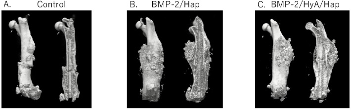
Representative 3D micro-computed tomography images of femurs 4 weeks after surgery. (A) Control, (B) Hap/BMP-2, and (C) IFGs-HyA/Hap/BMP-2-treated groups. IFGs-HyA: in situ-formed gels consisting of hyaluronan; Hap: hydroxyapatite; BMP-2: bone morphogenetic protein-2.

Bone volume and bone mineral content at sites of fracture 4 weeks after surgery. Analysis was conducted on micro-computed tomography images of femurs to examine new bone formation 4 weeks after surgery in the control (white bars), Hap/BMP-2 (gray bars), and IFGs-HyA/Hap/BMP2 (speckled bars) groups. (A) Bone volume, (B) bone mineral content. n = 10. ∗ P < 0.05.

Representative histological images of the fracture site 4 weeks after surgery. Sections through the femur were stained with hematoxylin and eosin to reveal new bone formation at the fracture site. (A) Control, (B) HA/BMP-2, and (C) IFGs-HyA/Hap/BMP2-treated groups. Areas enclosed by yellow dotted lines indicate newly formed bone. Scale bars indicate 500 μm. (D) Quantification of the area of new bone formed at fracture sites. Control (white bar), HA/BMP-2 (gray bar), and IFGs-HyA/Hap/BMP-2 (speckled bar) groups are shown. n = 10. ∗ P < 0.05.
A histological examination was also conducted four weeks after the fracture to assess bone union. Compared with the control group, mice treated with IFGs-HyA/Hap/BMP-2 showed larger calluses at the fracture site. The newly formed bone created a bridge around the perimeter of the fracture site (Fig. 4). In contrast, in the Hap/BMP-2 and control groups, we noted fibrous or cartilage callus tissue at the fracture site, with no indications of bone union (Table 1).
Bone union rates at graft sites 4 weeks after surgery
Bone union rates at graft sites 4 weeks after surgery
Micro-computed tomography images were analyzed for bone union rates 4 weeks following surgery in the control, and Hap/BMP-2- and IFGs-HyA/Hap/BMP2-treated groups.
Due to the osteoconductive nature of HA and its excellent biological affinity with bone tissue, HA has been widely used in medicine for many years. We previously showed that IFGs-HyA was capable of continuously releasing BMP-2 over 14 days in vitro. IFGs-HyA also had a significant effect on bone formation compared with Hap/BMP-2 composites, which rapidly released BMP-2 and showed limited efficacy for inducing bone formation [14]. Implantable or injectable HyA hydrogels have been shown to hold BMP-2 in vivo and promote bone formation during bone healing [16–22]. A previous study that implanted acrylated HyA hydrogel harboring BMP-2 and hydrazone-crosslinked Hap hydrogel harboring BMP-2 into rat calvarial defects to compare their ability to speed up bone repair showed that IFGs-HyA/Hap/BMP-2 composites have superior ability to induce bone formation and achieve bone union compared to Hap/BMP-2 [23]. The present study was designed to develop a material that is injectable and immediately cures and that can combine with IFGs-HyA and Hap. Using refractory fracture model mice, we showed that IFGs-HyA/Hap in conjunction with BMP-2 facilitated bone formation and bone union. Evidence suggests that IFGs-HyA, which has the combined advantage of being injectable and implantable, may improve the ability of bones to heal and reduce potential side effects related to diffusion from the HyA hydrogel into soft tissue. The combination of IFGs-HyA/Hap/BMP-2 may thus be an effective material to facilitate healing of refractory fractures in clinical settings.
However, as humans and rodents respond differently to BMP-2, our findings may not be translatable to the clinic [24,25]. Additional studies are required to validate our results in large animal models.
Conclusion
We investigated the effectiveness of HyA gel containing Hap harboring BMP-2 for facilitating bone formation and union in refractory fracture model mice. Injection of IFGs-HyA/Hap/BMP-2 into fracture sites significant improved BV, BMC, and bone union compared no treatment or injection of Hap/BMP-2. Therefore, IFGs-HyA/Hap/BMP-2 may be an effective treatment strategy for refractory fractures.
Footnotes
Acknowledgements
This investigation was supported in part by Grant-in-Aid for Scientific Research (B) No. 21H03059, Grant-in-Aid for Young Scientists (Start-up) No. 21K20978, and a Kitasato University Research Grant for Young Researchers.